. 2024 Nov 22:29:101361.
doi: 10.1016/j.mtbio.2024.101361. eCollection 2024 Dec.

An advanced chitosan based sponges dressing system with antioxidative, immunoregulation, angiogenesis and neurogenesis for promoting diabetic wound healing

Affiliations

An advanced chitosan based sponges dressing system with antioxidative, immunoregulation, angiogenesis and neurogenesis for promoting diabetic wound healing

Xianmou Fan et al. Mater Today Bio. .

Abstract

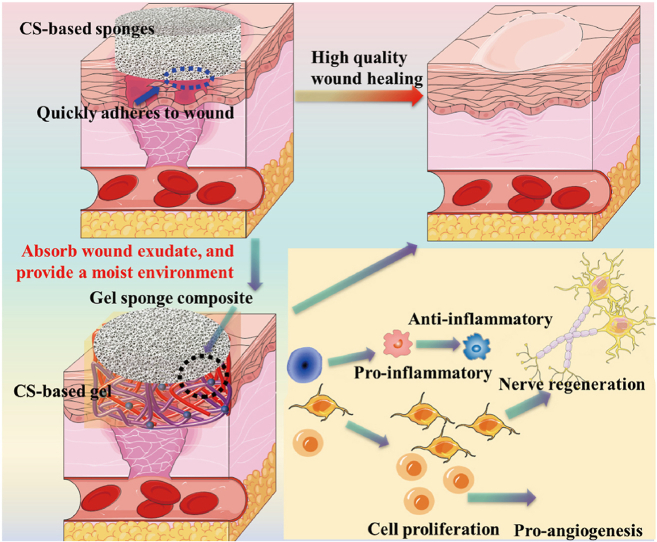
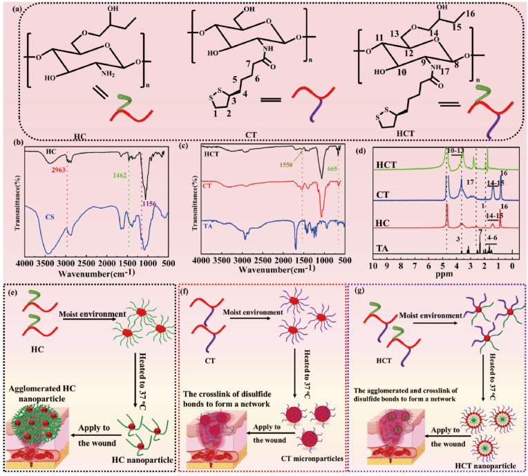
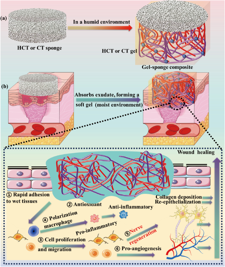
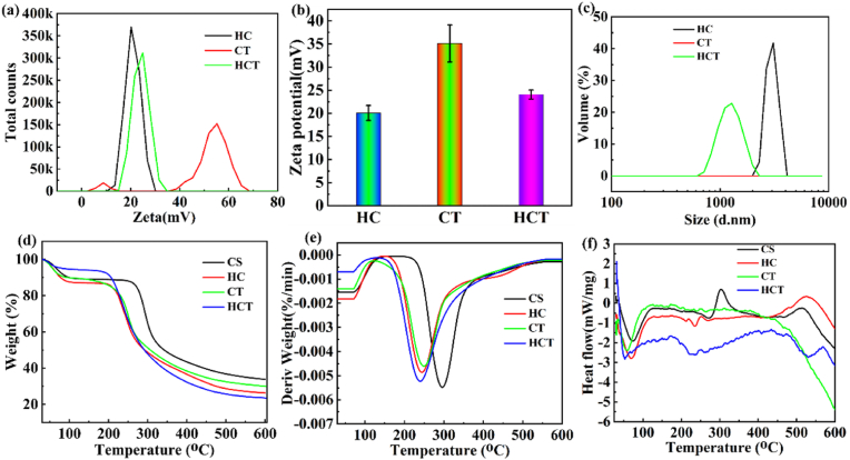
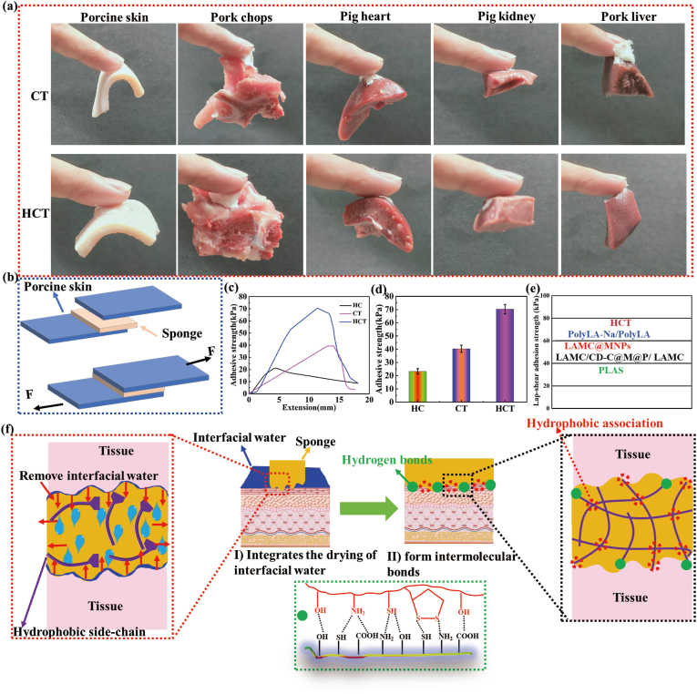
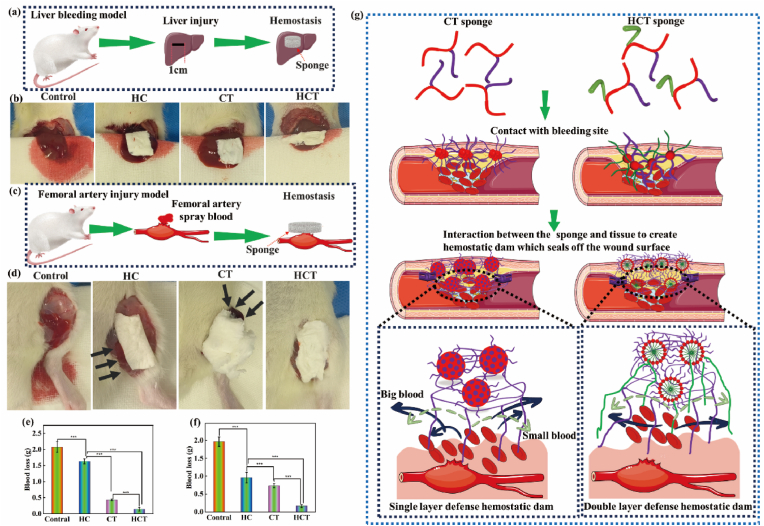
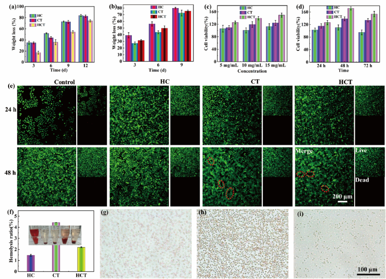
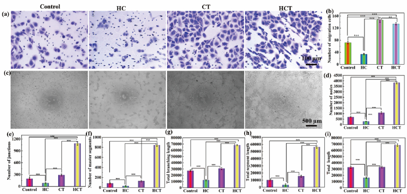
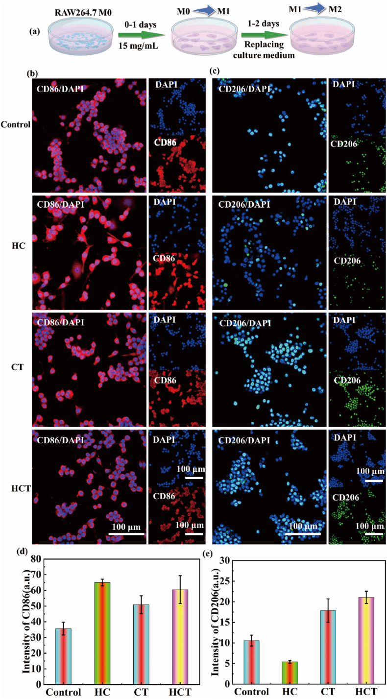
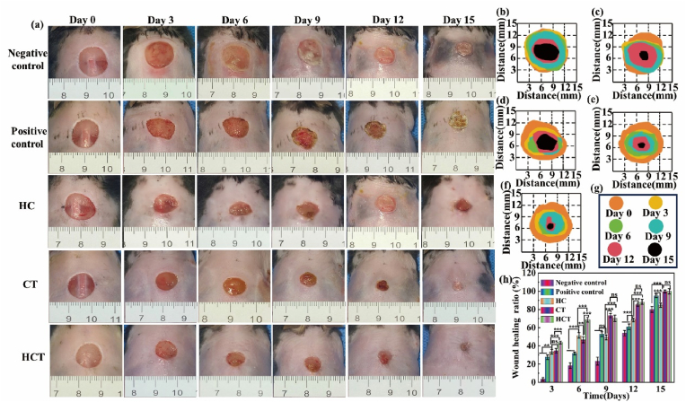
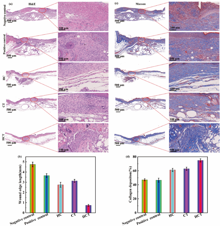
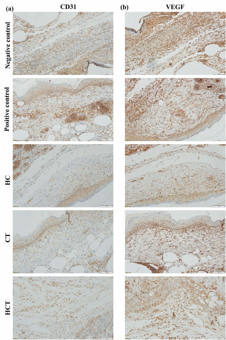
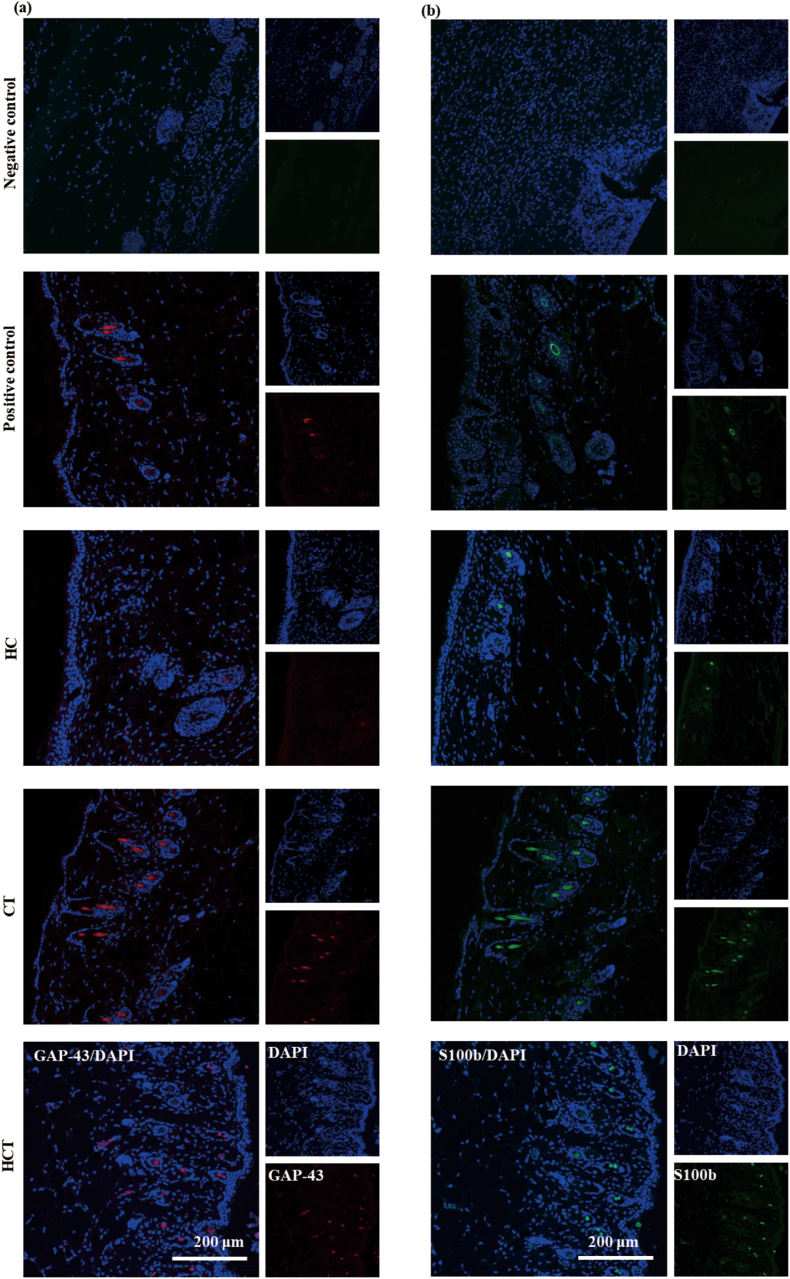
Promoting wound nerve regeneration and synchronously initiating angiogenesis are critical factors in the healing process of diabetic wounds. However, existing research on diabetic wounds mainly focuses on angiogenesis, bacterial infection and reactive oxygen species, often failing to coordinate neurogenesis and angiogenesis. To coordinate the symbiosis of nerves and blood vessels in the diabetic wounds, we successfully designed a multifunctional chitosan (CS)-based sponges by regulating the structure of CS specifically for diabetic wound healing. This sponge, which facilitates effective exudate transfer and modulates the wound microenvironment, was constructed using hydroxybutyl CS grafted with thioctic acid (TA), named as HCT sponge. When applied in a humid environment, the hydrophobic side chains of the HCT sponge interact with self-assembled hydrophobic domains, forming gel-sponge composite. Experimental results showed that the adhesion strength of the HCT sponge to wet porcine skin was 70.3 kPa. Additionally, the sponge exhibited favorable degradability, cytocompatibility and antioxidant properties. As it is shown in the experiments in vitro, sponge can not only promote cell proliferation, migration, and blood vessel formation, but also promote M2 macrophage polarization. Moreover, the rat liver and femoral artery injury model validated that the HCT sponge can effectively treat heavy bleeding from wounds efficacy through quickly sealing wounds and the formation of multiple hemostatic dams. In vivo studies indicated that the HCT sponge significantly accelerated the diabetic wound healing process compared to the recombinant bovine basic fibroblast growth factor gel, achieving a better recovery from the HCT sponge after 15 days. Pathological results show that the designed novel sponge holds considerable promise for treating diabetic wound, allowing regenerative neurogenesis and angiogenesis at the wound site, which provides a significant potential for further improving clinical applications.

Keywords: Angiogenesis; CS-based sponges; Diabetic wound healing; Nerve regeneration; Wet tissue adhesion.


Conflict of interest statement

The authors declare that no conflicts of financial interests or personal relationships have influenced the work reported in this paper.

Figures

Image 1
Graphical abstract
Fig. 1
Fig. 1
(a) Molecular formulas of HC, CT, and HCT. (b) FTIR spectra for CS and HC. (c) FTIR spectra of TA, CT and HCT. (d) 1HNMR spectra of TA, CT and HCT. (e–g) CS-based sponges were applied to diabetic wounds. From left to right are HC sponge (e), CT sponge (f), and HCT sponge (g).
Scheme 1
Scheme 1
Schematic illustration of CS-based sponges forms a gel-sponge composite that is easily applied to wounds in a diabetic mice model, along with the mechanism of the gel-sponge composite in diabetic wound treatment. (a) When CS-based sponges into contact with humid environment, it absorbs the liquid to form a gel-sponge composite. (b) The CS-based sponges promote diabetic wound healing though several mechanisms: ① Rapid adhesion to wet tissues: When the HCT or CT sponge contacts the wound, the hydrophobic segments of the sponge quickly absorb body fluids, inducing hydrophobic-hydrophilic interactions within the structure to form a gel-sponge composite. Meanwhile, the hydroxyl, carboxyl, amino, and sulfhydryl in gel-sponge composite interact with the tissue, improving the adhesion ability of wet tissue. ② Antioxidant properties: Both CT and HCT showed good antioxidant ability, accelerating diabetic wound healing by alleviating oxidative stress and protecting cells [20,21]. ③ Cell proliferation and migration: Owing to the antioxidant performance of TA, dressing containing TA can alleviate the oxidative damage, thereby preserving cell proliferation, migration efficiency [12]. ④ Polarization macrophage: Our previous studies have proved that PolyTA underwater tissue adhesive can regulate macrophage polarization [35]. ⑤ Nerve regeneration: It is well known that TA is one of the drugs of choice for the treatment of DPN [[11], [12], [13]]. The CT and HCT sponges contribute to improved nerve regeneration capacity of the wound through the TA part of their structure. ⑥ Pro-angiogenesis: CT and HCT promote angiogenesis by up-regulating angiogenic factors to improve HUVEC migration.
Fig. 2
Fig. 2
(a) and (b) Zeta potential of the HC, CT and HCT at water. (c) The particle size of HC, CT and HCT. (d) TGA and (e) DTG for HC, CT and HCT. (f) DSC thermograms for HC, CT and HCT.
Fig. 3
Fig. 3
Adhesive property of CS-based sponges. (a) The adhesiveness of CT and HCT sponges: porcine skin, pork chops, pig heart, pig kidney, and pork liver. (b) Schematic diagrams of the lap shear test of sponge-tissue adhesion. (c) Adhesive strength-displacement curves of the HC, CT, and HCT sponge. (d) Adhesive strength of the HC, CT, and HCT sponge. (e) Comparison of lap-shear adhesion strength among HCT sponge and various reported containing TA tissue adhesive materials. (f) Wet tissue adhesion mechanism of HCT sponge.
Fig. 4
Fig. 4
In vivo hemostatic performance of CS-based sponges. (a) Schematic illustration of rat liver bleeding model and sponge application. (b) Hemostasis of the CS-based sponges in SD rat liver bleeding model (step 1: liver was exposed under deep anesthesia with isoflurane; step 2: a linear wound of liver was created to mimic accidental hemorrhage; step 3: After bleeding for 2 s, the bleeding wound was treated using the sponge; step 4: hemostasis was achieved and the rats were sacrificed under deep isoflurane anesthesia). (c) Schematic representation of hemostasis in a rat femoral artery injury model. (d) Picture display of the rat femoral artery injury model was conducted and used to stop bleeding using CS-based sponges. The procedures of hemostasis in the femoral artery were basically the same as the hemostasis in the liver. Blood loss of different sponge in rat liver model (e) and in a rat femoral artery injury model (f). (g) The schematic diagram showing the hemostatic mechanism of CT and HCT sponges.
Fig. 5
Fig. 5
Biosafety evaluation of the CS-based sponges in vitro. Degradation properties of the CS-based sponges (a) in vitro and (b) in vivo. (c) Cell viability of HUVECs treated with different concentrations of the sponge for 24 h. (d)The cell viability of HUVECs after incubating with the sponge for 24, 48 and 72 h. (e) HUVECs stained with live (green)/dead (red) following 24 h and 48 h of varied treatments. (f) Hemolysis ratios of all sponge. The bar graph shows the hemolysis ratio and imaging depicting the hemolysis results of supernatant after centrifugation. The inset picture, from left to right, shows blood treated with deionized water, PBS, HC, CT, and HCT sponge. Red blood cell morphologies: (g) PBS, (h) HCT, and (i) deionized water. (For interpretation of the references to color in this figure legend, the reader is referred to the Web version of this article.)
Fig. 6
Fig. 6
Transwell migration and tube formation of HUVECs under various conditions. (a) Their respective images of HUVECs migration. (b) Statistical evaluation of cell migration numbers. (c) Typical image of tube formation of HUVECs incubated with different sponges for 4 h. Quantitative analysis of the number of nodes (d), number of junctions (e), number of master segments(f), total branching length (g), total segment length (h), and total length (i) under CS-based sponges from (c).
Fig. 7
Fig. 7
Effects of the CS-based sponges on macrophage polarization in vitro. (a) Flow chart of inducing macrophage phenotypic polarization test by RAW 264.7 cells in vitro. Representative immunofluorescence images and quantification of CD86 (Red fluorescence) (b and d) or CD206 (Green fluorescence) (c and e), and nuclei (blue) on RAW 264.7 cell treated with different treatments. (For interpretation of the references to color in this figure legend, the reader is referred to the Web version of this article.)
Fig. 8
Fig. 8
Diabetic wounds healing in vivo. (a) Representative digital photos of wounds treated with ordinary medical gauze (Negative control), recombinant bovine basic fibroblast growth factor for external use (Positive control), HC, CT, and HCT sponge. (b–g) Wound area over time in the five treatment groups: the sequence is Negative control, Positive control, HC, CT, and HCT sponge. (h) Quantification of the closed wound area percentage in the five treatment groups.
Fig. 9
Fig. 9
Histological analysis of healing wounds on day 15. Representative images of (a) H&E staining and (c) Masson staining of different groups. Histograms of quantification of average wound edge length (b) and collagen deposition (d) in each group.
Fig. 10
Fig. 10
Representative pictures of immunohistochemical staining of healing wounds on day 15. (a) CD 31 and (b) VEGF expression was determined by immunohistochemical staining in wounds on day 15.
Fig. 11
Fig. 11
Immunofluorescence staining of healing wounds on day 15. Representative images of immunofluorescence staining of axons (GAP-43), RSC 96 (S100b), and cell nuclei (DAPI) were stained red, green, and blue colors at the wound site on day 15 for each group. (For interpretation of the references to color in this figure legend, the reader is referred to the Web version of this article.)

Similar articles

Cited by

References

    1. Jaul E. Non-healing wounds: the geriatric approach. Arch. Gerontol. Geriatr. 2009;49(2):224–226. - PubMed
    1. Xiong Y., Lin Z., Bu P., Yu T., Endo Y., Zhou W., Sun Y., Cao F., Dai G., Hu Y., Lu L., Chen L., Cheng P., Zha K., Shahbazi M.A., Feng Q., Mi B., Liu G. A whole-course-repair system based on neurogenesis-angiogenesis crosstalk and macrophage reprogramming promotes diabetic wound healing. Adv. Mater. 2023;35(19) - PubMed
    1. Butenko S., Nagalla R.R., Guerrero Juarez C.F., Palomba F., David L.M., Nguyen R.Q., Gay D., Almet A.A., Digman M.A., Nie Q., Scumpia P.O., Plikus M.V., Liu W.F. Hydrogel crosslinking modulates macrophages, fibroblasts, and their communication, during wound healing. Nat. Commun. 2024;15(1):6820. - PMC - PubMed
    1. Han X., Saengow C., Ju L., Ren W., Ewoldt R.H., Irudayaraj J. Exosome-coated oxygen nanobubble-laden hydrogel augments intracellular delivery of exosomes for enhanced wound healing. Nat. Commun. 2024;15(1):3435. - PMC - PubMed
    1. Fan X., Huang J., Zhang W., Su Z., Li J., Wu Z., Zhang P. A multifunctional, tough, stretchable, and transparent curcumin hydrogel with potent antimicrobial, antioxidative, anti-inflammatory, and angiogenesis capabilities for diabetic wound healing. ACS Appl. Mater. Interfaces. 2024;16(8):9749–9767. - PubMed

LinkOut - more resources