. 2024 Nov 15;14(11):5286-5303.
doi: 10.62347/BVFO4627. eCollection 2024.

Bioinformatics- and quantitative proteomics-based identification of gastric adenocarcinoma-related proteins and analysis

Affiliations

Bioinformatics- and quantitative proteomics-based identification of gastric adenocarcinoma-related proteins and analysis

Wenbo Liu et al. Am J Cancer Res. .

Abstract

Background: The emergence of immune resistance and a lack of effective therapeutic targets have become significant challenges in immunotherapy, highlighting the urgent need for new molecular markers and treatment targets. Moreover, the significance and mechanisms of PGRN (Progranulin) in gastric cancer remain ambiguous.

Objective: To identify differentially expressed proteins in gastric cancer and elucidate the function and mechanism of PGRN.

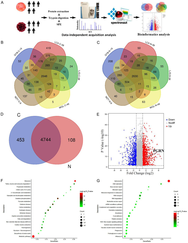
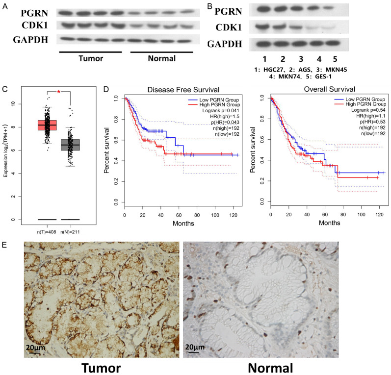
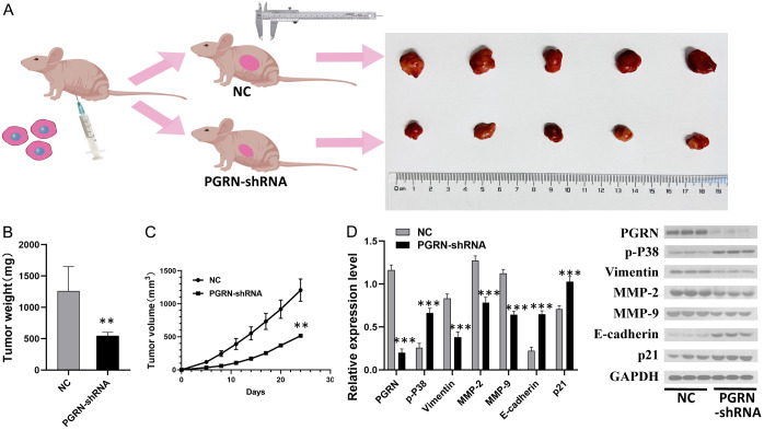
Methods: The data-independent acquisition proteomics was used to identify the differentially expressed proteins in gastric adenocarcinoma and the corresponding paraneoplastic tissues, providing a comprehensive dataset of gastric cancer-related proteins. The function and mechanism of PGRN in gastric cancer were further explored using a series of experiments, including RT-qPCR (Real Time-Quantitative Polymerase Chain Reaction), cell transfection, cell viability assays, cell scratch, immunohistochemistry and Transwell assays, Western blot, and a mouse tumor-bearing model. These investigations were combined with bioinformatics analyses to examine the relationship between PGRN expression and clinical-pathological characteristics, confirming its high expression of PGRN in gastric cancer tissues.

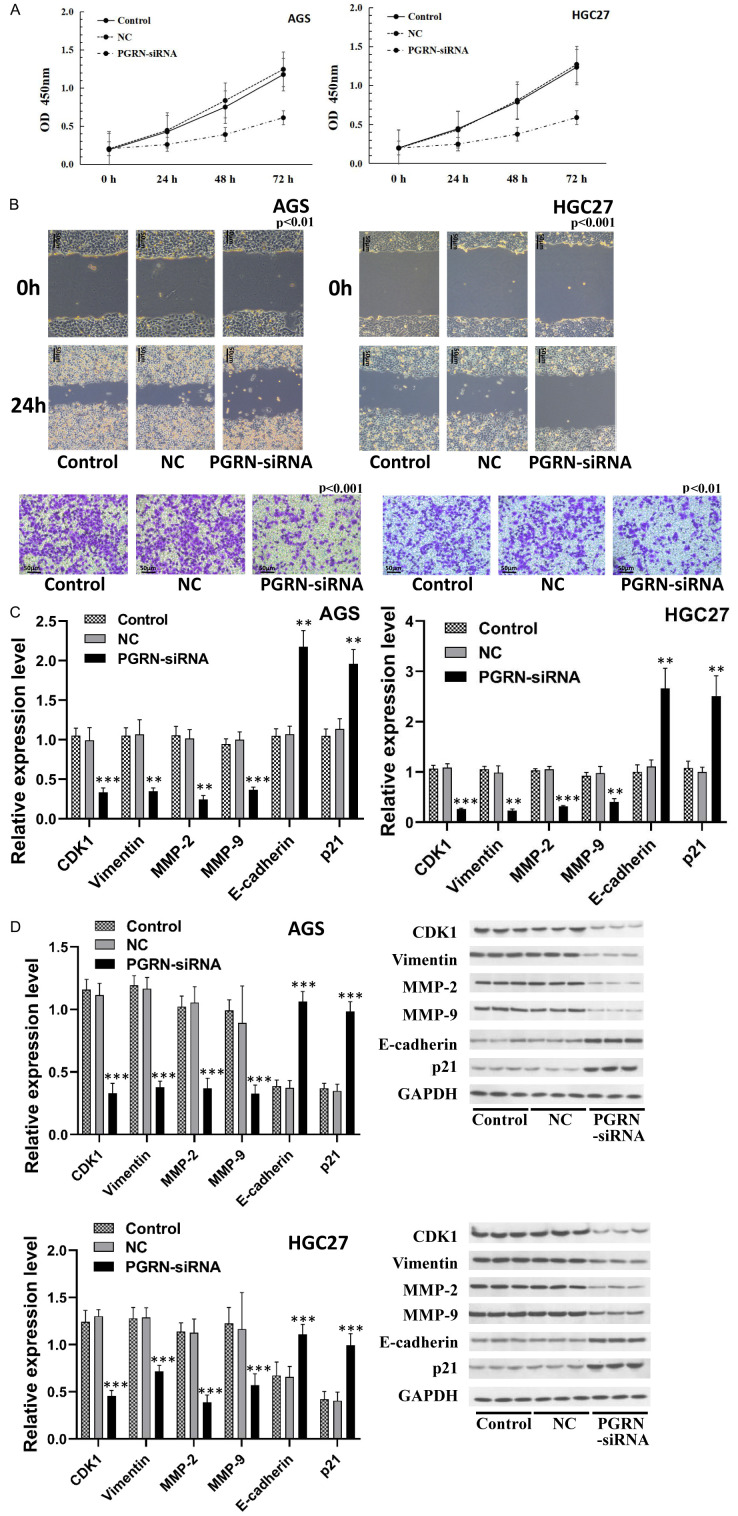
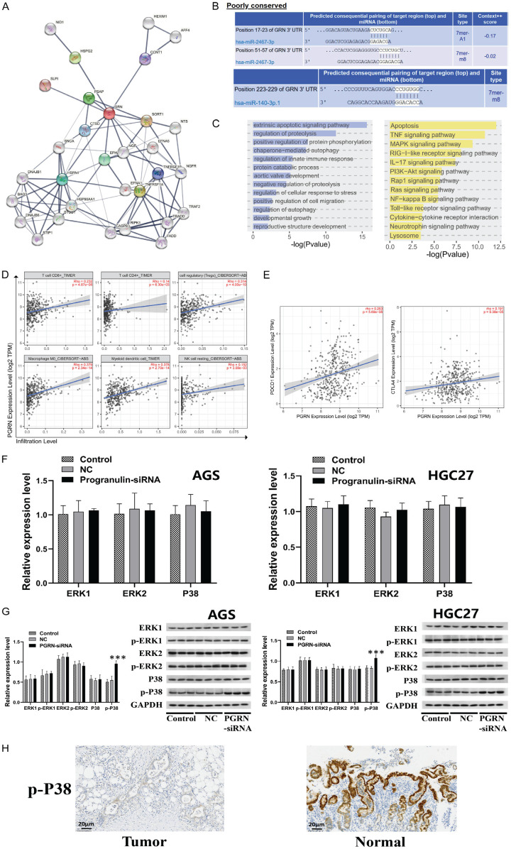
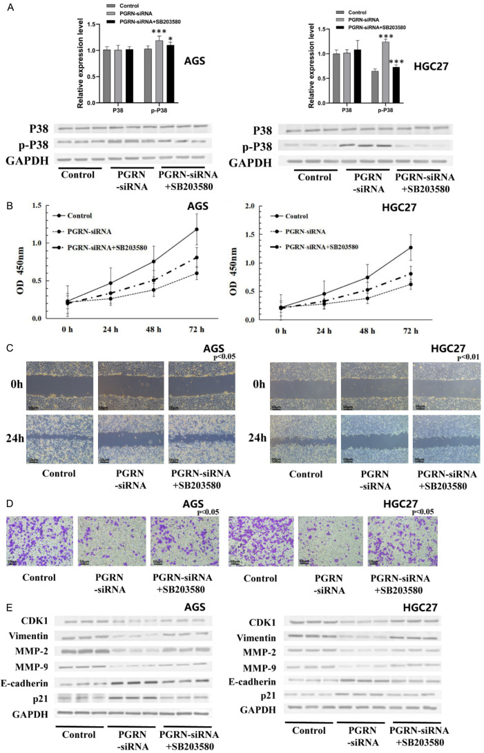
Results: We identified a large number of differentially expressed proteins between gastric cancer and adjacent tissues and conducted an initial functional analysis. Further studies on PGRN showed that it was associated with gastric cancer prognosis and lymph node metastasis. The inhibition of PGRN expression led to reduced cell viability, migration, and invasion, with corresponding changes in related genes and proteins. In a mouse tumor-bearing model, the tumor growth of the subcutaneously transplanted tumors in nude mice was reduced after the inhibition of PGRN expression. An in-depth functional analysis of PGRN was performed using bioinformatics to predict protein interactions, miRNA regulation, and relationships with multiple immune cell types. Enrichment analysis indicated that PGRN is involved in multiple signaling pathways, with the MAPK (Mitogen-Activated Protein Kinase) pathway selected for validation. In AGS and HGC27 cells, PGRN inhibition led to increased expression of phosphorylated p38 (p-p38) in the MAPK pathway, suggesting that PGRN may promote gastric cancer development by regulating p-p38.

Conclusions: This study identified significant differences in protein expression between gastric adenocarcinoma and adjacent tissues, with PGRN emerging as a key protein influencing gastric cancer proliferation, migration, and invasion. These findings suggest that PGRN could serve as a potential therapeutic target for gastric cancer.

Keywords: Gastric cancer; PGRN; bioinformatics; functional analysis; quantitative proteomics.


Conflict of interest statement

None.

Figures

Figure 1
Figure 1
Identification and quantification of proteins and preliminary analysis. A. Quantitative proteomic analysis of five paraneoplastic and five tumor tissue samples using the DIA (Data-independent acquisition) quantitative proteomics method. The figure illustrates the experimental workflow. B. In the five tumor tissue samples, 3,697, 4,521, 3,674, 3,270, and 4,514 proteins were identified, with 2,648 proteins at the intersection of these five datasets. C. In the five paraneoplastic tissue samples, 4,517, 3,090, 3,711, 4,133, and 3,816 proteins were identified, with 2,630 proteins at the intersection of these five datasets. D. 5,198 proteins were identified in tumor tissues, and 4,853 proteins were identified in paraneoplastic tissues, with 4,744 proteins intersecting between the two groups. E. Differentially expressed proteins were defined as proteins with a fold change of expression ≥ 2 or ≤ 0.5. The volcano plot includes 693 upregulated proteins and 1,006 downregulated proteins that were identified. F. The metabolic pathway was found to have the smallest p-value among the upregulated proteins, and other proteins involved in various signaling pathways are also depicted. G. The spliceosome pathway was found to have the smallest p-value among the downregulated proteins, and other proteins involved in various signaling pathways are also shown.
Figure 2
Figure 2
Relationship between PGRN (Progranulin) expression and prognostic, clinical, and pathological features. A. Western blotting results indicated that the expression levels of PGRN and CDK1 (Cyclin Dependent Kinase 1) were higher in gastric cancer tissues than in paraneoplastic tissues (n = 30). B. PGRN and CDK1 expression was lower in GES-1 cells than in any of the gastric cancer cell lines, with the cell lines in decreasing order of PGRN expression being HGC27, AGS, MKN45, MKN74, and GES-1 (n = 6). C. Analysis of differentially expressed proteins indicated that PGRN was highly expressed in gastric cancer tissues (P < 0.05). D. Survival analysis showed that PGRN expression correlated with the disease-free survival of patients (P < 0.05). E. Immunohistochemistry showed that PGRN expression was significantly higher in gastric cancer tissues than in paraneoplastic tissues (400×) (n = 30).
Figure 3
Figure 3
Relationship between PGRN expression and cell viability, migration, and invasion. (A) CCK-8 results showed that cell viability was significantly reduced after inhibition of PGRN expression in AGS and HGC27 cells (P < 0.05). (B) Scratch and Transwell invasion assays showed that the invasion and migration of cells were significantly reduced after inhibition of PGRN expression in AGS and HGC27 cells (200×) (P < 0.01). (C) RT-qPCR and (D) western blotting showed that genes and proteins associated with proliferation, invasion, and migration were significantly altered in AGS and HGC27 cells after PGRN inhibition (P < 0.01). All experiments were repeated three times.
Figure 4
Figure 4
Functional analysis and mechanism of action of PGRN. A. PPI network of PGRN (three-step neighbor method). B. Prediction of miRNA regulation of PGRN. C. Enrichment analysis of genes in the PPI network by GO and KEGG analyses. D. Analysis of correlation between PGRN expression and immune cell abundance. E. Analysis of the correlation between PGRN expression and immune checkpoint gene expression. F, G. RT-qPCR and WB showed that the expression of ERK1/2, p-ERK1/2, or P38-MAPK in the MAPK signaling pathway was not significantly changed after inhibiting the expression of PGRN in AGS and HGC27 cells; however, the expression of p-P38 was significantly increased (P < 0.001). H. Immunohistochemistry detection of p-P38 expression in tissues showed that p-P38 expression was significantly lower in gastric cancer tissues than in paraneoplastic tissues (200×). All experiments were repeated three times.
Figure 5
Figure 5
Effect of PGRN expression on subcutaneously transplanted tumor growth in nude mice. A. The transplanted tumors in the PGRN-shRNA transfected group was smaller than those in the empty vector-transfected group. B, C. The average weight of transplanted tumors in the PGRN-shRNA transfected group was significantly lower than that in the empty vector-transfected group, and the growth curve showed a delay (P < 0.01). D. Western blotting analysis of related proteins in both groups showed that the protein expression of MMP-2, MMP-9, and vimentin decreased, whereas P21 and E-cadherin expression increased in the PGRN-shRNA transfected group compared with those in the empty vector-transfected group (all P < 0.001) (n = 3). These results confirm that inhibiting PGRN expression can suppress tumor formation in gastric cancer cells in vivo.
Figure 6
Figure 6
SB203580 alleviated the effects of inhibiting PGRN expression on cell activity, migration, invasion, and related gene and protein expression by inhibiting p-P38 in MAPK pathway. A. The expression of p-P38 in the PGRN-siRNA group was higher than that in the control group, and the expression of p-P38 in the SB203580 combined with PGRN-siRNA group was lower than that in the PGRN-siRNA group. B-D. Compared with the PGRN-siRNA group, the SB203580 combined with PGRN-siRNA group showed increased cell activity, migration, and invasion abilities (200×). E. Compared with the PGRN-siRNA group, the expression of genes and proteins related to proliferation, invasion, and migration was altered after treatment with SB203580 combined with PGRN-siRNA in AGS and HGC27 cells (P < 0.05). All experiments were repeated three times.

Similar articles

References

    1. Sung H, Ferlay J, Siegel RL, Laversanne M, Soerjomataram I, Jemal A, Bray F. Global cancer statistics 2020: GLOBOCAN estimates of incidence and mortality worldwide for 36 cancers in 185 countries. CA Cancer J Clin. 2021;71:209–249. - PubMed
    1. Zheng R, Zhang S, Zeng H, Wang S, Sun K, Chen R, Li L, Wei W, He J. Cancer incidence and mortality in China, 2016. J Natl Cancer Cent. 2022;2:1–9. - PMC - PubMed
    1. Li Y, Feng A, Zheng S, Chen C, Lyu J. Recent estimates and predictions of 5-year survival in patients with gastric cancer: a model-based period analysis. Cancer Control. 2022;29:10732748221099227. - PMC - PubMed
    1. Dong L, Lu D, Chen R, Lin Y, Zhu H, Zhang Z, Cai S, Cui P, Song G, Rao D, Yi X, Wu Y, Song N, Liu F, Zou Y, Zhang S, Zhang X, Wang X, Qiu S, Zhou J, Wang S, Zhang X, Shi Y, Figeys D, Ding L, Wang P, Zhang B, Rodriguez H, Gao Q, Gao D, Zhou H, Fan J. Proteogenomic characterization identifies clinically relevant subgroups of intrahepatic cholangiocarcinoma. Cancer Cell. 2022;40:70–87. e15. - PubMed
    1. Puttamallesh VN, Deb B, Gondkar K, Jain A, Nair B, Pandey A, Chatterjee A, Gowda H, Kumar P. Quantitative proteomics of urinary bladder cancer cell lines identify UAP1 as a potential therapeutic target. Genes (Basel) 2020;11:763. - PMC - PubMed

LinkOut - more resources