Review
. 2024 Nov;85(6):1126-1140.
doi: 10.3348/jksr.2024.0037. Epub 2024 Nov 26.

MR Imaging for Ectopic Pregnancy

Review

MR Imaging for Ectopic Pregnancy

Jung Joo Hong et al. J Korean Soc Radiol. 2024 Nov.

Abstract

Ectopic pregnancy (EP) is diagnosed based on laboratory values and ultrasonography (US) findings. Evaluation for suspected EP should begin with a quantitative measurement of the serum β-human chorionic gonadotropin levels and transvaginal US. MR imaging is not preferentially performed in the evaluation of EP; however, if the findings of transvaginal US are uncertain, MR imaging can be used, as it has the advantages of superior soft-tissue contrast resolution and a wide scanning range. Identifying the exact location of implantation transfer using MR imaging can help in the diagnosis and establishment of treatment strategies for ectopic pregnancies, including laparoscopy. In particular, as the incidence of heterotopic pregnancy has increased with the recent increase in use of assisted reproductive technology, the scope of application of MR imaging is expected to expand further. This pictorial essay describes the various manifestations of EP and related conditions on MR imaging and US. Familiarity with the clinical setting and the US and MR imaging features of EP and associated conditions can lead to a more accurate diagnosis and treatment.

자궁 외 임신의 진단은 임상검사 및 초음파 소견을 바탕으로 이루어진다. 자궁 외 임신이 의심되는 상황에서 가장 먼저 시행하게 되는 검사는 혈중 사람 융모성 성선 자극 호르몬의 정량적 평가와 경질 초음파이다. 자궁 외 임신의 평가에 우선적으로 자기공명영상을 시행하지는 않지만, 경질 초음파의 소견이 불확실한 경우, 연조직 대비 해상도가 뛰어나고 스캔 범위가 넓다는 장점을 지니는 자기공명영상을 이용해 볼 수 있다. 자기공명영상을 통해 정확한 착상 위치를 파악한다면 진단과 더불어 복강경을 비롯한 자궁 외 임신의 치료 전략을 세우는데 도움이 될 수 있을 것이다. 특히, 최근 보조생식술의 시행이 증가하면서 이소성 임신의 빈도가 함께 증가함에 따라, 자기공명영상의 적용 범위는 더욱 넓어질 것으로 기대한다. 본 임상화보에서는 자궁 외 임신 및 관련 상태의 초음파 및 자기공명영상 소견에 대해 살펴보고자 한다. 임상적인 상황과 더불어 초음파 및 자기공명영상 소견에 익숙해진다면, 자궁 외 임신 및 관련 상태의 더욱 정확한 진단 및 치료가 가능할 것으로 기대한다.

Keywords: Ectopic Pregnancy; Heterotopic Pregnancy; Magnetic Resonance Imaging; Ultrasonography.


Conflict of interest statement

Conflicts of Interest: The authors have no potential conflicts of interest to disclose.

Figures

Fig. 1
Fig. 1. US feature of ectopic pregnancy: decidual cyst.
The sagittal transvaginal view of the uterus shows a small fluid collection at the junction of the endometrium and myometrium (arrow).
Fig. 2
Fig. 2. US feature of ectopic pregnancy: pseudosac.
A sagittal transvaginal scan of the uterus shows a small, centrally located fluid collection with internal blood and debris in the endometrial cavity.
Fig. 3
Fig. 3. US feature of ectopic pregnancy: live extrauterine embryo.
A, B. Transvaginal view with m-mode shows embryo (calipers) with normal cardiac activity (arrows).
Fig. 4
Fig. 4. US feature of ectopic pregnancy: tubal ring sign.
A, B. Transvaginal view of the uterus and right ovary shows an extrauterine mass (calipers) with thick echogenic wall. Note that the mass moves separately from the ovary.
Fig. 5
Fig. 5. US feature of EP: the “ring-of-fire” sign.
Color Doppler US shows increased blood flow around an EP. EP = ectopic pregnancy
Fig. 6
Fig. 6. US feature of ectopic pregnancy: tubal mass.
Transvaginal scan shows a nonspecific mass with heterogeneous echogenicity.
Fig. 7
Fig. 7. US feature of ectopic pregnancy: hemoperitoneum.
Transabdominal view of the pelvis shows large amount of free fluid with mixed echogenicity.
Fig. 8
Fig. 8. MR imaging findings in a 40-year-old woman with ectopic pregnancy.
A. Transverse T2-weighted imaging shows a thick-walled cyst (arrow), indicating extrauterine gestational sac and a “three-ring sign.” B, C. Diffusion-weighted imaging (B) and apparent diffusion coefficient map (C) show a “ring-of-restriction” sign (arrows).
Fig. 9
Fig. 9. MR imaging findings in a 42-year-old woman with chronic pelvic pain for 1 year and low level of β-human chorionic gonadotropin elevation.
A-C. Transverse T2WI (A), transverse contrast-enhanced-T1WI (B), and coronal T2WI (C) show a heterogeneous mass in the pelvis. The combined left tubal dilatation and enhancement are shown. A few chorionic villi in a hemorrhagic background at the left adnexa are consistent with ectopic gestation. WI = weighted imaging
Fig. 10
Fig. 10. MR imaging findings in a 32-year-old woman with sudden abdominal pain due to heterotopic pregnancy. She had undergone in vitro fertilization and embryo transfer and had a twin pregnancy (gestational age: 7 weeks).
A. Transvaginal sonogram shows normal IUP (calipers). B. Transverse T2WI shows IUP (P) and small hemoperitoneum (F). C, D. Transverse (C) and sagittal (D) T2WI show right-sided ectopic pregnancy (arrows) and small hemoperitoneum (F). IUP = intrauterine pregnancy, WI = weighted imaging
Fig. 11
Fig. 11. MR imaging findings in a 27-year-old woman with right lower quadrant pain for 1 day.
A. Transvaginal US demonstrates an intrauterine GS. B-D. Transverse and coronal T2-weighted imaging show an endometrial GS (P) and another GS (arrows) in the right interstitium of the uterus, suggesting interstitial and heterotopic pregnancy. A large amount of hemoperitoneum (*) at the right paracolic gutter is evident. MR imaging can detect EP and intrauterine pregnancy. E. Laparoscopy shows an EP (arrow) in the right interstitium of the uterus with active bleeding. Hemoperitoneum in the pelvis to the upper liver dome is also evident. EP = ectopic pregnancy, GS = gestational sac
Fig. 12
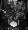
Fig. 12. MR imaging finding in a 28-year-old woman with abdominal EP.
Coronal T2-weighted imaging shows a well-defined cystic lesion with a thick peripheral wall (arrow) in the lateral aspect of the ascending colon, suggested to be the focus of the EP. Adapted from Kim et al. Abdom Radiol (NY) 2022;47:2254-2276, with permission of Springer Nature (20). EP = ectopic pregnancy

References

    1. Lin EP, Bhatt S, Dogra VS. Diagnostic clues to ectopic pregnancy. Radiographics. 2008;28:1661–1671. - PubMed
    1. Refaat B, Dalton E, Ledger WL. Ectopic pregnancy secondary to in vitro fertilisation-embryo transfer: pathogenic mechanisms and management strategies. Reprod Biol Endocrinol. 2015;13:30. - PMC - PubMed
    1. Levine D. Ectopic pregnancy. Radiology. 2007;245:385–397. - PubMed
    1. Lipscomb GH, Stovall TG, Ling FW. Nonsurgical treatment of ectopic pregnancy. N Engl J Med. 2000;343:1325–1329. - PubMed
    1. Eyvazzadeh AD, Levine D. Imaging of pelvic pain in the first trimester of pregnancy. Radiol Clin North Am. 2006;44:863–877. - PubMed

LinkOut - more resources