. 2025 Jan 31;207(1):e0040824.
doi: 10.1128/jb.00408-24. Epub 2024 Dec 11.

The histidine kinase NahK regulates denitrification and nitric oxide accumulation through RsmA in Pseudomonas aeruginosa

Affiliations

The histidine kinase NahK regulates denitrification and nitric oxide accumulation through RsmA in Pseudomonas aeruginosa

Danielle Guercio et al. J Bacteriol. .

Abstract

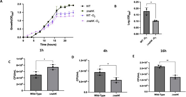
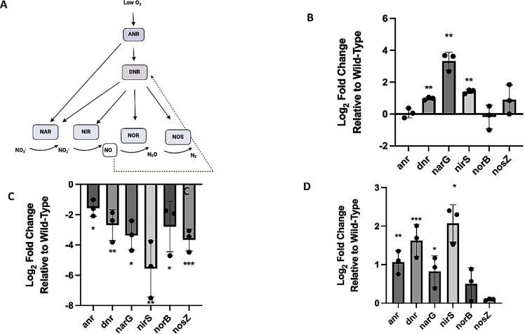
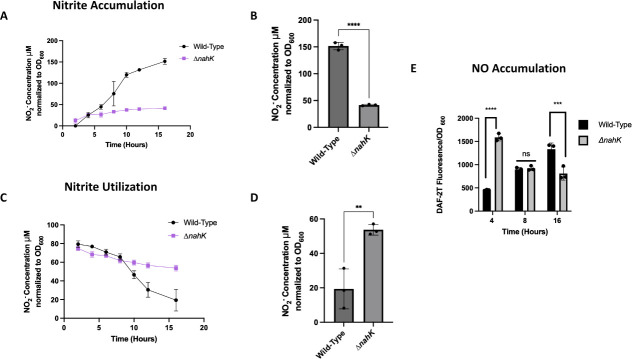
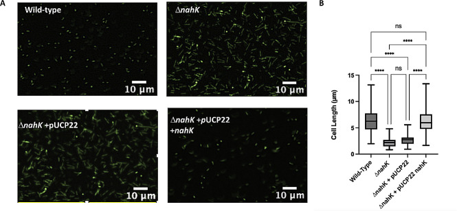
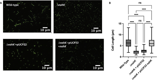
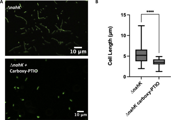
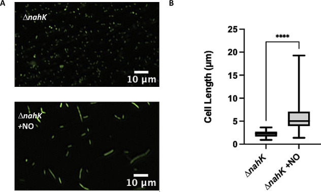
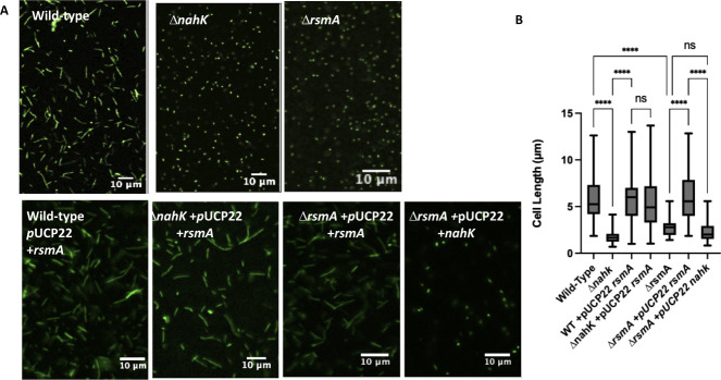
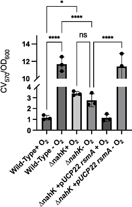
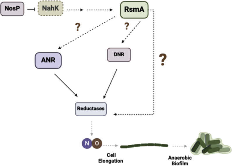
Pseudomonas aeruginosa have a versatile metabolism; they can adapt to many stressors, including limited oxygen and nutrient availability. This versatility is especially important within a biofilm where multiple microenvironments are present. As a facultative anaerobe, P. aeruginosa can survive under anaerobic conditions utilizing denitrification. This process produces nitric oxide (NO) which has been shown to result in cell elongation. However, the molecular mechanism underlying this phenotype is poorly understood. Our laboratory has previously shown that NosP is a NO-sensitive hemoprotein that works with the histidine kinase NahK to regulate biofilm formation in P. aeruginosa. In this study, we identify NahK as a novel regulator of denitrification under anaerobic conditions. Under anaerobic conditions, deletion of nahK leads to a reduction of growth coupled with reduced transcriptional expression and activity of the denitrification reductases. Furthermore, during stationary phase under anaerobic conditions, ΔnahK does not exhibit cell elongation, which is characteristic of P. aeruginosa. We determine the loss of cell elongation is due to changes in NO accumulation in ΔnahK. We further provide evidence that NahK may regulate denitrification through modification of RsmA levels.

Importance: Pseudomonas aeruginosa is an opportunistic multi-drug resistance pathogen that is associated with hospital-acquired infections. P. aeruginosa is highly virulent, in part due to its versatile metabolism and ability to form biofilms. Therefore, better understanding of the molecular mechanisms that regulate these processes should lead to new therapeutics to treat P. aeruginosa infections. The histidine kinase NahK has been previously shown to be involved in both nitric oxide (NO) signaling and quorum sensing through RsmA. The data presented here demonstrate that NahK is responsive to NO produced during denitrification to regulate cell morphology. Understanding the role of NahK in metabolism under anaerobic conditions has larger implications in determining its role in a heterogeneous metabolic environment such as a biofilm.

Keywords: NahK; NosP; RsmA; biofilm; cell elongation; denitrification; nitric oxide; quorum sensing.


Conflict of interest statement

The authors declare no conflict of interest.

Figures

Fig 1
Fig 1
Anaerobic growth is altered in ΔnahK. (A, B) Cultures with a starting OD of 0.005 were grown in Luria-Bertani (LB) media in the presence of oxygen, or in the absence of oxygen but in media supplemented with 25 mM nitrate (NaNO3). OD readings were measured as a function of time. (A) The average OD readings, ±1 standard deviation, from three independent biological replicates, are plotted as a function of time. (B) Log (OD600) at the 24-h time point was quantified in the anaerobic cultures. P-values were calculated using unpaired two-tailed t-test comparing ΔnahK log (OD600) to wild-type log (OD600); *P ≤ 0.05. (C–E) CFU values were calculated after 1 h (C), 4 h (D), or 16 h (E) of anaerobic growth (from the same culture) in LB media supplemented with 25 mM NaNO3. The plotted values represent the average CFUs, ± 1 standard deviation, from three independent biological replicates. P-values were calculated using unpaired, two-tailed t-test comparing ΔnahK CFU to wild-type CFU; *P ≤ 0.05, **P ≤ 0.01.
Fig 2
Fig 2
Transcription of the denitrification machinery is mis-regulated in ΔnahK. (A) Schematic of denitrification in P. aeruginosa. (B–D) Log2 of transcript levels in ΔnahK, relative to wild-type, as measured by qPCR, for the four denitrification reductases and two transcriptional regulators are plotted. Gyrase A (gyrA) was used as a housekeeping gene; gyrA Ct values were similar in aerobic and anaerobic growth in these experiments (Table S4). Cultures were grown in Luria-Bertani media in the presence of oxygen, or in the absence of oxygen but in media supplemented with 25 mM NaNO3. RNA was extracted at several time points and growth conditions: (B) after 4 h anaerobic growth (exponential stage); (C) after 16 h anaerobic growth (early stationary phase); (D) and at an OD of 1.0 during aerobic growth. Bars above and below the threshold represent up- and down-regulation in ΔnahK, relative to wild-type, respectively. The values represent the average ± 1 standard deviation of three independent biological replicates (each biological replicate consists of two technical replicates). P-values were calculated using unpaired, two-tailed t-tests comparing ΔnahK ΔCt values to wild-type ΔCt values; n = 3; *P ≤ 0.05, **P ≤ 0.01, ***P ≤ 0.005.
Fig 3
Fig 3
Denitrification activity is mis-regulated in ΔnahK. (A, B) Nitrite production by NAR during anaerobic denitrifying conditions as a function of time. (A) Cultures with a starting OD of 0.005 were grown in Luria-Bertani (LB) media supplemented with 25 mM NaNO3. At each time point, the absorbance from the Griess reagent was normalized to the OD600 and that value was used to calculate NO2- concentration using a NO2- calibration curve. The average nitrite concentration, ± 1 standard deviation, from three independent biological replicates (each biological replicate consists of three technical replicates) is plotted as a function of time. (B) The average nitrite concentration after 16 h of anaerobic growth (stationary phase), ± 1 standard deviation, is plotted. P-values were calculated using unpaired, two-tailed t-test comparing ΔnahK to wild-type; ****P ≤ 0.001. Initially, ΔnahK produces a little more nitrite than wild-type, but over time, nitrite production stagnates in the mutant while steadily increasing in the wild-type. (C, D) Nitrite utilization by NIR during anaerobic denitrifying conditions as a function of time. (C) Cultures with a starting OD of 0.005 were grown in LB media supplemented with 25 mM NaNO2. At each time point, the absorbance from the Griess reagent was normalized to the OD600 and that value was used to calculate the amount of NO2- remaining in the culture using a NO2- calibration curve. The average nitrite concentration, ± 1 standard deviation, from three independent biological replicates (each biological replicate consists of three technical replicates) is plotted as a function of time. (D) The average nitrite concentration after 16 h of anaerobic growth (stationary phase), ± 1 standard deviation, is plotted. P-values were calculated using unpaired, two-tailed t-test comparing ΔnahK to wild-type; **P ≤ 0.01. Initially, ΔnahK utilizes more nitrite than wild-type, but over time, nitrite utilization stagnates in the mutant while steadily continuing in the wild-type. (E) NO produced by NIR during anaerobic denitrifying conditions as a function of time. Cultures with a starting OD of 0.005 were grown in LB media supplemented with 25 mM NaNO3. NO concentration was measured using an NO-sensitive fluorescent dye. Fluorescence was normalized to OD600 at each time point. The average value, ± 1 standard deviation, is plotted. P-values were calculated using unpaired, two-tailed t-test comparing ΔnahK to wild-type; ***P ≤ 0.005, ****P ≤ 0.001. We observe initial accumulation of NO at early exponential phase (4 h) and then dissipation by stationary phase (16 h) in ΔnahK, while in contrast, NO is steadily produced in the wild-type strain.
Fig 4
Fig 4
ΔnahK prematurely elongates during early exponential phase, in comparison with the wild-type strain. (A) Micrographs of wild-type and ΔnahK, with and without plasmid-based expression of nahK, after 4 h of anaerobic growth in Luria-Bertani, supplemented with 25 mM NaNO3 are shown. Cells were fixed in 4% paraformaldehyde, stained with Syto9, and pipetted onto a microscope slide. Three independent cultures (biological replicates) were imaged in three to five random locations; representative images are shown. The premature elongation in ΔnahK is consistent with an initial increase in denitrification activity and NO accumulation. This phenotype reverts when ΔnahK is transformed with pUCP22 expressing nahK, but not empty pUCP22. (B) Quantification of cell length. The center line denotes the median cell length (50th percentile), the box contains the 25th to 75th percentiles of measured cell length, and the whiskers mark the maximum and minimum values measured. The select line tool was used to determine the cell length from scaled images using ImageJ. n = 75; P-values were calculated using one-way analysis of variance and a Tukey multiple comparisons test; ****P ≤ 0.001.
Fig 5
Fig 5
ΔnahK is not elongated during stationary phase, unlike the wild-type strain. (A) Micrographs of wild-type and ΔnahK, with and without plasmid-based expression of nahK, after 16 h of anaerobic growth in Luria-Bertani, supplemented with 25 mM NaNO3 are shown. Cells were fixed in 4% paraformaldehyde, stained with Syto9, and pipetted onto a microscope slide. Three independent cultures (biological replicates) were imaged in three to five random locations; representative images are shown. The wild-type strain elongates under these conditions; however, the early elongation observed in ΔnahK is absent at 16 h, suggesting that denitrification activity and NO accumulation is reduced in this mutant. Elongation is partially recovered when ΔnahK is transformed with pUCP22 expressing nahK, but not empty pUCP22. (B) Quantification of cell length. The center line denotes the median cell length (50th percentile), the box contains the 25th to 75th percentiles of measured cell length, and the whiskers mark the maximum and minimum values measured. The select line tool was used to determine the cell length from scaled images using ImageJ. n = 75; P-values were calculated using one-way analysis of variance and a Tukey multiple comparisons test; ****P ≤ 0.001.
Fig 6
Fig 6
ΔnahK does not elongate during early exponential phase in the absence of NO. (A) Micrographs of ΔnahK, after 4 h of anaerobic growth in Luria-Bertani, supplemented with 25 mM NaNO3 and 2 mM carboxy-PTIO (a NO scavenger), are shown. Cells were fixed in 4% paraformaldehyde, stained with Syto9, and pipetted onto a microscope slide. Three independent cultures (biological replicates) were imaged in three to five random locations; representative images are shown. Loss of elongation in ΔnahK suggests that elongation during exponential phase is due to accumulation of NO. (B) Quantification of cell length. The center line denotes the median cell length (50th percentile), the box contains the 25th to 75th percentiles of measured cell length, and the whiskers mark the maximum and minimum values measured. The select line tool was used to determine the cell length from scaled images using ImageJ. n = 75; P-values were calculated using an unpaired t-test; ****P ≤ 0.001.
Fig 7
Fig 7
ΔnahK elongates during stationary phase anaerobically in the presence of exogenous NO. (A) Micrographs of ΔnahK, after 16 h of anaerobic growth in Luria-Bertani, supplemented with 25 mM NaNO3 and 100 µM DETA-NONOate (a NO donor; ~100 nM NO), are shown. Cells were fixed in 4% paraformaldehyde, stained with Syto9, and pipetted onto a microscope slide. Three independent cultures (biological replicates) were imaged in three to five random locations; representative images are shown. Recovery of elongation in stationary phase in ΔnahK in the presence of NO supports our hypothesis that ΔnahK does not elongate in stationary phase due to a loss of NO accumulation at this time point. (B) Quantification of cell length. The center line denotes the median cell length (50th percentile), the box contains the 25th to 75th percentiles of measured cell length, and the whiskers mark the maximum and minimum values measured. The select line tool was used to determine the cell length from scaled images using ImageJ. n = 75; P-values were calculated using an unpaired t-test; ****P ≤ 0.001.
Fig 8
Fig 8
NahK regulates NO-dependent cell elongation through modulation of RsmA levels. (A) Micrographs of wild-type, ΔnahK, and ΔrsmA, with and without plasmid-based expression of rsmA or nahK, after 16 h of anaerobic growth in Luria-Bertani, supplemented with 25 mM NaNO3, are shown. Cells were fixed in 4% paraformaldehyde, stained with Syto9, and pipetted onto a microscope slide. Three independent cultures (biological replicates) were imaged in three to five random locations; representative images are shown. Overexpression of rsmA in ΔnahK or ΔrsmA restores elongation to wild-type levels. Overexpression of nahK in ΔrsmA has no effect on the elongation phenotype of ΔrsmA. These data suggest that RsmA is downstream of NahK and that RsmA levels are reduced in ΔnahK, and that the reduction of NO accumulation in ΔnahK is in part due this reduction in RsmA levels. (B) Quantification of cell length. The center line denotes the median cell length (50th percentile), the box contains the 25th to 75th percentiles of measured cell length, and the whiskers mark the maximum and minimum values measured. The select line tool was used to determine the cell length from scaled images using ImageJ. n = 75; P-values were calculated using one-way analysis of variance and a Tukey multiple comparisons test; ****P ≤ 0.001.
Fig 9
Fig 9
Anaerobic biofilm formation is mis-regulated in ΔnahK. A static biofilm assay was utilized to assess biofilm formation in P. aeruginosa strains grown 16 h in Luria-Bertani media in the presence of oxygen, or in the absence of oxygen but in media supplemented with 25 mM NaNO3. Anoxic incubation was achieved using Thermo Scientific AnaeroPack-Anaero Anaerobic Gas Generator system. Surface attached biofilms were stained using 0.1% crystal violet (CV) solubilized in 30% acetic acid. Biofilm mass was quantified by the absorption of CV (570 nm) normalized to cell optical density (600 nm). Average biofilm mass, ± 1 standard deviation, from three independent biological replicates (each biological replicate consists of three technical replicates) is plotted as a function of time. P-values were calculated using one-way analysis of variance and a Tukey multiple comparisons test; *P ≤ 0.05, ****P ≤ 0.001.
Fig 10
Fig 10
NahK regulates denitrification and anaerobic biofilm formation through the modulation of RsmA levels. Inhibition of NahK contributes to a reduction in RsmA signaling. This reduction reduces the expression of the reductases and reduces denitrification activity. This loss of denitrification activity contributes to reduced levels of NO accumulation resulting in a loss of cell elongation and biofilm formation under anaerobic conditions.

Similar articles

References

    1. Qin S, Xiao W, Zhou C, Pu Q, Deng X, Lan L, Liang H, Song X, Wu M. 2022. Pseudomonas aeruginosa: pathogenesis, virulence factors, antibiotic resistance, interaction with host, technology advances and emerging therapeutics. Signal Transduct Target Ther 7:199. doi:10.1038/s41392-022-01056-1 - DOI - PMC - PubMed
    1. Newman J, Floyd R, Fothergill J. 2022. Invasion and diversity in Pseudomonas aeruginosa urinary tract infections. J Med Microbiol 71:001458. doi:10.1099/jmm.0.001458 - DOI - PMC - PubMed
    1. Gonzalez MR, Fleuchot B, Lauciello L, Jafari P, Applegate LA, Raffoul W, Que YA, Perron K. 2016. Effect of human burn wound exudate on Pseudomonas aeruginosa virulence. mSphere 1:111–115. doi:10.1128/mSphere.00111-15 - DOI - PMC - PubMed
    1. Malhotra S, Hayes D Jr, Wozniak DJ. 2019. Cystic fibrosis and Pseudomonas aeruginosa: the host-microbe interface. Clin Microbiol Rev 32:e00138-18. doi:10.1128/CMR.00138-18 - DOI - PMC - PubMed
    1. Cutruzzolà F, Frankenberg-Dinkel N. 2016. Origin and impact of nitric oxide in Pseudomonas aeruginosa biofilms. J Bacteriol 198:55–65. doi:10.1128/JB.00371-15 - DOI - PMC - PubMed

MeSH terms

LinkOut - more resources