. 2024 Dec 17;121(51):e2410653121.
doi: 10.1073/pnas.2410653121. Epub 2024 Dec 11.

Activation of the proton-sensing GPCR, GPR65 on fibroblast-like synoviocytes contributes to inflammatory joint pain

Affiliations

Activation of the proton-sensing GPCR, GPR65 on fibroblast-like synoviocytes contributes to inflammatory joint pain

Luke A Pattison et al. Proc Natl Acad Sci U S A. .

Abstract

Inflammation is associated with localized acidosis, however, attributing physiological and pathological roles to proton-sensitive receptors is challenging due to their diversity and widespread expression. Here, agonists of the proton-sensing GPCR, GPR65, were systematically characterized. The synthetic agonist BTB09089 (BTB) recapitulated many proton-induced signaling events and demonstrated selectivity for GPR65. BTB was used to show that GPR65 activation on fibroblast-like synoviocytes (FLS), cells that line synovial joints, results in the secretion of proinflammatory mediators capable of recruiting immune cells and sensitizing sensory neurons. Intra-articular injection of BTB resulted in GPR65-dependent sensitization of knee-innervating neurons and nocifensive behaviors in mice. Stimulation of GPR65 on human FLS also triggered the release of inflammatory mediators and synovial fluid samples from human osteoarthritis patients were shown to activate GPR65. These results suggest a role of GPR65 in mediating cell-cell interactions that drive inflammatory joint pain in both mice and humans.

Keywords: GPCR; acidosis; arthritis; inflammatory pain; nociception.


Conflict of interest statement

Competing interests statement:The authors declare no competing interest.

Figures
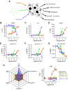
Fig. 1.
Fig. 1.
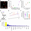
BTB recapitulates a similar intracellular signaling signature to protons at GPR65. (A) Intracellular signaling responses coordinated by mouse GPR65 were assessed in a CHO cell background, the ability of protons, BTB, or psychosine to coordinate (B) accumulation of cAMP (FSK, forskolin), (C) intracellular Ca2+ mobilization, (D) phosphorylation of ERK1/2 (PDBu, phorbol 12,13-dibutyrate), recruitment of (E) β-arrestin 1 or (F) β-arrestin 2 and (G) receptor internalization was assessed. (H) To compare the signaling profiles of each GPR65 agonist, the peak response of each agonist in each pathway was normalized to that achieved by proton stimulation. The Emax of psychosine in the cAMP pathway was set as 0. (I) The selectivity of BTB among other PS-GPCRs was assessed using stable cell lines and the cAMP assay. Data are from at least three independent experiments where each [agonist] was assayed in duplicate. *P-adj < 0.05, **P-adj < 0.01, ***P-adj < 0.001, ****P-adj < 0.0001: Two-way ANOVA followed by Bonferroni-corrected post hoc.
Fig. 2.
Fig. 2.
Intra-articular injection of BTB causes inflammation and pain-like behaviors in mice. (A) Schematic representation and experimental timeline. Mice received a unilateral injection of either 100 µM BTB, 0.1% (v/v) DMSO, 1 mg MIA or 10 µg CFA. (B) The ratio of the ipsilateral to contralateral knee width was calculated as a measure of the extent of inflammation. (C) Mechanical sensitivity of injected knee joints was determined by pressure application measurement. The (D) latency to dig, (E) time spent digging and (F) number of burrows dug were also measured across experimental time. *P-adj < 0.05, **P-adj < 0.01, ***P-adj < 0.001, ****P-adj < 0.0001: Repeated-measures ANOVA followed by Bonferroni-corrected post hoc; for ease of interpretation only statistical differences comparing the effect of BTB injection to the baseline measures of this group are annotated. N = 16 mice (8 female, 8 male) for BTB and DMSO groups, 6 mice (3 female, 3 male) for CFA group and 4 mice (1 female, 3 male) for the MIA group.
Fig. 3.
Fig. 3.
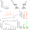
BTB-induced sensitization of sensory neurons depends upon cells resident in the joint. (A) Naïve lumbar (L2-L5) DRG neurons were cultured overnight with 100 µM BTB, 0.1% (v/v) DMSO or regular culture media, before electrophysiological characterization. (B) Representative current clamp recordings of neurons of comparable capacitance, showing action potentials evoked by ramp injection of current (0 to 1 nA, 1 s). (C) Stepwise current injections were used to determine the rheobase of sensory neurons. (D) Following retrograde labeling of knee-innervating sensory neurons (with Fast Blue), mice were injected with 100 µM BTB into one knee, 24-h postinjection DRG from the ipsilateral (Ipsi) and contralateral (Contra) were collected and cultured for electrophysiological characterization. (E) Representative current clamp recordings of neurons of comparable capacitance, showing action potentials evoked by ramp injection of current (0 to 1 nA, 1 s). (F) Stepwise current injections were used to determine the rheobase of sensory neurons. ***P < 0.001: (C) One-way ANOVA followed by Bonferroni-corrected post hoc, (F) unpaired t-test.
Fig. 4.
Fig. 4.
FLS express GPR65 and respond to BTB stimulation. (A) Mouse FLS express CDH-11 (Red: aCDH-11, Blue: Nuclear stain. (Scale bar, 50 µm.) (B) FLS gene expression was further interrogated via qPCR. (C) Intracellular FLS cAMP concentration following stimulation with pH 7.4 vehicle, BTB, or pH 6 solution. (D) FLS were cultured overnight with either BTB, DMSO, or regular culture media, 24-h poststimulation media were collected and later incubated with naïve DRG neurons overnight before electrophysiological characterization. (E) Representative current clamp recordings of neurons of comparable capacitance, showing action potentials evoked by ramp injection of current (0 to 1 nA, 1 s). (F) Stepwise current injections were used to determine the rheobase of sensory neurons. (G) BTB-induced fold change in detection of inflammatory cytokines in conditioned media from stimulated FLS. *P-adj < 0.05, **P-adj < 0.01: (F) One-way ANOVA followed by Bonferroni-corrected post hoc. (G) Two-way ANOVA followed by Bonferroni-corrected post hoc.
Fig. 5.
Fig. 5.
BTB exerts its pro-inflammatory effects via GPR65 expressing FLS. (A) Intracellular FLS cAMP concentration of FLS from GPR65 KO mice following stimulation with pH 7.4 vehicle, BTB, or pH 6 solution. (B) WT (closed symbols/solid lines) and KO FLS (open symbols/dashed lines) were cultured overnight with BTB, the conditioned media collected 24-h poststimulation was then incubated with naïve DRG neurons overnight before electrophysiological characterization. (C) Representative current clamp recordings of neurons of comparable capacitance, showing action potentials evoked by ramp injection of current (0 to 1 nA, 1 s). (D) Stepwise current injections were used to determine the rheobase of sensory neurons (E) Relative quantification of pro-inflammatory cytokines present in the conditioned media of WT and KO FLS following BTB stimulation. (F) phosphoCREB staining intensity of cultures of FLS from WT or KO animals following overnight stimulation with DMSO (blue), BTB (orange), or pH 6 (green). *P/P-adj < 0.05, **P/P-adj < 0.01, ***P./P-adj < 0.001: (D) unpaired t-test. (E and F) Two-way ANOVA followed by Bonferroni-corrected post hoc.
Fig. 6.
Fig. 6.
GPR65 KO mice do not develop joint inflammation or pain following intra-articular injection of BTB. (A) WT (WT, closed symbols) and GPR65 KO, open symbols) mice of either sex (females, square symbols; males, triangular symbols) received unilateral intra-articular injections of 100 µM BTB. (B) The ratio of the ipsilateral to contralateral knee width was calculated as a measure of the extent of inflammation. (C) Mechanical sensitivity of the injected knee joints was determined by pressure application measurement. The (D) latency to dig, (E) time spent digging and (F) number of burrows dug were also measured across experimental time. Following conclusion of the behavioral study, ipsilateral knee joints were collected and examined for histological changes. (G) Representative nuclei staining of an ipsilateral knee collected from a KO animal: i pink area represents the synovium, ii pink puncta represent automatically detected nuclei within the synovial perimeter (Scale bars, 100 µm.) The (H) synovium thickness and (I) percentage of synovial area occupied by nuclei was compared between WT and KO mice. *P < 0.05, ***P-adj < 0.001, ****P-adj < 0.0001: (BF) repeated measures ANOVA followed by Bonferroni-corrected post hoc. (H and I) unpaired t-test. (BF): N = 14 WT mice (7 female, 7 male) and 16 KO mice (8 female, 8 male). (H and I): N = 6 WT mice (3 female, 3 male) and 6 KO mice (3 female, 3 male).
Fig. 7.
Fig. 7.
Human FLS express GPR65 and arthritic synovial fluid samples activate GPR65. (A) Expression of GPR65 in synovium tissue from painful (pink) and nonpainful (green) sites of end-stage OA patients, data from Nanus et al. (51). (B) BTB-induced fold change in detection of inflammatory cytokines in conditioned media from stimulated human FLS. (C) Intracellular cAMP concentration of FlpIN CHO (open symbols) or mGPR65-CHO (closed symbols) cells, following stimulation with human OA synovial fluid samples, pH solutions, or BTB. *P-adj < 0.05, **P-adj < 0.01, ***P-adj < 0.001: (A) Published differentially expressed gene (DEG) data from Nanus et al. (51) was filtered to identify DEGs using two-group statistical comparison for >1.5 fold change and P < 0.05. (B) Two-way ANOVA followed by Bonferroni-corrected post hoc.
Fig. 8.
Fig. 8.
Activation of the proton-sensing GPCR, GPR65 on FLS contributes to inflammatory joint pain. Arthritic conditions are associated with localized acidosis of the joint environment and proliferation of FLS (synovitis). FLS express GPR65, are activated by the increased local concentration of protons (H+), leading to release of pro-inflammatory mediators capable of recruiting immune cells to the area, further driving inflammation. Mediators released by both FLS and immune cells act on sensory neurons that innervate the joint contributing to peripheral sensitization and the increased pain associated with arthritis.

References

    1. Ghouri A., Conaghan P. G., Treating osteoarthritis pain: Recent approaches using pharmacological therapies. Clin. Exp. Rheumatol. 37 Suppl 120, 124–129 (2019). - PubMed
    1. Radu A.-F., Bungau S. G., Management of rheumatoid arthritis: An overview. Cells 10, 2857 (2021). - PMC - PubMed
    1. Pattison L. A., et al. , Digging deeper into pain: An ethological behavior assay correlating well-being in mice with human pain experience. PAIN 165, 1761–1773 (2024). - PMC - PubMed
    1. Andersson S. E., Lexmüller K., Johansson A., Ekström G. M., Tissue and intracellular pH in normal periarticular soft tissue and during different phases of antigen induced arthritis in the rat. J. Rheumatol. 26, 2018–24 (1999). - PubMed
    1. Sasaki Y., Hada R., Nakajima H., Fukuda S., Munakata A., Improved localizing method of radiopill in measurement of entire gastrointestinal pH profiles: Colonic luminal pH in normal subjects and patients with Crohn’s disease. Am. J. Gastroenterol. 92, 114–8 (1997). - PubMed

LinkOut - more resources