. 2024 Dec 13;10(50):eado5914.
doi: 10.1126/sciadv.ado5914. Epub 2024 Dec 11.

Identification of the Wnt signal peptide that directs secretion on extracellular vesicles

Affiliations

Identification of the Wnt signal peptide that directs secretion on extracellular vesicles

Uxia Gurriaran-Rodriguez et al. Sci Adv. .

Abstract

Wnt proteins are hydrophobic glycoproteins that are nevertheless capable of long-range signaling. We found that Wnt7a is secreted long distance on the surface of extracellular vesicles (EVs) following muscle injury. We defined a signal peptide region in Wnts required for secretion on EVs, termed exosome-binding peptide (EBP). Addition of EBP to an unrelated protein directed secretion on EVs. Palmitoylation and the signal peptide were not required for Wnt7a-EV secretion. Coatomer was identified as the EV-binding protein for the EBP. Analysis of cocrystal structures, binding thermodynamics, and mutagenesis found that a dilysine motif mediates EBP binding to coatomer with a conserved function across the Wnt family. We showed that EBP is required for Wnt7a bioactivity when expressed in vivo during regeneration. Overall, our study has elucidated the structural basis and singularity of Wnt secretion on EVs, alternatively to canonical secretion, opening avenues for innovative therapeutic targeting strategies and systemic protein delivery.


Figures

Fig. 1.
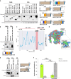
Fig. 1.. Muscle injury triggers secretion of Wnt7a on the surface of EVs.
(A) iTEM of anti-Wnt7a labeling of new regenerating myofibers at 96 hours after CTX injury from wild-type (WT) mice shows Wnt7a secretion on ILVs contained in a MVB. Scale bars, 500 and 100 nm. (B) iTEM of anti-Wnt7a labeling of injured TA muscle from WT mice shows the presence of Wnt7a on EVs infiltrated on the injured tissue that surrounds the new regenerating myofibers. Scale bar, 500 and 100 nm. (C) Immunoblot analysis of EVs fraction from muscle showing Wnt7a expression. (D) Schematic representation of mouse strains used to generate conditional Wnt7a floxed in Myf5-expressing cells. (E) Immunofluorescence confirmation of Wnt7a expression abrogation in Myf5(Cre/+):Wnt7a(fl/fl) injured TA at 96 hours after CTX injury. Scale bar, 50 μm. (F) iTEM of anti-Wnt7a labeling of EVs showing abrogation of Wnt7a expression in EVs from Myf5(Cre/+): Wnt7a(fl/fl) mice muscle explants. Scale bar, 100 nm. (G) Immunoblot verification of Wnt7a expression abrogation in EVs isolated from Myf5(Cre/+): Wnt7a(fl/fl) hindlimb muscle at 96 hours after CTX injury. (H) pMHC immunofluorescence representative images of hypertrophied myotubes after muscle EVs stimulation containing Wnt7a (n = 3). Scale bar, 50 μm. Experiments are representative of n = 4 biological replicates. (I) Hypertrophy assay of murine primary myotubes treated with EVs from muscle decreases hypertrophy after Wnt7a deletion. Wnt7a recombinant protein was used as a positive control. n = 3 mice each with eight technical replicates, means ± SEM. P values were determined by two-sided Student’s t test (*P < 0.05, **P < 0.005, and ***P < 0.0005). iTEM, immunogold transmission electron microscopy; MVBs, multivesicular bodies; pMHC, pan myosin heavy chain.
Fig. 2.
Fig. 2.. Wnt7a secretion on EVs is regulated by an internal peptide sequence.
(A) Immunoblot analysis shows interruption of EV secretion following deletion beyond position 100 amino acid but not after position 300. (B) Immunoblot analysis showing the internal 100– to 300–amino acid sequence of Wnt7a is sufficient for EV secretion. (C) ΔGFoldX of Wnt7a indicates Δ1–49 and Δ301–349 do not affect folding—ΔΔG < 0 respect to WT protein—and function is not lost. Δ1–212 affects protein folding. Δ251–349 does not affect folding, but function is lost because a region of the EBP is truncated. (D) Surface of Wnt7a with negative charged residues in red, positive charged residues in blue, and hydrophobic residues in green. The EBP is positively charged. (E) Immunoblot analysis, demonstrates replacement of EBP with a GSGS linker abrogates EV secretion in favor of non-EV Wnt7a secretion. FP, free protein. (F) Quantification of Wnt7a expression on EVs and non-EV protein fractions with and without EBP. Experiments are representative of three independent biological replicates performed in HEK293T cells transfected with different Wnt7a-HA tagged truncates. n = 3 biological replicates, means ± SEM. P values were determined by two-sided Student’s t test (***P < 0.0005). HA, hemagglutinin.
Fig. 3.
Fig. 3.. The EBP is sufficient to confer targeting to EVs.
(A) Restoration of EV secretion by inserting EBP into an upstream domain of Wnt7a truncate that is not secreted on EVs. The insertion does not perturb the stability of the full-length protein (Wnt7a-Δ3aa*GSG versus Wnt7a-Δ3aa*EBP). Insertion of EBP to this site (Wnt7a-Δ213-249*EBP) restores EVs localization to Wnt7a-Δ213-249. (B) C-terminal linked EBP but not N-terminal linked confers targeting of Wnt7a-Δ213-249 onto EVs. (C) Linking of EBP HALO protein results in EV secretion. (D) iTEM images of EVs with anti-HALO immunostaining showing the expression of HALO*EBP on the surface of EVs. Scale bars, 100 μm. (E) Detection of HALO by fluorescence inside HEK293T cells following treatment with HALO*EBP EVs versus HALO EVs derived from HEK293T cells transfected with different Wnt7a-HA and HALO tagged truncates. n = 3 biological replicates.
Fig. 4.
Fig. 4.. Wnt7a secretion on EVs appears independent of the canonical pathway and requires Coatomer.
(A) SP is not required for EVs-Wnt7a secretion in transfected HEK293T cells. (B) Mutant Wnt7a_S206A, lacking the palmitoylation site, is secreted on EVs in transfected HEK293T cells. (C) Drug inhibition of PORCN does not affect secretion of Wnt7a on EVs. (D) Knockdown of WLS in siRNA partially affects secretion of Wnt7a on EVs but does abolish secretion of non-EV Wnt7a. (E) BirA constructs for BioID analysis. (F) Heatmap displaying fold change (log2 scale) of enriched proteins in mass spectrometry (ESP_BirA:BirA and Wnt7a_BirA:BirA). Shown are enrichment of >50% [log2 (FC) > 0.5849] on EBP and a positive enrichment [log2 (FC) > 0] on Wnt7a. (G) Wnt7a: COPA PLA (red) performed in murine primary myotubes either expressing Wnt7a-BirA or BirA. GM310 in green and 4′,6-diamidino-2-phenylindole (DAPI) in blue. Scale bars, 10 μm. (H) Wnt7a:COPA PLA (orange) performed in RPTEC- hTERT1 cells. Wheat Germ Agglutinin (WGA) in green and DAPI in blue. CTR-neg is control without Wnt7a antibody. Scale bars, 10 μm. (I) Wnt7a is secreted on EVs derived from RPTEC-hTERT1 cells. HEK293T cells lacking Wnt7a were used as a negative control. (J) Wnt7a:COPA PLA (red) performed in HEK293T cells expressing Wnt7a-FL, Wnt7a_ΔEBP*GSGS, or Wnt7a_ΔSP. GM310 in green and DAPI in blue. Scale bars, 10 μm. CTR-neg is control without Wnt7a antibody. (K) Wnt7a-HA interacts with COPA and COPB2. HEK293T cells overexpressing Wnt7a-HA were immunoprecipitated with COPB2 antibody or (L) immunoprecipitated with HA antibody. (M) Immunoblot EVs secretion analysis of Wnt7a after siRNA knockdown of COPA and COPB2 shows disruption of Wnt7a-EV secretion in transfected HEK293T cells. n = 3 biological replicates. IgG, immunoglobulin G; WGA, Wheat Germ Agglutinin.
Fig. 5.
Fig. 5.. Wnt7a binds Coatomer proteins.
(A) Predicted Wnt7a structure in AlphaFold (blue) with the EBP region highlighted in orange and the three positively charged motifs (red) within the EBP. Note that EBP is a solvent-exposed region. (B) Wnt7a-ESP*Scramble mutant maintains the dilysine motif and exhibits no impairment in EV secretion. (C) ITC measurements for COPB21–304 binding to potential dilysine/arginine motifs within the EBP. WT COPB21–304 binds to the LKIKKP subregion. (D to F) views of the KxKx motif of Wnt7a bound to COPB21–304. (D) Top view of the WD-repeat domain of COPB1–304 (green) with the LKIKKP peptide (orange) in ribbon representation. (E) Close-up view of the LKIKKP peptide with a difference electron density map calculated by omitting the peptide and contoured at 3σ (blue mesh). COPB2 surface is colored by electrostatic potential ranging from −5 kT/e (red) to 5 kT/e (blue). (F) Lateral view of the binding motif with hydrogen bonds and distances. (G) Structure-based point mutations confirm the molecular recognition of the KxKx motif in ITC assays. (H) Double lysine mutation of K253 and K255 by alanine disrupts Wnt7a-EV secretion. (I) Replacement of Wnt7a-EBP by either Wnt10a-EBP or Wnt16 EBP containing KR and RR (right). Replacement with Wnt10a-EBP or Wnt16 EBP rescues Wnt7a-EV secretion. (J) EV secretion analysis of Wnt10b after EBP removal or double arginine mutation within its EBP (right). Double arginine mutation disrupts secretion of Wnt10b on EVs to the same extent as removal of the entire Wnt10b EBP sequence. (K) Secretion analysis of Wnt3a after EBP removal or mutation of the entire positively charged motifs RPR, KHR, and KH within its EBP (right). Only concomitant mutation of RPR and KHR motifs disrupts secretion of Wnt3a on EVS to the same extent as removal of the entire Wnt3a EBP sequence. n = 3 biologic replicates.
Fig. 6.
Fig. 6.. Wnt7a interaction with Coatomer is required for the muscle regenerative response.
(A) Knockdown of COPA and COPB2 using siRNA disrupts Wnt7a-EV secretion in Wnt7a stably expressing primary myoblasts. (B) Knockdown of COPA and COPB2 decreases hypertrophy in Wnt7a-transfected myotubes. Conversely, knockdown did not affect hypertrophy in myotubes not expressing Wnt7a. (C) Representative images after simultaneously siRNA knockdown of COPA and COPB2 in myotubes. Scale bars, 50 μm. (D) Schematic representation of in vivo workflow. (E) Myofiber caliber distribution comparing TA electroporated with Wnt7a (green) versus Wnt7a_ΔEBP*GSGS (gray). (F) Average minimal fiber feret comparing TA electroporated with Wnt7a (green) versus Wnt7a_ΔEBP*GSGS (gray). (G) Quantification of fiber number comparing TA electroporated with Wnt7a (green) versus Wnt7a_ΔEBP*GSGS (gray). (H) Quantification of muscle area comparing TA electroporated with Wnt7a (green) versus Wnt7a_ΔEBP*GSGS (gray). (I) Section of TA muscles showing reduced myofiber caliber an increased number of myofibers after electroporation of Wnt7a_ΔEBP*GSGS into TA muscle of mdx mice. Scale bars, 100 μm. TA, tibialis anterior. In vitro experiments are representative of three independent biological replicates performed in murine primary myoblasts. In vivo experiments are representative n = 6 mice, means ± SEM. P value was determined by two-sided Student’s t test (*P < 0.05 and **P < 0.005).

Update of

References

    1. Wellenstein M. D., Coffelt S. B., Duits D. E. M., van Miltenburg M. H., Slagter M., de Rink I., Henneman L., Kas S. M., Prekovic S., Hau C.-S., Vrijland K., Drenth A. P., de Korte-Grimmerink R., Schut E., van der Heijden I., Zwart W., Wessels L. F. A., Schumacher T. N., Jonkers J., de Visser K. E., Loss of p53 triggers WNT-dependent systemic inflammation to drive breast cancer metastasis. Nature 572, 538–542 (2019). - PMC - PubMed
    1. Willert K., Nusse R., Wnt proteins. Cold Spring Harb. Perspect. Biol. 4, a007864 (2012). - PMC - PubMed
    1. Polesskaya A., Seale P., Rudnicki M. A., Wnt signaling induces the myogenic specification of resident CD45+ adult stem cells during muscle regeneration. Cell 113, 841–852 (2003). - PubMed
    1. Logan C. Y., Nusse R., The Wnt signaling pathway in development and disease. Annu. Rev. Cell Dev. Biol. 20, 781–810 (2004). - PubMed
    1. Nile A. H., Hannoush R. N., Fatty acylation of Wnt proteins. Nat. Chem. Biol. 12, 60–69 (2016). - PubMed

LinkOut - more resources