. 2024 Dec 12;13(12):764-778.
doi: 10.1302/2046-3758.1312.BJR-2024-0136.R2.

Long-term hypoxic atmosphere enhances the stemness, immunoregulatory functions, and therapeutic application of human umbilical cord mesenchymal stem cells

Affiliations

Long-term hypoxic atmosphere enhances the stemness, immunoregulatory functions, and therapeutic application of human umbilical cord mesenchymal stem cells

Qi-Ming Huang et al. Bone Joint Res. .

Abstract

Aims: Mesenchymal stem cells (MSCs) are usually cultured in a normoxic atmosphere (21%) in vitro, while the oxygen concentrations in human tissues and organs are 1% to 10% when the cells are transplanted in vivo. However, the impact of hypoxia on MSCs has not been deeply studied, especially its translational application.

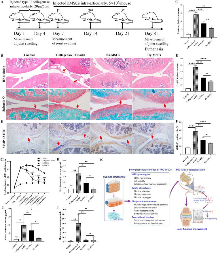
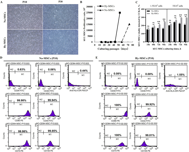
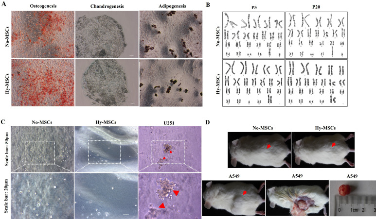
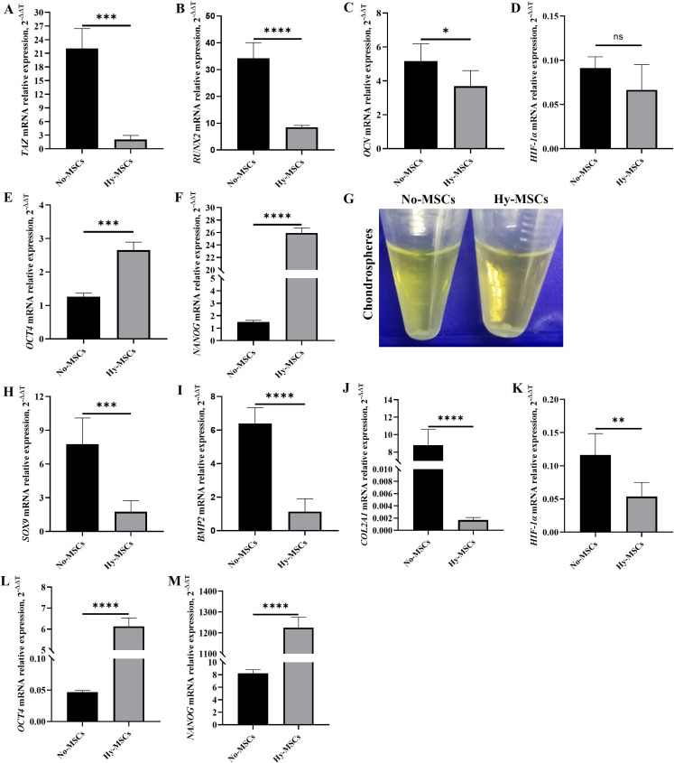
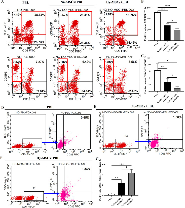
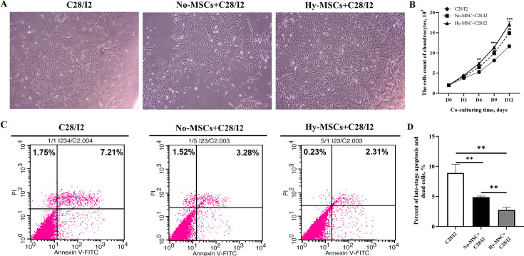
Methods: In the present study, we investigated the characterizations of human umbilical cord-derived mesenchymal stem cells (hUC-MSCs) in hypoxic (1%) and normoxic (21%) atmospheres with a long-term culture from primary to 30 generations, respectively. The comparison between both atmospheres systematically analyzed the biological functions of MSCs, mainly including stemness maintenance, immune regulation, and resistance to chondrocyte apoptosis, and studied their joint function and anti-inflammatory effects in osteoarthritis (OA) rats constructed by collagenase II.

Results: We observed that long-term hypoxic culture surpassed normoxic atmosphere during hUC-MSCs culture in respect of promoting proliferation, anti-tumorigenicity, maintaining normal karyotype and stemness, inhibiting senescence, and improving immunoregulatory function and the role of anti-apoptosis in chondrocytes. Furthermore, we demonstrated that the transplantation of long-term hypoxic hUC-MSCs (Hy-MSCs) had a better therapeutic effect on OA rats compared with the hUC-MSCs cultured in the normoxic atmosphere (No-MSCs) in terms of the improved function and swelling recovery in the joints, and substantially inhibited the secretion of pro-inflammatory factors, which effectively alleviated cartilage damage by reducing the expression of matrix metallopeptidase 13 (MMP-13).

Conclusion: Our results demonstrate that Hy-MSCs possess immense potential for clinical applications via promoting stemness maintenance and enhancing immunoregulatory function.


Conflict of interest statement

The authors report grants from the National Key Research and Development Program of China (No. 2022YFA1104300), the National Nature Science Foundation of China (No. 82270302, 82260173, 81970256), the Key Research Project (No. 20192BBH80015, 20202BBG73028), and the Project for Leading Talent of department of science and technology, Jiangxi Province (No. 20204BCJ22035, 20212BDH81020), all related to this study.

Figures

Fig. 1
Fig. 1
Long-term hypoxic human umbilical cord-derived mesenchymal stem cells (hUC-MSCs) better represented joint function repair and anti-inflammatory effects in osteoarthritis (OA) rats. a) The scheme of modelling and MSC transplantation in OA rat. b) The pathological analysis of joint tissues in OA rats by haematoxylin and eosin (H&E) and Safranin O staining, scale bar: 50 μm. c) and d) Analysis of joint function before and after hUC-MSC transplantation in OA rats using Mankin scale and Osteoarthritis Research Society International (OARSI) scale evaluation (n = 5). e) Matrix metallopeptidase 13 (MMP-13) analysis of joint tissues in OA rats by immunohistochemistry (IHC), scale bar: 50 μm. f) Statistical analysis of MMP-13 positive cells percentage in joint tissues of OA rats. g) Measurement of joint swelling diameter before and after hUC-MSC transplantation in OA rats, including one, two, three, and four weeks (n = 5), and comparison with OA model. h) to j) Measurement and statistical analysis of pro-inflammatory factor, i.e. interleukin (IL)-1β, tumour necrosis factor alpha (TNF-α), and IL-6 (n = 5). k) Overview on biological characteristics and translational potential of hUC-MSCs in hypoxic atmosphere. Data are presented as mean (SD). *p < 0.05, **p < 0.01, ***p < 0.001, ****p < 0.0001; one-way analysis of variance (ANOVA). Hy-MSCs, hypoxic hUC-MSCs; No-MSCs, hUC-MSCs cultured with normoxic atmosphere; ns, non-significant.
Fig. 2
Fig. 2
Proliferative ability and phenotypes of human umbilical cord-derived mesenchymal stem cells (hUC-MSCs) in long-term normoxic and hypoxic atmospheres. a) The growth morphology of hUC-MSCs in normoxic and hypoxic atmospheres. Scale bar: 50 μm. b) Proliferative times of hUC-MSCs were detected by cell count continuously. c) Proliferative viability of hUC-MSCs (left: 1.5 × 10³ cells; right: 3 × 10³ cells) was detected by Cell Counting Kit-8 (CCK-8) assay. d) and e) Phenotypic assay of hUC-MSCs was detected by flow cytometry. Data are presented as mean (SD). **p < 0.01, one-way analysis of variance (ANOVA). Hy-MSCs, hypoxic hUC-MSCs; No-MSCs, hUC-MSCs cultured with normoxic atmosphere; ns, non-significant.
Fig. 3
Fig. 3
The multilineage differentiation, tumorigenicity, and karyotypes of human umbilical cord-derived mesenchymal stem cells (hUC-MSCs) in long-term hypoxic atmosphere. a) The differentiative potential of both hUC-MSCs cultured with normoxic atmosphere (No-MSCs) (P5) and hypoxic hUC-MSCs (Hy-MSCs) (P5) was shown through staining by Alizarin Red, Oil Red-O, and Alcian Blue, respectively, including in osteoblasts, chondrocytes, and adipocytes. Scale bar: 50 μm. b) The karyotypes of hUC-MSCs (P5 and P20) were measured by G-banding assay. c) Both No-MSCs and Hy-MSCs (P5) were analyzed for tumour formation in soft agar cloning experiments, and glioma cell line (U251) was used as a control. d) Both No-MSCs and Hy-MSCs (P5) were analyzed for tumour formation in immunodeficient mice (NOG), and lung cancer cell line (A549) was used as a control. The experiments were repeated three times independently, and the data of one representative experiment were shown.
Fig. 4
Fig. 4
The pluripotency and ability to low-differentiation of human umbilical cord-derived mesenchymal stem cells (hUC-MSCs) in a long-term hypoxic atmosphere. a) to c) The expression of specific genes of osteoblast differentiation was detected by quantitative real-time polymerase chain reaction (qRT-PCR) assay, i.e. TAZ, Runx2, and OCN. d) HIF-1α messenger RNA (mRNA) expression was detected in the osteoblast differentiation of hUC-MSCs. e) and f) The expression of stemness genes was measured in the osteoblast differentiation of hUC-MSCs, including OCT-4 and NANOG. g) The chondrospheres were formed in hUC-MSC chondrogenic differentiation at day 4. h) to j) The expression of related genes was detected in chondroblast differentiation, i.e. Sox-9, BMP2 and COL2A1. k) HIF-1α mRNA expression was detected in the chondroblast differentiation of hUC-MSCs. l) to m) The expression of stemness genes was measured in the chondroblast differentiation of hUC-MSCs, including OCT-4 and NANOG. Data are presented as mean (SD). *p < 0.05, **p < 0.01, ***p < 0.001, ****p < 0.0001; one-way analysis of variance (ANOVA).
Fig. 5
Fig. 5
The senescence level and stemness characteristics of human umbilical cord-derived mesenchymal stem cells (hUC-MSCs) in a long-term hypoxic atmosphere. a) The senescence level of both hUC-MSCs cultured with normoxic atmosphere (No-MSCs) and hypoxic hUC-MSCs (Hy-MSCs) were stained by β-galactosidase staining (P10). Scale bars: 100 μm and 50 μm. b) The positive β-galactosidase staining cells were counted and compared between No-MSCs and Hy-MSCs. c) and d) The expression of senescence genes of MSCs was detected by quantitative real-time polymerase chain reaction (qRT-PCR) assay, i.e. p16INK4a and p21WAF1. e) Telomerase activity of hUC-MSCs (P10, P20, and P30) was analyzed at normoxic and hypoxic atmospheres by PCR-ELISA assay. f) to h) The expression of stemness genes of hUC-MSCs was detected by qRT-PCR assay, i.e. OCT-4, SOX2, and NANOG. i) The expression of HIF-1α was tested in both No-MSCs and Hy-MSCs. Data are presented as mean (SD). *p < 0.05, **p < 0.01, ***p < 0.001, ****p < 0.0001, one-way analysis of variance (ANOVA). ELISA, enzyme-linked immunosorbent assay; PCR, polymerase chain reaction.
Fig. 6
Fig. 6
Human umbilical cord-derived mesenchymal stem cells (hUC-MSCs) promoted the abilites of immunoregulation and proliferation and inhibited apoptosis of chondrocytes in a hypoxic atmosphere. a) The phenotypes of peripheral blood lymphocytes (PBLs) were detected by flow cytometry analysis. b) and c) Statistical analysis of different lymphocyte subsets, including CD3+ CD8+ T cells and CD3-CD56/16+ natural killer (NK) cells (n = 4). d) to f) The production of regulatory T (Treg) cells was measured after coculturing with both hUC-MSCs cultured with normoxic atmosphere (No-MSCs) and hypoxic hUC-MSCs (Hy-MSCs). g) Statistical analysis of Treg cells percentage, i.e. CD4+ CD25+ Foxp3+ Treg (n = 4). Data are presented as mean (SD). *p < 0.05, **p < 0.01, ***p < 0.001; one-way analysis of variance (ANOVA). ns, non-significant.
Fig. 7
Fig. 7
Human umbilical cord-derived mesenchymal stem cells (hUC-MSCs) better inhibited apoptosis of chondrocytes in a hypoxic atmosphere. a) The growth state of chondrocytes after co-culturing with hUC-MSCs cultured with normoxic atmosphere (No-MSCs) and hypoxic hUC-MSCs (Hy-MSCs) for three days. b) The statistical analysis of the chondrocytes count after co-culturing with No-MSCs and Hy-MSCs by comparing with control C28/I2. c) Flow cytometry detection of apoptosis levels on chondrocytes after co-culturing with No-MSCs and Hy-MSCs for three days. d) The comparative analysis of late-stage and death apoptosis levels on chondrocytes after co-culturing with No-MSCs and Hy-MSCs. Data are presented as mean (SD). *p < 0.05, **p < 0.01, ***p < 0.001, one-way analysis of variance (ANOVA).

Similar articles

Cited by

References

    1. Gopalarethinam J, Nair AP, Iyer M, Vellingiri B, Subramaniam MD. Advantages of mesenchymal stem cell over the other stem cells. Acta Histochem. 2023;125(4):152041. doi: 10.1016/j.acthis.2023.152041. - DOI - PubMed
    1. Liau LL, Ruszymah BHI, Ng MH, Law JX. Characteristics and clinical applications of Wharton’s jelly-derived mesenchymal stromal cells. Curr Res Transl Med. 2020;68(1):5–16. doi: 10.1016/j.retram.2019.09.001. - DOI - PubMed
    1. Prasopthum A, Cooper M, Shakesheff KM, Yang J. Three-dimensional printed scaffolds with controlled micro-/nanoporous surface topography direct chondrogenic and osteogenic differentiation of mesenchymal stem cells. ACS Appl Mater Interfaces. 2019;11(21):18896–18906. doi: 10.1021/acsami.9b01472. - DOI - PubMed
    1. Joel MDM, Yuan J, Wang J, et al. MSC: Immunoregulatory effects, roles on neutrophils and evolving clinical potentials. Am J Transl Res. 2019;11(6):3890–3904. - PMC - PubMed
    1. Parvin Nejad S, Lecce M, Mirani B, et al. Serum- and xeno-free culture of human umbilical cord perivascular cells for pediatric heart valve tissue engineering. Stem Cell Res Ther. 2023;14(1):96. doi: 10.1186/s13287-023-03318-3. - DOI - PMC - PubMed