. 2025 Feb;638(8052):1054-1064.
doi: 10.1038/s41586-024-08489-4. Epub 2024 Dec 11.

Discovery and engineering of the antibody response to a prominent skin commensal

Affiliations

Discovery and engineering of the antibody response to a prominent skin commensal

Djenet Bousbaine et al. Nature. 2025 Feb.

Abstract

The ubiquitous skin colonist Staphylococcus epidermidis elicits a CD8+ T cell response pre-emptively, in the absence of an infection1. However, the scope and purpose of this anticommensal immune programme are not well defined, limiting our ability to harness it therapeutically. Here, we show that this colonist also induces a potent, durable and specific antibody response that is conserved in humans and non-human primates. A series of S. epidermidis cell-wall mutants revealed that the cell surface protein Aap is a predominant target. By colonizing mice with a strain of S. epidermidis in which the parallel β-helix domain of Aap is replaced by tetanus toxin fragment C, we elicit a potent neutralizing antibody response that protects mice against a lethal challenge. A similar strain of S. epidermidis expressing an Aap-SpyCatcher chimera can be conjugated with recombinant immunogens; the resulting labelled commensal elicits high antibody titres under conditions of physiologic colonization, including a robust IgA response in the nasal and pulmonary mucosa. Thus, immunity to a common skin colonist involves a coordinated T and B cell response, the latter of which can be redirected against pathogens as a new form of topical vaccination.


Conflict of interest statement

Competing interests: M.A.F. is a cofounder of Kelonia and Revolution Medicines, a member of the scientific advisory boards of the Chan Zuckerberg Initiative, NGM Biopharmaceuticals and TCG Laboratories/Soleil Laboratories, and an innovation partner at The Column Group. D.B., Y.E.C., K.D.B., P.V.L., M.I.M., C.O.B., Y.B. and M.A.F. are inventors on patent applications submitted by Stanford University and the Chan Zuckerberg Biohub that cover methods for using engineered bacteria to elicit antigen-specific immune cells.

Figures

Extended Data Figure 1:
Extended Data Figure 1:. Colonization with S. epidermidis induces a systemic B cell response.
(a) Colony forming units (CFUs) per ear for the experiment shown in Fig. 1a. Two independent experiments, all mice shown. (b) S. epidermidis LM088 was stained with serum from mice that were colonized (blue) or not (naïve, grey) with the same strain for 6 weeks and analyzed by flow cytometry. Representative of three independent experiments. Bacteria were gated on Syto9 positive cells. (c) CFUs for the experiment shown in Fig. 1b. Two independent experiments, all mice shown. (d) CFUs for experiment shown in Fig. 1g. Two independent experiments, all mice shown. (e) Gating strategy for germinal center B cells as shown in Fig. 1e.
Extended Data Figure 2:
Extended Data Figure 2:. Antibody response against the native microbiome.
(a) Representative example of a dot blot analysis using serum from mice housed at Charles River (CR) room H44, Taconic CC facility or at Jackson against a panel of strains isolated from the same mice. (b) Bacterial ELISA of Staphylococcus xylosus and Streptococcus sp. isolated from Jackson and Taconic (Tac) GT mice respectively against a panel of serum from CR, Tac and Jackson. N=5–15, one to three independent cages pooled. (c) SPF mice (Taconic SD) were colonized for five weeks with murine strains of S. xylosus, Staphylococcus nepalensis or Staphyloccocus/Mammaliicoccus lentus and serum antibody titers were measured by ELISA. Colonization efficiency was confirmed at the experimental endpoint by CFUs (data not shown). (d) Same as (b) but against two isolates of S./M. lentus isolated from Tac GT or CR H44. (e) ELISA using serum from 11 healthy donors against a panel of commensals and environmental strains. (f) Same as (e) but using the serum of 10 healthy non-human primates (NHP). (e-f) One representative of three independent experiments. (e-f) P values were calculated using one-way ANOVA with Tukey correction for multiple comparisons. Uncropped dot blot images can be found in Supplementary Fig. 2a.
Extended Data Figure 3:
Extended Data Figure 3:. Identification of the minimal epitope in the B domain of Aap.
(a) Schematic of LM088 Aap protein truncations for expression in E. coli. (b) Immunoblot analysis of the constructs shown in (a) using anti-HA antibody to assess expression (induced by IPTG), using serum from naïve mice or mice colonized with LM088. (1= amino acid repeat domain, 2= parallel β-helix domain, and 3=one repeat of the B domain). (c) Schematic of the B domain truncations fused to superfolder GFP (sGFP) for expression in E. coli. (d) Immunoblot analysis of the truncations shown in (c) using anti-HA antibody or the serum of mice colonized with LM088. (e) Minimal epitope identified in the B domain and the peptides synthesized and tested in (f). (f) Dot blot analysis of the biotinylated peptides shown in (e) using streptavidin-HRP or the serum of mice colonized with LM088. All blots are representative of three independent experiments. (g) Immunoblot of cell lysate (L) and culture supernatant (S) of LM088 and LM088 Δaap using serum from mice colonized with LM088 or LM088 Δaap (from the mice shown in Fig. 2j). Representative of two independent experiments. Uncropped immunoblot and dot blot images can be found in Supplementary Fig. 2b–d and 3a–b.
Extended Data Figure 4:
Extended Data Figure 4:. Redirecting the B cell response to S. epidermidis using engineered strains.
(a) 6–10-week-old SPF mice were colonized for 6 weeks with S. epidermidis strain recombinantly expressing different tetanus toxin fragment C (TTFC) fusion proteins. The constructs were built in either WT LM087 (black) or LM088 Δaap (purple) background strain. (b) Serum titers against TTFC six weeks post colonization and (c) TTFC specific IgA in nasal washes (undiluted washes). N=8/group, two independent experiments pooled. Of note, for clarity, panel (b) shows medians and panel (c) displays means. The dashed lines show the corresponding median (b) and mean (c) values for strain Δaap + Aap-TTFC (wTTFC) (see Fig. 3d, which was run together with the data shown in Extended Data Fig. 4). (d-e) Colony forming units (CFUs) at the experimental endpoint for the constructs described in (a) on BHI (d) or BHI chlor (e). p=peptidoglycan targeting (i.e. LysM-2x TTFC). LOD= limit of detection.
Extended Data Figure 5:
Extended Data Figure 5:. Neonatal precolonization does not prevent a subsequent response to the same strain engineered to express TTFC.
(a) Neonate SPF mice were colonized, or not, with the background strain Δaap every other day starting at day 7 for 1 week and subsequently once per week until day 42. The mice were rested for 2 weeks and subsequently colonized with wTTFC as adults following the typical schedule (every other day for 1 week following by one boost per week for a total of 6 weeks). At the experimental endpoint (day 98), serum was harvested and tested for the presence of TTFC-specific antibodies by ELISA (b). (c) Colony forming units at the end point on BHI (left) and BHI chlor (right). Of note, from day 7–21 the whole body of neonates and their mothers was colonized. After day 21, only the head was colonized. P values were calculated by unpaired two-sided Student t tests.
Extended Data Figure 6:
Extended Data Figure 6:. The SpyCatcher system can be used to conjugate proteins to the surface of S. epidermidis.
(a) Coomassie staining of purified superfolder GFP (sGFP)-SpyTag003. (b) S. epidermidis LM087 was engineered to express either a fusion between Aap and SpyCatcher (Aap-sc, yellow) or a catalytically dead version of SpyCatcher (Aap-sc*, grey). Both strains were incubated with 0.1, 0.3 or 1 mg/ml of sGFP-SpyTag for 2, 5, 15 or 60 min and visualized by flow cytometry. (c) Surface expression of the transgene Aap-sc or Aap-sc* using a HA tag present on the construct for two different colonies (col) each. (d) Coomassie staining of purified tetanus toxin fragment C (TTFC)-SpyTag003. (e,f) Surface expression of Aap-sc and Aap-TTFC transgene on the corresponding strains as visualized on a flow plot (left) or the mean fluorescence intensity for four different colonies (right). (a,d) One representative gel of at least three independent experiments. Uncropped Coomassie gels can be found in Supplementary Fig. 3c–d.
Extended Data Figure 7:
Extended Data Figure 7:. Quantification of the conjugation efficiency of TTFC to the Aap-sc strain.
(a) Gating strategy for bacterial flow cytometry analysis. (b) Schematic of Aap-sc* and conjugated Aap-sc-TTFC used in this figure. Both strains contain a HA tag fused to the SpyCatcher domain and TTFC-SpyTag003 a FLAG tag. (c) Show the conjugation efficiency to TTFC-SpyTag003 for Aap-sc and Aap-sc* as measured by flow cytometry. (d) Representative quantification experiment: (left) quantification beads and (right) four different colonies of Aap-sc conjugated to TTFC-SpyTag003. (e) Quantification of the number of TTFC-SpyTag003 conjugated per bacteria and total amount of antigen given to mice per colonization for 6 colonies pooled from two different experiments. (f) Formula used to calculate the total quantity of TTFC given per colonization using the #TTFC/bacterium calculated in (d, e).
Extended Data Figure 8:
Extended Data Figure 8:. Aap-sc-TTFC elicit potent antibody response against TTFC systemically and at mucosal surface.
(a) Experimental design. To ensure that conjugation of TTFC to the bacteria (i.e. Aap-sc) was necessary to induce antibodies against TTFC, we tested whether application of TTFC topically alone was sufficient to induce a response against TTFC. To identify the right amount of TTFC to apply, we first quantified the total amount of antigen given to mice when provided as Aap-sc-TTFC (see Extended Data Fig. 7). We estimated that each Aap-sc bacterium was conjugated to 30–60,000 TTFC molecules, corresponding to ~1 μg of TTFC per mouse per colonization. For topical administration, we thus used 5 μg of TTFC per application to be conservative. (b) from top to bottom: TTFC-specific IgG titers in the serum, IgG in bronchoalveolar lavage (BAL) fluid, IgA in BAL fluid, and IgA in nasal washes for 13 (left), 3 (middle) or 2 (right) colonizations. (c) Colony forming units (CFUs) for the mice shown above in (b) and in Fig. 4f. All mice shown from two independent experiments.
Extended Data Figure 9:
Extended Data Figure 9:. Serum from Aap-sc-TTFC-colonized mice is protective against tetanus toxin challenge but not pure TTFC alone.
(a) Experimental design. Mice received 5 μg of topical TTFC per application 13, 3 or 2 times, or were injected with mi3-TTFC intramuscularly twice. After 6 weeks, all mice received a lethal dose of tetanus toxin (150 ng/kg). (b) Survival after challenge for the experiment described in (a). (c) Experimental design. Mice were injected with a lethal dose of tetanus toxin (110 ng/kg) preincubated with serum from mice colonized with either Aap-sc-TTFC or Aap-sc for 6 weeks. (d) survival curves for the experiment described in (c). P values were calculated with log-rank tests. All mice shown for two independent experiments.
Extended Data Figure 10:
Extended Data Figure 10:. A reduced number of colonizations with Aap-sc-TTFC still elicits protective responses against tetanus toxin.
(a) Experimental design. SPF mice were colonized with either Aap-sc or Aap-sc-TTFC 13 times (bacterial culture at OD600 = 6) on the head or 2, 5, or 6 times (bacterial culture at OD600 = 12) on the whole body. (b) Antibody titers against TTFC in the nasal washes and bronchoalveolar lavage (BAL) fluid. (c) Colony forming units (CFUs) for mice shown in (b) and in Fig. 4i. (d) Experimental design for the challenge experiment shown in (e). (e) Survival curves after receiving 1000 ng/kg of tetanus toxin. Of note, ~170 h post injection, 1/8 mice in the wTTFC group (5 and 6 colonizations) develop mild tetanus symptoms which never reached the humane endpoint (see *). P values were calculated log-ranked tests. All mice shown from two independent experiments.
Figure 1:
Figure 1:. Colonization with Staphylococcus epidermidis induces a potent B cell response.
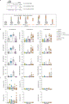
(a) Specific pathogen-free (SPF) mice were colonized with S. epidermidis strain LM087 or LM088. After 6 weeks, serum was harvested and antibody titers measured against the colonist by ELISA (n=24, 23, 6 or 3, one to three independent experiments pooled). (b-d) SPF mice were colonized with LM088 for 2–9 weeks; serum and nasal washes were tested for the presence of α-LM088 antibodies by ELISA. The graphs show all mice analyzed (n=5–9/timepoint, two independent experiments pooled). (e) Frequency of germinal center B cells among live B cells (Live+CD19+CD4) in the draining cervical lymph nodes (dLNs) of one representative mouse/group (left) and all mice analyzed (right) (n=3–8 mice/group, one to two independent experiments pooled). (f) Phylogenetic analysis of a set of S. epidermidis strains. (g) SPF mice were colonized with the indicated strains; five weeks later, the serum was tested for α-S. epidermidis antibodies and their strain specificity (n=4–8/group, two independent experiments pooled). (h) Bacterial IgG ELISA against a panel of Staphylococcus and Corynebacterium human skin isolates using serum from 11 healthy human donors. The data are displayed by bacterial strain (top) or by donor (bottom). All donors were tested in one experiment. (i) Bacterial IgG ELISA against the panel of Staphylococcus isolates used in (h) with either an unrelated monoclonal antibody or polyclonal antibodies isolated from human sera. We assumed that the concentration of IgG in human serum was 16 mg/ml and serially diluted the antibody mixture similarly to (h). (j) Bacterial IgG ELISA using serum from 10 non-human primates (NHPs) against S. epidermidis strains isolated from their skin. P-values were calculated using one-way ANOVA (e) with Tukey’s multiple comparisons test and two-sided paired Student t test (i). LOD: limit of detection. All graphs show mean +/− SEM.
Figure 2:
Figure 2:. The predominant target of the antibody response is Aap.
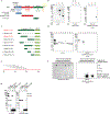
(a) Electron micrograph (and zoom) showing LM088 coated with IgG from the serum of mice colonized with LM088 for 6 weeks (n=1 experiment). (b) Schematic of the S. epidermidis cell surface mutants used in (c). (c) Bacterial ELISA using serum from LM087-colonized mice against a panel of cell surface mutants (n=23, three independent experiments pooled). (d) Immunoblot of cell lysate (L) and culture supernatant (S) from LM087 and LM088 using serum from LM088-colonized mice. Representative of three experiments. (e) Schematic showing predicted sortase substrates from S. epidermidis LM087 (purple), LM088 (blue) or both strains (grey). (f) Immunoblot against LM088 (blue), LM088 Δaap (purple) or LM088 Δaap + plasmid-borne accumulation-associated protein (Aap) (black) using serum from LM088-colonized mice. (g) ELISA against LM088, LM088 Δaap, or LM088 Δaap expressing plasmid-borne Aap using serum from LM088-colonized mice (n=24, three experiments pooled). (h) Schematic of Aap in LM088 (left) and predicted structure of its two main domains using AlphaFold3 (right). (i) Immunoblot against the lysate of E. coli expressing superfolder-GFP (sGFP) fused to the minimal epitope identified in the B domain of Aap (aa 20–83) using serum from LM087- or LM088-colonized mice. One representative of three experiments. (j) SPF mice were colonized for 6 weeks with LM088 or LM088 Δaap. The plot shows one representative mouse per group. The graphs display the percent of germinal center (GC) B cells among live B cells (Live+CD19+CD4) in the cervical draining lymph nodes (middle) and the colony forming units (CFUs) recovered (right) (n=7–8 for flow, n=6 for CFUs, two experiments pooled). Graphs were analyzed using one-way ANOVA with Dunnett’s correction (c, g) and unpaired two-sided Student t tests (j). All serum samples are from mice colonized for 6 weeks. LOD: limit of detection. All graphs show mean +/− SEM. Uncropped immunoblot images are in Supplementary Fig. 1.
Figure 3:
Figure 3:. Redirecting the B cell response against a non-native immunogen.
(a) Schematic of the approach used to redirect the B cell response toward a non-native antigen of interest. (b) Antibody titers against LM088, Δaap, and Δaap expressing plasmid-borne Aap (Δaap+Aap) in mice colonized for 5 weeks with LM087 harboring an empty vector or expressing Aap on a plasmid. (c) Strategy used to express a non-native immunogen in S. epidermidis: an engineered version of Aap is expressed in which the parallel β-helix domain is replaced with the antigen. (d) SPF mice were colonized for 6 weeks with S. epidermidis LM088 Δaap engineered to express tetanus toxin fragment C (TTFC), a model immunogen. TTFC was either targeted for secretion (sTTFC) or attached to the cell wall by fusion to Aap (wTTFC) and the quantity of TTFC-specific antibodies were measured in the serum and in nasal washes (undiluted, absorbance at 450nm). (n=8/group, two independent experiments pooled). (e) IgG serum titers specific for diphtheria toxin (DT) in mice colonized with LM088 Δaap + Aap-TTFC (wTTFC) or + Aap fused to a catalytic mutant version of DT, Aap-DT (wDT) for 6 weeks. (n=8/group, two independent experiments pooled). (f) Mice colonized with either wTTFC or wDT were challenged with a lethal dose of tetanus toxin (150 ng/kg) 6 weeks later. (n=7–8/group, two independent experiments pooled). (g) Mice were injected with a lethal dose of tetanus toxin (110 ng/kg) preincubated with the serum of mice colonized with either wTTFC or wDT (n=10/group, serum shown in (d, e)). Of note, ~120 h post injection, 4/10 mice in the wTTFC group developed mild tetanus symptoms which never reached the humane endpoint (see *). P values were calculated with one-way ANOVA with Dunnett’s (b, e) and Tukey (d) multiple comparison tests and log-rank tests (f, g). LOD: limit of detection. All graphs show mean +/− SEM.
Figure 4:
Figure 4:. Generation of immunity using enzymatic conjugation.
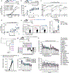
(a) Schematic representing the difference in approaches developed to elicit an antibody response against a non-native antigen using engineered S. epidermidis. (b) Chemical reaction between SpyCatcher (sc) and SpyTag-containing proteins. (c) Schematic representing the attachment of a non-native antigen to a strain expressing an Aap-SpyCatcher (Aap-sc) fusion. (d) S. epidermidis LM087 was engineered to express either a fusion between Aap and SpyCatcher (Aap-sc, yellow) or a catalytically dead version of SpyCatcher (Aap-sc*, grey). Both strains were incubated with 0.1 mg/ml of sGFP-SpyTag for 2 min and visualized by flow cytometry. (e, f) Mice were colonized with either Aap-sc conjugated with TTFC-SpyTag003 (Aap-sc-TTFC), the unconjugated strain (Aap-sc), or wTTFC; antibody titers specific for TTFC. Another group of mice were immunized intramuscularly with SpyCatcher-based nanoparticles (mi3) conjugated with TTFC. Antibody titers were measured at 6 weeks. BAL: bronchoalveolar lavage. (n=10/group). (g) Mice colonized with LM087, Aap-sc, wTTFC or Aap-sc-TTFC were challenged 6 weeks later with a lethal dose of tetanus toxin (1000 ng/kg) (n=8/group). (h) Mice were colonized with either Aap-sc or Aap-sc-TTFC. One group of mice received 13 colonizations on the head with a bacterial culture at OD600 = 6; the other groups received 2, 5 or 6 colonizations on the whole body from an OD600 = 12 culture. (n=7–8/group). (i-j) For the experiment described in (h), serum IgG titers against TTFC (i) and survival curve after challenge with a lethal dose of tetanus toxin (1000 ng/kg) for the mice colonized 13 or 2 times (j). P values were calculated with one-way ANOVA with Tukey’s multiple comparison tests (f), log-rank tests (g, j) or two-sided unpaired Student t test (i). Panel (d) is representative of three independent experiments and panels (e-j) show two independent experiments pooled. LOD: limit of detection. All graphs show mean +/− SEM.

Update of

References

    1. Naik S et al. Commensal-dendritic-cell interaction specifies a unique protective skin immune signature. Nature 520, 104–108 (2015). - PMC - PubMed
    1. Ivanov II et al. Induction of intestinal Th17 cells by segmented filamentous bacteria. Cell 139, 485–498 (2009). - PMC - PubMed
    1. Lathrop SK et al. Peripheral education of the immune system by colonic commensal microbiota. Nature 478, 250–254 (2011). - PMC - PubMed
    1. Cervantes-Barragan L et al. Lactobacillus reuteri induces gut intraepithelial CD4+CD8αα+ T cells. Science 357, 806–810 (2017). - PMC - PubMed
    1. Bousbaine D et al. A conserved Bacteroidetes antigen induces anti-inflammatory intestinal T lymphocytes. Science 377, 660–666 (2022). - PMC - PubMed

ADDITIONAL REFERENCES

    1. Naik S et al. Compartmentalized control of skin immunity by resident commensals. Science 337, 1115–1119 (2012). - PMC - PubMed
    1. Bjerre RD et al. Effects of sampling strategy and DNA extraction on human skin microbiome investigations. Sci. Rep. 9, 17287 (2019). - PMC - PubMed
    1. Coryell MP, Sava RL, Hastie JL & Carlson PE Application of MALDI-TOF MS for enumerating bacterial constituents of defined consortia. Appl. Microbiol. Biotechnol. 107, 4069–4077 (2023). - PMC - PubMed
    1. Ersching J et al. Germinal Center Selection and Affinity Maturation Require Dynamic Regulation of mTORC1 Kinase. Immunity 46, 1045–1058.e6 (2017). - PMC - PubMed
    1. Pirazzini M et al. Exceptionally potent human monoclonal antibodies are effective for prophylaxis and treatment of tetanus in mice. The Journal of Clinical Investigation (2021). - PMC - PubMed

MeSH terms

LinkOut - more resources