. 2025 Apr;18(2):312-325.
doi: 10.1016/j.mucimm.2024.12.001. Epub 2024 Dec 9.

IL-33 is associated with alveolar dysfunction in patients with viral lower respiratory tract disease

Affiliations

IL-33 is associated with alveolar dysfunction in patients with viral lower respiratory tract disease

Ian C Scott et al. Mucosal Immunol. 2025 Apr.

Abstract

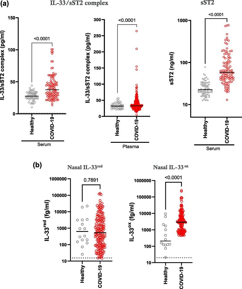
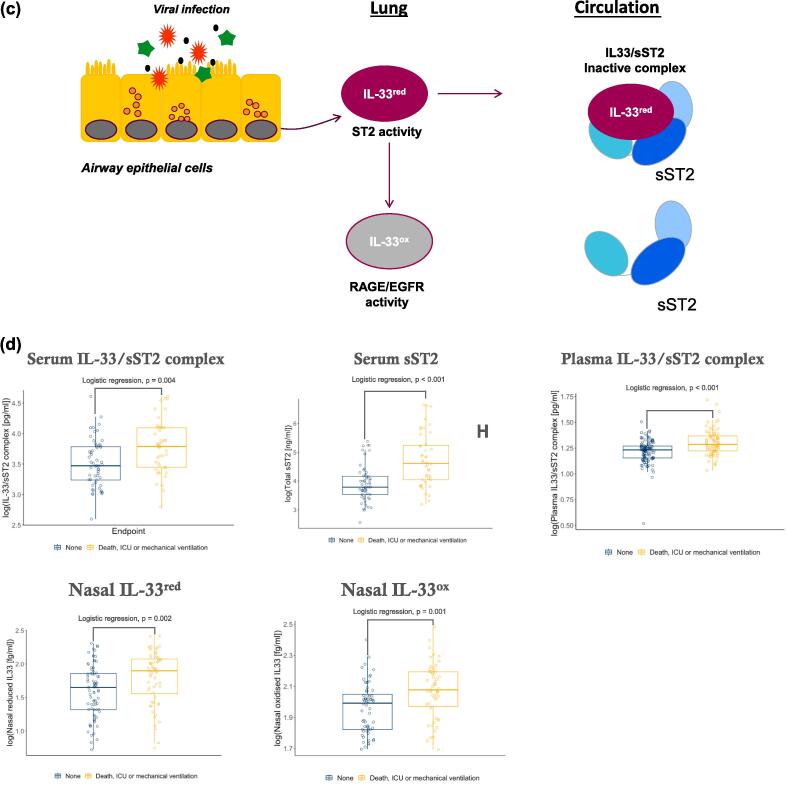
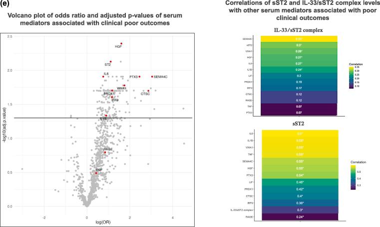
Interleukin (IL)-33 is released following tissue damage, causing airway inflammation and remodelling via reduced IL-33 (IL-33red)/serum stimulation-2 (ST2) and oxidised IL-33 (IL-33ox)/receptor for advanced glycation end products (RAGE)/epidermal growth factor receptor (EGFR) pathways. This study aimed to identify associations of IL-33 with clinical outcomes and pathological mechanisms during viral lower respiratory tract disease (LRTD). Ultra-sensitive immunoassays were developed to measure IL-33red, IL-33ox and IL-33/sST2 complexes in samples from patients hospitalised with COVID-19. Immunohistochemistry and multiomics were used to characterise lung samples. Elevated IL-33 in the airway and IL-33/sST2 complex in the circulation correlated with poor clinical outcomes (death, need for intensive care or mechanical ventilation). IL-33 was localised to airway epithelial and endothelial barriers, whereas IL1RL1 was expressed on aerocytes, alveolar endothelial cells specialised for gaseous exchange. IL-33 increased expression of mediators of neutrophilic inflammation, immune cell infiltration, interferon signalling and coagulation in endothelial cell cultures. Endothelial IL-33 signatures were strongly related with signatures associated with viral LRTD. Increased IL-33 release following respiratory viral infections is associated with poor clinical outcomes and might contribute to alveolar dysfunction. Although this does not show a causal relationship with disease, these results provide a rationale to evaluate pathological roles for IL-33 in viral LRTD.

Keywords: Aerocyte; Alarmin; Alveolar; COVID-19; IL-33; Viral LRTD.


Conflict of interest statement

Declarations of competing interests ICS, NVZ, VAN, KT, HK, ZL, CM, DGR, EE, MG, KH, ESC, ZB, DS, AF and AP are employees of AstraZeneca and may hold stock or stock options in AstraZeneca. JAC is a former employee of AstraZeneca and may hold stock or stock options in AstraZeneca. PJMO has received fees for scientific advisory boards from GSK, Moderna, Seqirus, Janssen and Sanofi Pasteur. MGS has received grants from the Department of Health and Social Care National Institute for Heath and Care Research, MRC, HPRU in Emerging and Zoonotic Infections, and the University of Liverpool during the conduct of the study; and has received other grants from Integrum Scientific LLC and Greensboro outside the submitted work. TW has received grants and fees from AstraZeneca, Bergenbio, Boehringer Ingelheim, Chiesi, GSK, Janssen, Olam, MMH, Synairgen, Union Chimique Belge and Valneva. DM, RT and JKB have no conflict of interests.

Figures

None
Graphical abstract
Fig. 1
Fig. 1
Summary of samples and experimental methods used in this study. COVID-19 = coronavirus disease 2019; MLF = mucosal lining fluid.
Fig. 2
Fig. 2
IL-33 and sST2 release are elevated in patients with COVID-19 and are associated with poor clinical outcomes. (A) measurement of IL-33/sST2 complex in serum (left panel) and plasma (middle panel) from healthy individuals (serum, n = 56; plasma, n = 40) and patients hospitalised with COVID-19 (serum, n = 100; plasma, n = 203). Measurement of serum sST2 (right panel) from healthy individuals (n = 76) and patients hospitalised with COVID-19 (n = 100). (B) measurement of IL-33red (left panel) and IL-33ox (right panel) in nasal mucosal lining fluids from healthy individuals (n = 16 and 14, respectively) and patients with COVID-19 (n = 151 and 130, respectively). Individual patient data points are shown. Median bar, p values are shown (Mann Whitney test) (A and B) (C) Schematic diagram of the different forms of IL-33 in the lung and in the circulation. IL-33red released from virally infected airway epithelial cells can be rapidly oxidized (IL-33ox) locally. IL-33red released into the circulation forms an inactive complex with sST2 (IL-33/sST2 complex) (D) measurement of serum IL-33/sST2 complex, serum sST2, plasma IL-33/sST2 complex, nasal IL-33red and nasal IL-33ox in patients who did or did not meet the composite clinical endpoint. (E) volcano plot (left panel) of associations of serum proteins with poor clinical outcomes adjusted for age, gender and ethnicity; the horizontal line represents adjusted p < 0.05; selected proteins are highlighted by red dots. Heat maps (right panel) showing the correlations between IL-33/sST2 complex and sST2 levels with selected other serum mediators; r values are shown; *adjusted p values < 0.05. COVID-19 = coronavirus disease 2019; IL = interleukin; IL-33ox = oxidised IL-33; IL-33red = reduced IL-33; ns = not significant; OR = odds ratio; r = correlation coefficient; sST2 = soluble serum stimulation-2. (For interpretation of the references to colour in this figure legend, the reader is referred to the web version of this article.)
Fig. 2
Fig. 2
IL-33 and sST2 release are elevated in patients with COVID-19 and are associated with poor clinical outcomes. (A) measurement of IL-33/sST2 complex in serum (left panel) and plasma (middle panel) from healthy individuals (serum, n = 56; plasma, n = 40) and patients hospitalised with COVID-19 (serum, n = 100; plasma, n = 203). Measurement of serum sST2 (right panel) from healthy individuals (n = 76) and patients hospitalised with COVID-19 (n = 100). (B) measurement of IL-33red (left panel) and IL-33ox (right panel) in nasal mucosal lining fluids from healthy individuals (n = 16 and 14, respectively) and patients with COVID-19 (n = 151 and 130, respectively). Individual patient data points are shown. Median bar, p values are shown (Mann Whitney test) (A and B) (C) Schematic diagram of the different forms of IL-33 in the lung and in the circulation. IL-33red released from virally infected airway epithelial cells can be rapidly oxidized (IL-33ox) locally. IL-33red released into the circulation forms an inactive complex with sST2 (IL-33/sST2 complex) (D) measurement of serum IL-33/sST2 complex, serum sST2, plasma IL-33/sST2 complex, nasal IL-33red and nasal IL-33ox in patients who did or did not meet the composite clinical endpoint. (E) volcano plot (left panel) of associations of serum proteins with poor clinical outcomes adjusted for age, gender and ethnicity; the horizontal line represents adjusted p < 0.05; selected proteins are highlighted by red dots. Heat maps (right panel) showing the correlations between IL-33/sST2 complex and sST2 levels with selected other serum mediators; r values are shown; *adjusted p values < 0.05. COVID-19 = coronavirus disease 2019; IL = interleukin; IL-33ox = oxidised IL-33; IL-33red = reduced IL-33; ns = not significant; OR = odds ratio; r = correlation coefficient; sST2 = soluble serum stimulation-2. (For interpretation of the references to colour in this figure legend, the reader is referred to the web version of this article.)
Fig. 3
Fig. 3
IL1RL1, AGER and EGFR are expressed in alveolar tissues in the lungs from patients with COVID-19. (A) representative images of haematoxylin and eosin-stained tissue slides of lungs from patients with COVID-19 pneumonia. Peri-vascular pathology, including lymphohistiocytic and granulocytic vasculitis in the small arteriole (top left panel). Immune cells were localised in the lumina of vessels in non-pneumonia lungs (top middle panel). Alveolar walls disrupted by microthrombi, loss of pneumocytes and endothelium, abundant fibrin and mixed inflammatory infiltrate (top right panel). Representative images of immunohistochemical-stained COVID-19 pneumonia lung for neutrophil elastase (bottom left panel, yellow stain), EPX (bottom middle panel, brown stain) and mast cell tryptase (bottom right panel, brown stain). Semi-quantitative analysis of immunostaining scores in post-mortem lung tissues from COVID-19 pneumonia (49 samples from 15 patients), non-COVID pneumonia (15 samples from 10 patients) and non-pneumonia lung (10 samples from 5 participants) for neutrophil elastase (left panel), EPX (middle panel), mast cell tryptase (right panel). Median bars and p values are shown (one-way ANNOVA tests). (B) representative images of immunohistochemical-stained slides for IL-33 in COVID-19 (left panels) and non-COVID pneumonia (right panels) lungs; large blood vessel (brown arrows, top panels), vascular smooth muscle cells (blue arrows, top panels), small blood vessels (black arrows, middle panels) and basal epithelial cells of bronchi and bronchioles (purple arrows, bottom panels). Semi-quantitative analysis of endothelial IL-33 (left panel), vascular smooth muscle cell IL-33 (middle panel), and epithelial IL-33 (right panel). Median bars and p values are shown (one-way ANNOVA tests). (C) UMAP dimensionally reduction plots showing the cell states identified in the complete dataset and the expression levels (log normalised) of IL1RL1, AGER, EGFR and IL-33 in post-mortem lungs from patients with COVID-19 and non-COVID participants (11 6313 total cells). (D) dot plots of log-normalised expression levels of IL1RL1 (left panels) and IL-33 (right panels) in broad cell types (top panels) and in endothelial cell states (bottom panels) in lungs from patients with COVID-19 (n = 19) and patients without COVID-19 (n = 7). (E) dot plots of normalised scaled expression profiles of IL1RL1, IL1R1, IL18R1 and IL1RL2 in endothelial cell types in the lungs from patients with COVID-19 and patients without COVID-19. COVID-19 = coronavirus disease 2019; EPX = eosinophil peroxidase; IL = interleukin; UMAP = uniform manifold approximation and projection. (For interpretation of the references to colour in this figure legend, the reader is referred to the web version of this article.)
Fig. 3
Fig. 3
IL1RL1, AGER and EGFR are expressed in alveolar tissues in the lungs from patients with COVID-19. (A) representative images of haematoxylin and eosin-stained tissue slides of lungs from patients with COVID-19 pneumonia. Peri-vascular pathology, including lymphohistiocytic and granulocytic vasculitis in the small arteriole (top left panel). Immune cells were localised in the lumina of vessels in non-pneumonia lungs (top middle panel). Alveolar walls disrupted by microthrombi, loss of pneumocytes and endothelium, abundant fibrin and mixed inflammatory infiltrate (top right panel). Representative images of immunohistochemical-stained COVID-19 pneumonia lung for neutrophil elastase (bottom left panel, yellow stain), EPX (bottom middle panel, brown stain) and mast cell tryptase (bottom right panel, brown stain). Semi-quantitative analysis of immunostaining scores in post-mortem lung tissues from COVID-19 pneumonia (49 samples from 15 patients), non-COVID pneumonia (15 samples from 10 patients) and non-pneumonia lung (10 samples from 5 participants) for neutrophil elastase (left panel), EPX (middle panel), mast cell tryptase (right panel). Median bars and p values are shown (one-way ANNOVA tests). (B) representative images of immunohistochemical-stained slides for IL-33 in COVID-19 (left panels) and non-COVID pneumonia (right panels) lungs; large blood vessel (brown arrows, top panels), vascular smooth muscle cells (blue arrows, top panels), small blood vessels (black arrows, middle panels) and basal epithelial cells of bronchi and bronchioles (purple arrows, bottom panels). Semi-quantitative analysis of endothelial IL-33 (left panel), vascular smooth muscle cell IL-33 (middle panel), and epithelial IL-33 (right panel). Median bars and p values are shown (one-way ANNOVA tests). (C) UMAP dimensionally reduction plots showing the cell states identified in the complete dataset and the expression levels (log normalised) of IL1RL1, AGER, EGFR and IL-33 in post-mortem lungs from patients with COVID-19 and non-COVID participants (11 6313 total cells). (D) dot plots of log-normalised expression levels of IL1RL1 (left panels) and IL-33 (right panels) in broad cell types (top panels) and in endothelial cell states (bottom panels) in lungs from patients with COVID-19 (n = 19) and patients without COVID-19 (n = 7). (E) dot plots of normalised scaled expression profiles of IL1RL1, IL1R1, IL18R1 and IL1RL2 in endothelial cell types in the lungs from patients with COVID-19 and patients without COVID-19. COVID-19 = coronavirus disease 2019; EPX = eosinophil peroxidase; IL = interleukin; UMAP = uniform manifold approximation and projection. (For interpretation of the references to colour in this figure legend, the reader is referred to the web version of this article.)
Fig. 4
Fig. 4
IL-33 drives type I inflammation and coagulation pathways in endothelial cells. (A) heat map (left panel) depicting differential gene expression (log2FC, fdr < 0.05) in HUVECs following treatment for 6 h with IL-33 versus vehicle control (left column) and IL-33/tozorakimab versus IL-33/isotype control (right column). GO pathway enrichment analysis (right panel) number of biological processes for IL-33/tozorakimab-driven gene expression changes (6 h and 24 h, fdr < 0.05 and abs[logFC] > 1). (B) schematic representation of endothelial IL-33 transcriptomic signatures, disease signature mapping and downstream pathways and network analyses (top panel) and KEGG pathway enrichment analysis of disease associations of endothelial IL-33 signatures (bottom panel). (C) GO pathway enrichment analysis of biological processes for serum proteins (p < 0.05) associated with poor clinical outcomes in patients hospitalised with COVID-19. (D) integrated omics and network proximity analyses of associations of IL-33/tozorakimab endothelial signatures with serum mediators in COVID-19 patients and quantification of the weighted average distance between serum mediators and endothelial signatures in COVID-19 compared with a randomly distributed network (p < 0.01). COVID-19 = coronavirus disease 2019; fdr = false discovery rate; GO = Gene Ontology; KEGG = Kyoto Encyclopaedia of Genes and Genomes; IL = interleukin.
Fig. 4
Fig. 4
IL-33 drives type I inflammation and coagulation pathways in endothelial cells. (A) heat map (left panel) depicting differential gene expression (log2FC, fdr < 0.05) in HUVECs following treatment for 6 h with IL-33 versus vehicle control (left column) and IL-33/tozorakimab versus IL-33/isotype control (right column). GO pathway enrichment analysis (right panel) number of biological processes for IL-33/tozorakimab-driven gene expression changes (6 h and 24 h, fdr < 0.05 and abs[logFC] > 1). (B) schematic representation of endothelial IL-33 transcriptomic signatures, disease signature mapping and downstream pathways and network analyses (top panel) and KEGG pathway enrichment analysis of disease associations of endothelial IL-33 signatures (bottom panel). (C) GO pathway enrichment analysis of biological processes for serum proteins (p < 0.05) associated with poor clinical outcomes in patients hospitalised with COVID-19. (D) integrated omics and network proximity analyses of associations of IL-33/tozorakimab endothelial signatures with serum mediators in COVID-19 patients and quantification of the weighted average distance between serum mediators and endothelial signatures in COVID-19 compared with a randomly distributed network (p < 0.01). COVID-19 = coronavirus disease 2019; fdr = false discovery rate; GO = Gene Ontology; KEGG = Kyoto Encyclopaedia of Genes and Genomes; IL = interleukin.

References

    1. Cayrol C., Girard J.P. Interleukin-33 (IL-33): A critical review of its biology and the mechanisms involved in its release as a potent extracellular cytokine. Cytokine. 2022;156 - PubMed
    1. Rabe K.F., Celli B.R., Wechsler M.E., et al. Safety and efficacy of itepekimab in patients with moderate-to-severe COPD: a genetic association study and randomised, double-blind, phase 2a trial. Lancet Respir Med. 2021;9:1288–1298. - PubMed
    1. Wechsler M.E., Ruddy M.K., Pavord I.D., et al. Efficacy and Safety of Itepekimab in Patients with Moderate-to-Severe Asthma. N Engl J Med. 2021;385:1656–1668. - PubMed
    1. Robinson K.M., Ramanan K., Clay M.E., McHugh K.J., Rich H.E., Alcorn J.F. Novel protective mechanism for interleukin-33 at the mucosal barrier during influenza-associated bacterial superinfection. Mucosal Immunol. 2018;11:199–208. - PMC - PubMed
    1. Brunner T.M., Serve S., Marx A.F., et al. A type 1 immunity-restricted promoter of the IL-33 receptor gene directs antiviral T-cell responses. Nat Immunol. 2024;25:256–267. - PMC - PubMed