Review
. 2025 May 1;32(5):649-659.
doi: 10.5551/jat.65298. Epub 2024 Dec 10.

A Rare Case of Autoimmune-Mediated Lecithin:Cholesterol Acyltransferase Insufficiency Manifesting as the Acute Onset of Extremely Hypo-High-Density Lipoprotein-Cholesterolemia and Spontaneous Improvement: A Case Report with a Review of the Literature

Affiliations
Review

A Rare Case of Autoimmune-Mediated Lecithin:Cholesterol Acyltransferase Insufficiency Manifesting as the Acute Onset of Extremely Hypo-High-Density Lipoprotein-Cholesterolemia and Spontaneous Improvement: A Case Report with a Review of the Literature

Atsuko Tamaki et al. J Atheroscler Thromb. .

Abstract

A 59-year-old Japanese woman was referred for an extremely low level of circulating high-density lipoprotein cholesterol (HDL-C). The serum HDL-C level had long been within the normal range but suddenly decreased asymptomatically to 7 mg/dL. She had no typical symptoms associated with familial lecithin, cholesterol acyltransferase deficiency (FLD), including proteinuria, anemia, and corneal opacity. The circulating level of ApoA-1 was also markedly decreased at 48 mg/dL, and the proportion of esterified cholesterol to free cholesterol was irregularly low at 26%. Whole-genome sequencing revealed no apparent pathological mutations in the LCAT gene. Notably, anti-LCAT antibodies were detected in the serum at 146±1.7 ng/mL, resulting in her being diagnosed with acquired LCAT insufficiency (ALCATI) caused by anti-LCAT antibodies. Five years after her HDL-C levels spontaneously decreased, they increased without any identifiable cause. To our knowledge, only six cases of ALCATI caused by anti-LCAT antibodies have been reported to date. In contrast to the present case, previously reported cases of ALCATI manifested proteinuria that improved with steroid therapy. The unique clinical course in the present case highlights the heterogeneity of ALCATI, warranting further research to clarify the molecular pathophysiology of FLD and ALCATI.

Keywords: Autoantibody; Hypo-high-density cholesterolemia; Lecithin-cholesterol acyltransferase; Spontaneous improvement.


Conflict of interest statement

Atsuko Tamaki, Ken Yonaha, Yohei Ishiki, Moriyuki Uehara, Yoshiro Nakayama, Ken-ichiro Honma, Rei Chinen, Tsugumi Uema, Shiki Okamoto, Junko Miyoshi, Mika Kirinashizawa, Kazuki Sato, Tsutomu Aohara, Misato Yamamoto, and Hiroaki Masuzaki have not been disclosed. Masayuki Kuroda received patent royalty and licensing fees from CellGenTech, Inc. Yoshiro Maezawa received clinical research funding from NTT Docomo, Japan. Koutaro Yokote has received honoraria from MSD K.K., Kowa Company, Ltd., Sanofi K.K., Sumitomo Pharma Co., Ltd., Daichi Sankyo Company, Limited, Taisho Pharmaceutical Co., Ltd., Mitsubishi Tanabe Pharma Corporation, Boehringer Ingelheim International GmbH., Novartis Pharma K.K., Novo Nordisk Pharma Ltd., Bayer Yakuhin, Ltd., Pfizer Japan Inc., and received clinical research funding from CellGenTech, Inc. KY also received scholarship grants from Abbot Japan LLC, Eisai Co., Ltd., Otsuka Pharmaceutical Co., Ltd., Kowa Company, Ltd., Sumitomo Pharma Co., Ltd., Taisho Pharmaceutical Co., Ltd., Takeda Pharmaceutical Company Limited, Mitsubishi Tanabe Pharma Corporation, TEIJIN PHARMA LIMITED, Eli Lily Japan K.K., Boehringer Ingelheim International GmbH, MOCHIDA PHARMACEUTICAL Co., Ltd..

Figures

Fig.1. Schematic illustration of phenotypic differences between FLD and ALCATI
FLD is caused by a mutation in LCAT, whereas ALCATI is caused by acquired antibodies against LCAT. Both conditions commonly lead to a dysfunction of LCAT activity, causing a decrease in HDL-C and LDL-C in circulation, as well as the appearance of abnormal lipoproteins. FLD often presents as corneal opacity, corneal ring, hemolysis, and renal impairment with proteinuria due to lipid deposition in the glomerular capillary walls. In contrast, ALCATI represents proteinuria associated with membranous nephropathy. Renal impairment with FLD takes a couple of decades to develop, whereas renal impairment with ALCATI occurs within a few years of onset. LCAT, lecithin:cholesterol acyltransferase; HDL-C, high-density lipoprotein cholesterol; LDL-C, low-density lipoprotein cholesterol.
Fig.2. Time course of serum lipid profile in the patient
The circulating level of HDL-C had been around 67-100 mg/dL until 5 years ago, when it suddenly decreased to 7 mg/dL 4 years ago without symptoms and remained low around 2-3 mg/dL thereafter. Just after the diagnosis and four years after the onset, the circulating level of HDL-C spontaneously increased without any treatments or apparent triggers. After the initiation of the administration of pemafibrate, a selective peroxisome proliferator-activated receptor α modulator (SPPARM-α), the serum lipid profile markedly improved to the normal range. The circulating level of LDL-C slightly fluctuated in synchrony with that of HDL-C. In contrast, the circulating levels of TG fluctuated inversely proportional to HDL-C and LDL-C levels. An HPLC analysis demonstrated that the serum TG levels in fractions 7 to 10, corresponding to a large LDL amount, decreased after the improvement of HDL-C (Fig. 4B), suggesting that impaired LDL hydrolysis contributed to the variation in serum TG levels. HDL-C, high-density lipoprotein cholesterol; LDL-C, low-density lipoprotein cholesterol; TG, triglyceride; HPLC, high-performance liquid chromatography.
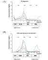
Fig.3. Profile of HPLC-GFC analyses at the diagnosis (A) and after the spontaneous improvement (B)
At the diagnosis, TC, TG, FC, and PL were extremely low in fractions 14 to 20 related to HDL at 8.33 mg/dL, 5.61 mg/dL, 7.34 mg/dL, 77.1 mg/dL, respectively (A). After spontaneous improvement, TC, TG, FC, and PL were increased in fractions 14 to 20, at 24.6 mg/dL, 11.1 mg/dL, 4.48 mg/dL, and 112.8 mg/dL, respectively. The peaks were observed at fraction 18, which was identical in normal subjects (B). Regarding fractions related to LDL, the peaks of TC, TG, FC, and PL were observed in fraction 8, whereas these peaks were typically observed in fraction 9 in healthy subjects (A). After the spontaneous improvement, although the peaks of these lipids remained in fraction 8, the content of TC in fractions 7 to 10, corresponding to large LDL, increased from 9.35% to 28.8%, and TG in fractions 7 to 10 decreased from 42.0% to 19.8%, suggesting that the hydrolysis of LDL was improved (B). HPLC-GFC, high-performance liquid chromatography with a gel filtration column; TC, total cholesterol; TG, triglyceride; FC, free cholesterol; PL, phospholipid; HDL, high-density lipoprotein; LDL, low-density lipoprotein.
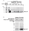
Fig.4. Western blot analyses following immunoprecipitation: (A) Serum from the patient and a healthy subject
To confirm that the serum from the patient was duly immunoprecipitated with recombinant human LCAT (rhLCAT), we combined the serum and rhLCAT and performed immunoprecipitation and subsequent western blot analyses. we used normal pooled serum (Kohjin bio, Japan) as a negative control (lane 2, 3) and the normal pooled serum with anti-human LCAT rabbit monoclonal antibodies as a positive control (lane 4, 5). Serum from the patient was immunoprecipitated with rhLCAT (lane 7, 9). Furthermore, the patient-derived LCAT proteins were detected when serum from the patient was precipitated with magnetic beads (lane 6, 8). (B) serum from the patient with adjusted anti-LCAT antibodies and protein A-agarose beads amounts. A significantly greater amount of LCAT protein was detected after adding anti-LCAT antibodies to the serum from the patient upon improvement. Despite an increased amount of the beads, however, the amount of LCAT protein was not increased. LCAT, lecithin:cholesterol acyltransferase.

References

    1. Pavanello C, Calabresi L: Genetic, biochemical, and clinical features of LCAT deficiency: update for 2020. Curr Opin Lipidol, 2020; 31: 232-237 - PubMed
    1. Jansen M, Pfaffelhuber P, Hoffmann MM, Puetz G, Winkler K: In silico modeling of the dynamics of low density lipoprotein composition via a single plasma sample. Journal of Lipid Research, 2016; 57: 882-893 - PMC - PubMed
    1. Kuroda M, Bujo H, Yokote K, Murano T, Yamaguchi T, Ogura M, Ikewaki K, Koseki M, Takeuchi Y, Nakatsuka A, Hori M, Matsuki K, Miida T, Yokoyama S, Wada J, Harada-Shiba M: Current Status of Familial LCAT Deficiency in Japan. J Atheroscler Thromb, 2021; 28: 679-691 - PMC - PubMed
    1. Ishibashi R, Takemoto M, Tsurutani Y, Kuroda M, Ogawa M, Wakabayashi H, Uesugi N, Nagata M, Imai N, Hattori A, Sakamoto K, Kitamoto T, Maezawa Y, Narita I, Hiroi S, Furuta A, Miida T, Yokote K: Immune-mediated acquired lecithin-cholesterol acyltransferase deficiency: A case report and literature review. J Clin Lipidol, 2018; 12: 888-897 e882 - PubMed
    1. Takahashi S, Hiromura K, Tsukida M, Ohishi Y, Hamatani H, Sakurai N, Sakairi T, Ikeuchi H, Kaneko Y, Maeshima A, Kuroiwa T, Yokoo H, Aoki T, Nagata M, Nojima Y: Nephrotic Syndrome Caused by Immune-Mediated Acquired LCAT Deficiency. Journal of the American Society of Nephrology, 2013; 24: 1305-1312 - PMC - PubMed

MeSH terms

Substances