Clinical Trial
. 2025 Jan;637(8048):1218-1227.
doi: 10.1038/s41586-024-08305-z. Epub 2024 Dec 11.

Neutralizing GDF-15 can overcome anti-PD-1 and anti-PD-L1 resistance in solid tumours

Ignacio Melero  1   2 Maria de Miguel Luken  3 Guillermo de Velasco  4 Elena Garralda  5 Juan Martín-Liberal  6 Markus Joerger  7 Guzman Alonso  8 Maria-Elisabeth Goebeler  9 Martin Schuler  10   11 David König  12 Reinhard Dummer  13 Maria Reig  14   15 Maria-Esperanza Rodriguez Ruiz  16 Emiliano Calvo  3 Jorge Esteban-Villarrubia  4 Arjun Oberoi  5 Paula Sabat  6 Juan José Soto-Castillo  6 Kira-Lee Koster  7 Omar Saavedra  8 Cyrus Sayehli  9 Tanja Gromke  10   11 Heinz Läubli  12 Egle Ramelyte  13 Marta Fortuny  14   15 Ana Landa-Magdalena  16 Irene Moreno  3 Javier Torres-Jiménez  4 Alberto Hernando-Calvo  5 Dagmar Hess  7 Fabricio Racca  8 Heike Richly  10   11 Andreas M Schmitt  12 Corinne Eggenschwiler  13 Marco Sanduzzi-Zamparelli  14   15 Anna Vilalta-Lacarra  16 Jörg Trojan  17 Christine Koch  17 Peter R Galle  18 Friedrich Foerster  18 Zlatko Trajanoski  19 Hubert Hackl  19 Falk Gogolla  19 Florestan J Koll  20 Peter Wild  21 Felix Kyoung Hwan Chun  20 Henning Reis  21 Peter Lloyd  22 Matthias Machacek  23 Thomas F Gajewski  24 Wolf H Fridman  25 Alexander M M Eggermont  26   27 Ralf Bargou  28 Sandra Schöniger  29 Josef Rüschoff  29 Anastasiia Tereshchenko  29 Carina Zink  30 Antonio da Silva  31 Felix S Lichtenegger  32 Julia Akdemir  32 Manfred Rüdiger  32 Phil L'Huillier  32 Aradhana Dutta  32 Markus Haake  32 Alexandra Auckenthaler  32 Ana Gjorgjioska  32 Bernhard Rössler  32 Frank Hermann  32 Mara Liebig  32 Daniela Reichhardt  32 Christine Schuberth-Wagner  32 Jörg Wischhusen  33 Petra Fettes  32 Marlene Auer  32 Kathrin Klar  32 Eugen Leo  34
Affiliations
Clinical Trial

Neutralizing GDF-15 can overcome anti-PD-1 and anti-PD-L1 resistance in solid tumours

Ignacio Melero et al. Nature. 2025 Jan.

Erratum in

  • Author Correction: Neutralizing GDF-15 can overcome anti-PD-1 and anti-PD-L1 resistance in solid tumours.
    Melero I, de Miguel Luken M, de Velasco G, Garralda E, Martín-Liberal J, Joerger M, Alonso G, Goebeler ME, Schuler M, König D, Dummer R, Reig M, Rodriguez Ruiz ME, Calvo E, Esteban-Villarrubia J, Oberoi A, Sabat P, Soto-Castillo JJ, Koster KL, Saavedra O, Sayehli C, Gromke T, Läubli H, Ramelyte E, Fortuny M, Landa-Magdalena A, Moreno I, Torres-Jiménez J, Hernando-Calvo A, Hess D, Racca F, Richly H, Schmitt AM, Eggenschwiler C, Sanduzzi-Zamparelli M, Vilalta-Lacarra A, Trojan J, Koch C, Galle PR, Foerster F, Trajanoski Z, Hackl H, Gogolla F, Koll FJ, Wild P, Chun FKH, Reis H, Lloyd P, Machacek M, Gajewski TF, Fridman WH, Eggermont AMM, Bargou R, Schöniger S, Rüschoff J, Tereshchenko A, Zink C, da Silva A, Lichtenegger FS, Akdemir J, Rüdiger M, L'Huillier P, Dutta A, Haake M, Auckenthaler A, Gjorgjioska A, Rössler B, Hermann F, Liebig M, Reichhardt D, Schuberth-Wagner C, Wischhusen J, Fettes P, Auer M, Klar K, Leo E. Melero I, et al. Nature. 2025 Mar;639(8054):E18. doi: 10.1038/s41586-025-08827-0. Nature. 2025. PMID: 40000750 Free PMC article. No abstract available.

Abstract

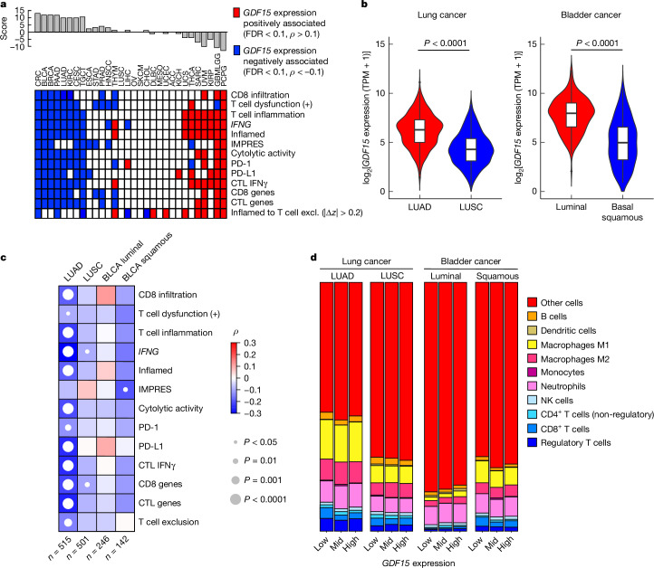
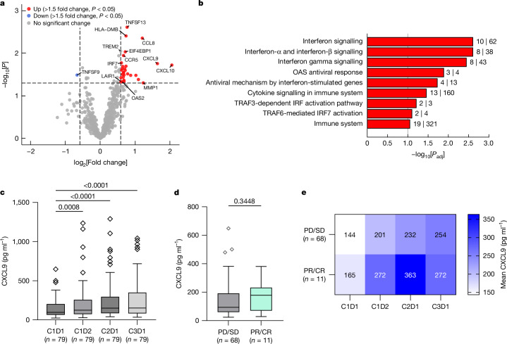
Cancer immunotherapies with antibodies blocking immune checkpoint molecules are clinically active across multiple cancer entities and have markedly improved cancer treatment1. Yet, response rates are still limited, and tumour progression commonly occurs2. Soluble and cell-bound factors in the tumour microenvironment negatively affect cancer immunity. Recently, growth differentiation factor 15 (GDF-15), a cytokine that is abundantly produced by many cancer types, was shown to interfere with antitumour immune response. In preclinical cancer models, GDF-15 blockade synergistically enhanced the efficacy of anti-PD-1-mediated checkpoint inhibition3. In a first-in-human phase 1-2a study (GDFATHER-1/2a trial, NCT04725474 ), patients with advanced cancers refractory to anti-PD-1 or anti-PD-L1 therapy (termed generally as anti-PD-1/PD-L1 refractoriness) were treated with the neutralizing anti-GDF-15 antibody visugromab (CTL-002) in combination with the anti-PD-1 antibody nivolumab. Here we show that durable and deep responses were achieved in some patients with non-squamous non-small cell lung cancer and urothelial cancer, two cancer entities identified as frequently immunosuppressed by GDF-15 in an in silico screening of approximately 10,000 tumour samples in The Cancer Genome Atlas database. Increased levels of tumour infiltration, proliferation, interferon-γ-related signalling and granzyme B expression by cytotoxic T cells were observed in response to treatment. Neutralizing GDF-15 holds promise in overcoming resistance to immune checkpoint inhibition in cancer.


Conflict of interest statement

Competing interests: M.H. and J.W. are co-founders and stock owners of CatalYm. A.d.S., M. Rüdiger, P.L’H., M.H., J.A., A.D., A.A., A.G., F.H., M.A., M.L., D.R., C.S.-W., F.S.L., P.F., K.K., F.H., B.R. and E.L. are current or former employees of and/or hold stock options in Catalym. R.D., M.H., J.W., K.K., E.L., F.H., M. Rüdiger and C.S.-W. are inventors on patents related to visugromab and anti-GDF-15 treatment (WO2014049087, WO2015144855, WO2017055613, WO2022101263, WO2024052532, WO2024126808 and unpublished). I. Melero, H.H., R.B., R.D., J.W., F.J.K., A.M.M.E., W.H.F., T.F.G., P.L., M.M., S.S., A.T. and J.R. have received research funding or consulting or advisory fees from CatalYm. Several authors and investigators declare potentially competing interests (grants or contracts and/or royalties or licences, stock or stock options, consulting fees, honoria, travel or meeting support, or advisory or data safety monitoring board participation) apart from their relationship with CatalYm: I. Melero (Agenus, Alligator, Allmiral, AstraZeneca, Biontech, BMS, Boehringer Ingelheim, Bright Peak, Crescendo Biologics, Curon, Dynamicure, F-Star, Genmab, Highlight Therapeutics, HotSpot, Merck Serono, MSD, Merus, Mestag, Numab, PharmaMar, Pieris, Pierre Fabre, Pioneering Medicines, Roche, Sanofi and Servier); G.d.V. (Astellas, AstraZeneca, Bayer, BMS, Ipsen, Janssen MSD, Merck Serono, Pfizer and Roche); E.G. (Anaveon, BeiGene, Boehringer Ingelheim, Ellipses Pharma, F-Star Therapeutics, Hengrui, Incyte, Janssen Global Services, MabDiscovery, Medscape, MSD, Novartis, Seattle Genetics, Sanofi, Roche, SeaGen, Taiho and Thermo Fisher); J.M.-L. (Astellas, BMS, Highlight Therapeutics, Merck Serono, MSD, Novartis, Ipsen, Roche, Sanofi and Pierre Fabre); M.J. (Adoram, AstraZeneca, Debiopharm, Novartis, Roche, Sanofi and Takeda); M.-E.G. (BMS, Janssen, Novartis and Roche); M.S. (Amgen, AstraZeneca, Blueprint Medicines, Boehringer Ingelheim, Bristol Myers Squibb, GlaxoSmithKline, Janssen, MSD, Novartis, Roche, Sanofi and Tacalyx); D.K. (AstraZeneca, Amgen, BMS, Merck Serono, MSD, Mirati, PharmaMar, Sanofi and Swiss Oncology in Motion); R.D. (Alligator, Amgen, BMS, MSD, Novartis, Pierre Fabre, Roche, Sun Pharma, Takeda, Sanofi, Second-Genome, Regeneron, T3 Pharma, MaviVax, Pfizer, Simcere and touchIME); M. Reig (AstraZeneca, Bayer, BMS, Boston Scientific, Biotoscana Pharma, Engitix Therapeutics, Eli Lilly, Geneos, Gilead, Ipsen, Merck, Roche and Universal DC); M.-E.R.R. (BMS, Imcore-Roche, Roche and Highlight Therapeutics); E.C. (Achilles, Adcendo, Alkermes, Amunix, Anaveon, Amcure, AstraZeneca, BeiGene, BMS, Boehringer, Chugai, Debiopharm, Diaccurate, Ellipses, Incyte, iTeos, Janssen, Merus, MonTa, MSD, Nanobiotix, Nouscom, Novartis, OncoDNA, PharmaMar, PsiOxus Therapeutics, Roche/Genentech, Sanofi, Servier, Shionogi, Syneos Health, TargImmune, T-knife and Tolremo); J.E.-V. (BMS, MSD and Pfizer); J.J.S.-C. (Eisai, Novartis, Pfizer and Seagen); K.-L.K. (Janssen and Takeda); O.S. (Affimed); C.S. (BMS, Eli Lilly and Boehringer Ingelheim); E.R. (BMS, Eli Lilly, Kyowa Kirin, Leo Pharma, MSD, Pierre Fabre and Sanofi); A.L.-M. (Incyte, Merck, PharmaMar, Pfizer, Roche, Rovi and Sanofi); I. Moreno (Digicore Initiative, Fondacion Intheos, Exafield, Guidepoint and Ellipses Pharma); A.H.-C. (BMS, Gilead, Kyowa Kirin, Merus and MSD); D.H. (Novartis and Roche); M.S.-Z. (Bayer, Instituto de Salud Carlos III (grant FI19/00222), BTG, MSD and Roche); A.V.-L. (AstraZeneca, BMS, Health in Code and Roche); J.T. (Amgen, AstraZeneca, Bristol Myers Squibb, Eisai, EXACT Therapeutics, Institut für Qualitätssicherung und Transparenz im Gesundheitswesen, Institut für Qualität und Wirtschaftlichkeit im Gesundheitswesen, Ipsen, Lilly ImClone, medupdate, Merck Serono, Merck Sharp & Dome, Oncolytics Biotech, onkowissen.de, Roche, Servier and streamedup!); C.K. (AstraZenca, BMS, DGVS, Incyte, MCI Deutschland, MSD, Servier, Ipsen, Merck Serono and Roche); P.R.G. (AstraZeneca, Bayer, BMS, Boston Scientific, Eisai, Guerbet, Ipsen, MSD, Lilly, Roche and Sirtex); F.F. (AstraZeneca, BMS, Eisai, Merck Serono, Servier and Roche); Z.T. (Boehringer Ingelheim); H.H. (Secarna); F.J.K. (German Cancer Aid (Deutsche Krebshilfe)); H. Reis (AstraZeneca, BMS, Boehringer Ingelheim, Chop GmbH, Diaceutics, GSK, HUeG, Janssen, MCI, Merck, Novartis, Roche Pharma and Sanofi); M.M. (LYO-X AG and TATAA Biocenter); T.G. (Allogene, Bicara, BMS, Merck, Pyxis and Samyang); W.H.F. (Anaveon, Atreca, Incendia, Ichnos Sciences, Genenta, OSE Immunotherapeutics, Oxford Biotherapeutics, Mestag, Novartis, Roche and Tabby); A.M.M.E. (Acetra, Agenus, BMS, Boehringer Ingelheim, BioInvent, BioNTech, Ellipses, Galecto, GSK, IO Biotech, IQVIA, Isa Pharmaceuticals, Merck & Co, MSD, Pfizer, Pierre Fabre, Sairopa, Sellas, SkylineDX, TigaTx and Trained Immunity); R.B. (Amgen, Avencell); J.R. (Astellas, AstraZeneca, BMS, Daiichi Sankyo, GSK, MSD, Merck and Sanofi); F.H. (Thermosome); P.F. (Curevac); J.W. (Bayrische Forschungsstiftung (FORTiTher) and the German Ministry for Education & Research (Transcan-3)). The remaining authors declare no competing interests.

Figures

Fig. 1
Fig. 1. Phase 1 dose escalation of visugromab in combination with nivolumab.
a, Swimmer plot for 25 patients with mixed solid tumours treated with escalating doses (DLs) of visugromab (0.3, 1.0, 3.0, 10 and 20 mg kg−1, once every other week) and nivolumab in last-line treatment, with relapsed or refractory cancer under prior anti-PD-1 or anti-PD-L1 treatment. Data show time course and remission status, with tumour regression (blue; greater than −5%) and PR (light green) as per RECIST 1.1. Broken lines indicate not evaluable (NE). MES, mesothelioma; UCEC, uterine corpus endometrial carcinoma; OMEL, ocular melanoma; CCA, cholangiocarcinoma; MEL, melanoma; RCC, renal cell carcinoma; CUP, cancer of unknown primary origin; OVA, ovarian cancer; TNBR, triple-negative breast cancer; CER, cervical cancer; H&N, head and neck squamous cell carcinoma; CRC, colorectal cancer; BF, backfill; PD, progressive disease; SD, stable disease; RT, radiotherapy; RFA, radiofrequency ablation. b, Fold change of CD4+FOXP3, CD8+, CD3+Ki67+ and CD3+GZMB+ T cells in evaluable, sequential biopsies of the above patients at day 14 (visugromab monotherapy) and day 28 (combination) compared to baseline (Bsl). Patients with >2-fold increase are depicted in black and patients with <2-fold increase are depicted in light grey. c, Representative immunohistochemical and immunofluorescence images of tumour area (green square 400 × 400 µm) demonstrating baseline (day 0; upper panel) versus monotherapy (day 14; middle panel) and combination therapy impact (day 28; lower panel), showing increase in CD8+ (purple label; n = 8), CD4+ (yellow label; n = 7), in the left panel, and CD3+Ki67+ (red label; n = 8) and increase in GZMB synthesis (pink label; n = 5), in the right panel. Source Data
Fig. 2
Fig. 2. Correlation of GDF15 with suppression of immune signatures in data from TCGA.
a, Immune-related analyses and relation to GDF15 mRNA expression ranked by a consensus score. IFNγ, interferon-γ. Two-sided Spearman’s rank correlation (coefficient ρ) test was performed. P values were adjusted for multiple testing for each indication across tested signature scores on the basis of the false discovery rate (FDR) according to the Benjamini–Hochberg method. Significant positive (FDR < 0.1 and ρ > 0.1) and negative (FDR < 0.1 and ρ < 0.1) relationships between GDF15 expression and immune signature score are indicated in red and blue, respectively. For inflamed to T cell exclusion (excl.), an absolute value of the difference in z-scores (|∆z|) > 0.2 was used as the cutoff (z-scores were calculated using Fisher’s z-transformation on ρ). ACC, adrenocortical carcinoma; BLCA, bladder urothelial carcinoma; BRCA, breast invasive carcinoma; CESC, cervical squamous cell carcinoma and endocervical adenocarcinoma; CHOL, cholangiocarcinoma; DLBC, diffuse large B-cell lymphoma; ESCA, oesophageal carcinoma; GBMLGG, glioblastoma and low-grade gliomas; HNSC, head and neck squamous cell carcinoma; KICH, kidney chromophobe; KIRC, kidney renal clear cell carcinoma; LUSC, lung squamous cell carcinoma; MESO, mesothelioma; OV, ovarian serous cystadenocarcinoma; PAAD, pancreatic adenocarcinoma; PCPG, pheochromocytoma and paraganglioma; PRAD, prostate adenocarcinoma; SARC, sarcoma; SKCM, skin cutaneous melanoma; STAD, stomach adenocarcinoma; TGCT, testicular germ cell tumours; THCA, thyroid carcinoma; THYM, thymoma; UCEC, uterine corpus endometrial carcinoma; UCS, uterine carcinosarcoma; UVM, uveal melanoma. b, Normalized GDF15 mRNA expression in LUAD (n = 515), lung squamous cell carcinoma (n = 501; left panel), luminal UC subtypes (luminal, luminal infiltrated, luminal papillary, n = 246) and basal squamous UC (n = 142, right panel). TPM, transcripts per million. Lower and upper hinges, first and third quartile (interquartile range (IQR)); thick line, median. The whiskers extend to the smallest and largest values no further than 1.5 × IQR. P values are from two-sided Wilcoxon rank sum test. c, Correlation analyses of normalized GDF15 expression in lung and bladder cancer subtypes with immune-related signatures; Spearman rank-based correlation coefficient rho (ρ) is indicated according to the legend. P values are from two-sided Spearman’s rank correlation test and not adjusted for multiple comparisons. IMPRES, immuno-predictive score. CTL, cytotoxic T lymphocytes. d, Distribution of averaged cell fractions according to GDF15 expression level (low, mid, high). NK, natural killer. Source Data
Fig. 3
Fig. 3. Visugromab in combination with nivolumab induces remissions in a fraction of anti-PD-1/PD-L1 relapsed or refractory last-line NSCLC and UC.
a,b, Swimmer plot for patients with NSCLC (a) and UC (b) illustrating response assessment according to RECIST 1.1 and time on study. Patients with PR or CR are highlighted in light and dark green, respectively; patients with SD but decrease in target lesion size (>5%; tumour shrinkage) are highlighted in blue. Light blue indicates SD, and PD is depicted in grey (X indicates detection of PD). Patients who received radiotherapy on treatment are not evaluable (NE (post RT)) anymore, which is indicated by a hatched fill. Scans were performed approximately every 8 weeks.
Fig. 4
Fig. 4. Induction of interferon-γ signalling in a fraction of anti-PD-1 and PD-L1 relapsed or refractory last-line cancer.
a, Volcano plot presenting differentially expressed genes in biopsies on day 14 of visugromab monotherapy compared to pretreatment biopsies in participants with >2-fold increase in CD8+ and CD4+ T cells (n = 5). P values are from two-sided moderated t-test using limma. Dashed vertical lines, −1.5 and +1.5 fold change. Dashed horizontal line, P = 0.05. b, Reactome pathway analysis shows that significantly upregulated genes from the PanCancer IO 360 panel in a are part of important inflammation-related pathways. P values are from two-sided Fisher’s exact test and adjusted according to the Benjamini–Hochberg method (Padj). Numbers of PanCancer IO 360 panel genes in the respective pathway (second number to right of each bar) and of significantly upregulated genes in the respective pathway (first number to right of each bar) are indicated. Grey vertical line, Padj = 0.1. OAS, oligoadenylate synthetases. c, Significant induction and maintenance of CXCL9 following treatment for 6 weeks in the blood of participants with NSCLC, HCC and UC in the phase 2a study. P values are reported from repeated-measures one-way analysis of variance (ANOVA) with Geisser–Greenhouse correction, corrected for multiple comparisons by Dunnett’s test. d, Baseline CXCL9 levels of participants with best overall response according to RECIST 1.1. P values are reported from two-tailed Mann–Whitney test. e, Heat map representing higher mean values (pg ml−1) of CXCL9 following treatment in participants with best overall response of PR and CR (lower row) compared to participants with best overall response of PD and SD (upper row). For the box plots in c,d: centre line, median; box, IQR; whiskers, 1.5 × IQR; outliers are depicted individually. C1D1, cycle 1 day 1; C1D2, cycle 1 day 2; C2D1, cycle 2 day 1; C3D1, cycle 3 day 1. Source Data
Fig. 5
Fig. 5. GDF-15 correlates with immune-cell density in the tumour at baseline across different tumour types of all patients enrolled in phase 1 and 2a expansions.
a, CD4+FOXP3+ T cell density, CD4+FOXP3 T cell density and CD3+Ki67+ T cell density are significantly reduced in tumour biopsies from patients in phase 1 and 2 with serum GDF-15 (sGDF-15) of >1.5 ng ml−1. P values are from two-sided Mann–Whitney test. b, In baseline tumour tissue with very high CD8+ T cell infiltration from patients in phase 1 and 2, low (pro-)GDF-15 expression is observed. P value is reported from two-sided Mann–Whitney test. c, TME characterization by PD-L1 tumour proportion score (TPS) and CD8+ T cell density (cells mm−2) in biopsies from patients in phase 1 and 2 before treatment (n = 97); PR and CR are highlighted in green. Source Data
Extended Data Fig. 1
Extended Data Fig. 1. PK/PD data observed in phase 1 is consistent with population-based model and predicts neutralization of free GDF-15 in the tumour.
(a) Observed concentrations of total visugromab and total GDF-15 (free and drug-bound GDF-15) determined in serum of phase 1 participants across all tested dose levels within 8 weeks of treatment and at end of treatment. Mean and individual data points. (b) A population PK/PD model was derived from NHP studies and updated with clinical phase 1 data to describe GDF-15 neutralization in tumour micro-vasculature. Free GDF-15 is predicted for serum GDF-15 baseline levels of 0.5 ng/mL (left panel), 2 ng/mL (middle panel) and 10 ng/mL (right panel) and dosing schemes (upper panel = q2wk, middle panel = q3wk, lower panel = q4wk) as well as different concentrations (red, solid line = 3 mg/kg, green dashed line = 10 mg/kg and blue dashed line = 20 mg/kg) of visugromab. The threshold indicates the average physiological serum GDF-15 level (0.5 ng/mL). Source Data
Extended Data Fig. 2
Extended Data Fig. 2. Visugromab in combination with nivolumab induces remissions in a fraction of anti-PD-1/-L1 relapsed/refractory last-line NSCLC and urothelial cancer.
(a, b) Spider plots of NSCLC (a) and UC (b) patients illustrating changes in tumour size, response assessment was done according to investigator-assessed RECIST1.1, and time on study. Patients with partial or complete response (PR and CR) are highlighted in light and dark green respectively, arrow indicates ongoing treatment. Source Data
Extended Data Fig. 3
Extended Data Fig. 3. Pharmacodynamic effects of visugromab monotherapy in phase 1.
(a) Exhaustion markers PDCD1, HAVCR2 and LAG3 are not differentially expressed in pre-(baseline) (n = 6) and on-treatment (D14) (n = 5) biopsies following visugromab therapy in trial participants with >2-fold (IMM) and participants with <2-fold (NOIMM) increase in CD8+ and CD4+ T cells. Boxplots black line, median; red line, mean; box, IQR; ns, not significant, (P > 0.5 from two-sided Wilcoxon rank sum test). (b) Volcano plot presenting differentially expressed genes (NS = no significant change) in biopsies on day 14 of visugromab monotherapy compared to pre-treatment biopsies in participants with <2-fold (NOIMM) increase in CD8+ and CD4 + T cells. P values are from moderated t-test using limma. Source Data
Extended Data Fig. 4
Extended Data Fig. 4. Impact of induction of Interferon-γ signalling in a fraction of anti-PD-1/PD-L1 relapsed/refractory last-line cancer on CXCL10 levels.
(a) Significant induction and maintenance of chemokine CXCL-10 following treatment for six weeks in the blood of phase 2a NSCLC, HCC and UC study participants. P values are reported from repeated measures ANOVA with Geisser-Greenhouse correction followed by Dunnett’s test corrected for multiple comparisons. (b) Baseline CXCL-10 levels between participant with best overall response (BOR) according to RECIST 1.1 of PD/SD (grey) and PR/CR (green). P values are reported from two-tailed Mann-Whitney test. (c) Heatmap representing higher mean values [pg/ml] of CXCL-10 following treatment in participants with BOR of PR or CR (lower row) compared to participants with BOR of PD or SD (upper row). (a-c) Boxplots centre line, median; box, IQR; whiskers, 1.5 x IQR; outliers are depicted individually. Source Data
Extended Data Fig. 5
Extended Data Fig. 5. Serum and intra-tumoural (pro-)GDF-15 inversely correlate with immune cell numbers in UC and NSCLC.
(a) Early-line, treatment-naïve UC (MIBC, NMIBC, mUC and UTUC) patients with high (n = 9) vs. low (n = 31) serum GDF-15 levels show reduced CD8+ T cell density, CD45+Ki67+ cell density and PD-L1+ tumour cell density in the tumour. P values were calculated by two-sided Mann-Whitney test. (b) Serum GDF-15 levels at baseline in phase 2a NSCLC and UC patients. The box bounds the inter-quartile range (IQR) divided by the median, and Tukey-style whiskers extend to a maximum of 1.5 x IQR beyond the box. Individual data points represent outliers. P value is reported from two-sided Mann-Whitney test. (c) Association of pro-GDF-15 H score and CD8+ T cell density in tumour tissue in NSCLC patients (left panel, n = 30) and UC patients (right panel, n = 19) of phase 2a. PR/CR with available baseline biopsies of the respective indications are highlighted in green. Best-fit of simple linear regression is depicted as red line with 95% confidence interval as light grey shaded area. Source Data
Extended Data Fig. 6
Extended Data Fig. 6. Serum GDF-15 at baseline is not predictive for response, but positively associated with weight gain in phase 2a patients.
(a) Serum GDF-15 levels at baseline of phase 2a NSCLC, UC and HCC patients according to RECIST 1.1 response assessment. (b) In NSCLC, UC and HCC patients with elevated GDF-15 (n = 17) serum levels at baseline weight gain is significantly higher than in patients with low GDF-15 (n = 21). Boxplots center line, median; box, IQR; whiskers, 1.5 x IQR; outliers are depicted individually. P values are reported from two-sided Mann-Whitney test. Source Data
Extended Data Fig. 7
Extended Data Fig. 7. CT and PET-CT scans of responding tumours.
(a-d) Participant with mesothelioma and large, bulky disease (phase 1 part). RECIST 1.1 response under treatment with partial response (a) and (c) show baseline tumour assessment with bulky disease in upper mediastinum at two levels. (b) and (d) show respective significant tumour reduction at corresponding scan level at Cycle 8, Day 1 (week 16). (e, f) Participant with metastatic non-squamous NSCLC, Baseline (e) vs. Cycle 12 (f) demonstrating partial response as per RECIST 1.1. (g, h) Participant with metastatic non-squamous NSCLC (phase 2a part). Baseline (g) vs. Cycle 35 with partial response (h) as per RECIST 1.1. (i-l) Participant with metastatic urothelial cancer (phase 2a part), shown is large retroperitoneal lesion. RECIST 1.1 response with −100% reduction (complete response). (i) Baseline assessment at Screening with lesion marked by red arrow and red line. (j) Cycle 5 Day 1 assessment: Partial response. (k) PET-CT at Cycle 28 Day 1 demonstrating complete metabolic response. (l) CT scan at Cycle 45 Day 1 with continued complete response.

Similar articles

Cited by

References

    1. Robert, C. A decade of immune-checkpoint inhibitors in cancer therapy. Nat. Commun.11, 3801 (2020). - PMC - PubMed
    1. Santiago-Sánchez, G. S., Fabian, K. P. & Hodge, J. W. A landscape of checkpoint blockade resistance in cancer: underlying mechanisms and current strategies to overcome resistance. Cancer Biol. Ther.25, 2308097 (2024). - PMC - PubMed
    1. Haake, M. et al. Tumor-derived GDF-15 blocks LFA-1 dependent T cell recruitment and suppresses responses to anti-PD-1 treatment. Nat. Commun.14, 4253 (2023). - PMC - PubMed
    1. Batlle, E. & Massagué, J. Transforming growth factor-β signaling in immunity and cancer. Immunity.50, 924–940 (2019). - PMC - PubMed
    1. Lodyga, M. & Hinz, B. TGF-β1 - a truly transforming growth factor in fibrosis and immunity. Semin. Cell Dev. Biol.101, 123–139 (2020). - PubMed

Publication types

MeSH terms