

Current implications of EEG and fNIRS as functional neuroimaging techniques for motor recovery after stroke
- PMID: 39664080
- PMCID: PMC11629311
- DOI: 10.1515/mr-2024-0010
Current implications of EEG and fNIRS as functional neuroimaging techniques for motor recovery after stroke
Abstract
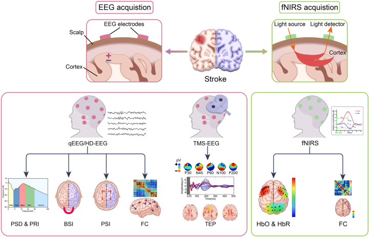
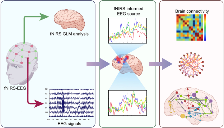
Persistent motor deficits are highly prevalent among post-stroke survivors, contributing significantly to disability. Despite the prevalence of these deficits, the precise mechanisms underlying motor recovery after stroke remain largely elusive. The exploration of motor system reorganization using functional neuroimaging techniques represents a compelling yet challenging avenue of research. Quantitative electroencephalography (qEEG) parameters, including the power ratio index, brain symmetry index, and phase synchrony index, have emerged as potential prognostic markers for overall motor recovery post-stroke. Current evidence suggests a correlation between qEEG parameters and functional motor outcomes in stroke recovery. However, accurately identifying the source activity poses a challenge, prompting the integration of EEG with other neuroimaging modalities, such as functional near-infrared spectroscopy (fNIRS). fNIRS is nowadays widely employed to investigate brain function, revealing disruptions in the functional motor network induced by stroke. Combining these two methods, referred to as integrated fNIRS-EEG, neural activity and hemodynamics signals can be pooled out and offer new types of neurovascular coupling-related features, which may be more accurate than the individual modality alone. By harnessing integrated fNIRS-EEG source localization, brain connectivity analysis could be applied to characterize cortical reorganization associated with stroke, providing valuable insights into the assessment and treatment of post-stroke motor recovery.
Keywords: electroencephalography; functional near-infrared spectroscopy; functional neuroimaging; motor function; stroke.
© 2024 the author(s), published by De Gruyter, Berlin/Boston.
Conflict of interest statement
Competing interests: Authors state no conflict of interest.
Figures
References
-
- Johnson CO, Nguyen M, Roth GA, Nichols E, Alam T, Abate D, et al. . GBD 2016 Stroke Collaborators Global, regional, and national burden of stroke, 1990–2016: a systematic analysis for the Global Burden of Disease Study 2016. Lancet Neurol. 2019;18:439–58. doi: 10.1016/s1474-4422(19)30034-1. - DOI - PMC - PubMed
Publication types
LinkOut - more resources
Full Text Sources