. 2024 Dec 11;14(53):39112-39121.
doi: 10.1039/d4ra05126b. eCollection 2024 Dec 10.

Black phosphorus/silk fibroin films hamper filamentous and invasive growth of Candida albicans

Affiliations

Black phosphorus/silk fibroin films hamper filamentous and invasive growth of Candida albicans

Martina Alunni Cardinali et al. RSC Adv. .

Abstract

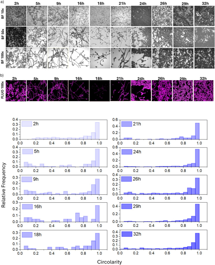
The ability of fungi and bacteria to form biofilms on surfaces poses a serious threat to health and a problem in industrial settings. In this work, we investigated how the surface stiffness of silk fibroin (SF) films is modulated by the interaction with black phosphorus (BP) flakes, quantifying the morphogenesis of C. albicans cells. Raman and infrared (IR) spectroscopies, along with scanning transmission electron microscopy, allowed us to quantify the thickness and diameter of BP flakes dispersed in the SF matrix (e.g., 5.5 nm in thickness and 20 μm in diameter), as well as an increase in beta-sheet secondary structures, resulting in the mesoscopic formation of a globular and nanofibrous surface. The formation of β-sheet crystals in the SF/BP film was correlated with a higher surface stiffness, influencing the shape of C. albicans cells and suppressing their filamentous growth. Raman spectroscopy analysis ultimately suggests an overall reduction in cell vitality and filmogenic capability of cells grown on fibroin-based films containing BP. Our results suggest that the conformational properties of SF can be suitably tuned to design optimized bioselective coatings for biomedical applications.


Conflict of interest statement

There are no conflicts to declare.

Figures

Fig. 1
Fig. 1. (a) STEM dark field images of BP flakes with SF. Top: a thin folded BP flake, bottom: a multilayer flake is observed; (b) optical image of the surface of SF/BP film, percentage fraction covered by BP flakes, and the ratio of relative Raman intensities of A1g and B2g bands; (c) Raman spectra (top) and ATR-FTIR (bottom) of SF and SF/BP films; (d) STEM bright field images of SF/BP films redissolved in PBS. The red circle shows the BP flake covered by SF redissolved in PBS at different magnifications. (e) Force/indentation depth and elastic modulus in SF and SF/BP films.
Fig. 2
Fig. 2. Optical images of C. albicans cells grown on SF, FTO and SF/BP substrates after (a) 2 and (b) 24 hours of incubation; (c) photograph of the FTO glass support with on top the SF film after crystal violet staining and box plots reporting the relative surface covered by C. albicans cells for SF, FTO and SF/BP substrates. Statistical significance (S) is assigned to a p-value ≤0.05.
Fig. 3
Fig. 3. (a) Optical images of C. albicans deposited on SF, SF/BP and on FTO (from top to bottom) recorded using a 3D Hirox digital microscope (HRX-01) and (b) histograms showing the relative frequency of individual forms of C. albicans cells.
Fig. 4
Fig. 4. (a) Optical images of C. albicans cells on the FTO substrate at different times of incubation and with different magnifications (BF10×, BF50×, BF100×), (b) fluorescence images (FLUO100×) obtained after crystal violet staining using the 100× objective and (c) histograms showing the relative frequency of individual forms of C. albicans cells on the FTO substrate.
Fig. 5
Fig. 5. (a) Raman spectra of C. albicans cells at 2 h deposition on FTO (black) and SF film (blue). This latter was obtained by subtracting pure SF contribution from the total profile of sample of C. albicans cells deposited on the SF substrate. The single asterisk marks the bands that assigned to proteins. (b) Raman spectra of C. albicans cells at 2 h deposition on SF (blue), and SF/BP (red) films. This latter was obtained by subtracting pure SF/BP contribution from the total profile of sample of C. albicans cells deposited on the SF/BP substrate. Double asterisks and “O” mark the bands assigned to cytochrome c and glycoproteins respectively.

References

    1. Qi Y. Wang H. Wei K. Yang Y. Zheng R. Y. Kim I. S. Zhang K. Q. Int. J. Mol. Sci. 2017;18:237. - PMC - PubMed
    1. Zhang W. Chen L. Chen J. Wang L. Gui X. Ran J. Xu G. Zhao H. Zeng M. Ji J. Qian L. Zhou J. Ouyang H. Zou X. Adv. Healthcare Mater. 2017;6:1700121. - PubMed
    1. Choudhury A. J. Gogoi D. Chutia J. Kandimalla R. Kalita S. Kotoky J. Chaudhari Y. B. Khan M. R. Kalita K. Surgery. 2016;159:539–547. - PubMed
    1. Schnaider L. Toprakcioglu Z. Ezra A. Liu X. Bychenko D. Levin A. Gazit E. Knowles T. P. Nano Lett. 2020;20:1590–1597. - PubMed
    1. Fei X. Jia M. Du X. Yang Y. Zhang R. Shao Z. Zhao X. Chen X. Biomacromolecules. 2013;14:4483–4488. - PubMed

LinkOut - more resources