. 2024 Jul;31(2):78-85.
doi: 10.18787/jr.2024.00012. Epub 2024 Jul 31.

Salvaging Vision: A Study of Non-Traumatic Optic Neuropathies

Affiliations

Salvaging Vision: A Study of Non-Traumatic Optic Neuropathies

Hetal Marfatia et al. J Rhinol. 2024 Jul.

Abstract

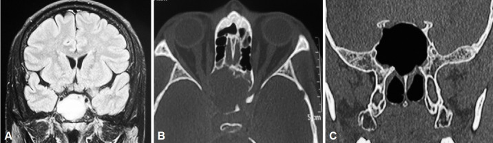
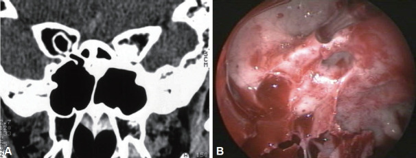
Background and objectives: Various ear, nose, and throat (ENT) conditions can result in vision loss. The purpose of this study is to identify the etiologies, presentations, and radiological findings associated with impaired vision in the context of ENT. Additionally, this article discusses management protocols, including optic nerve decompression and orbital decompression.

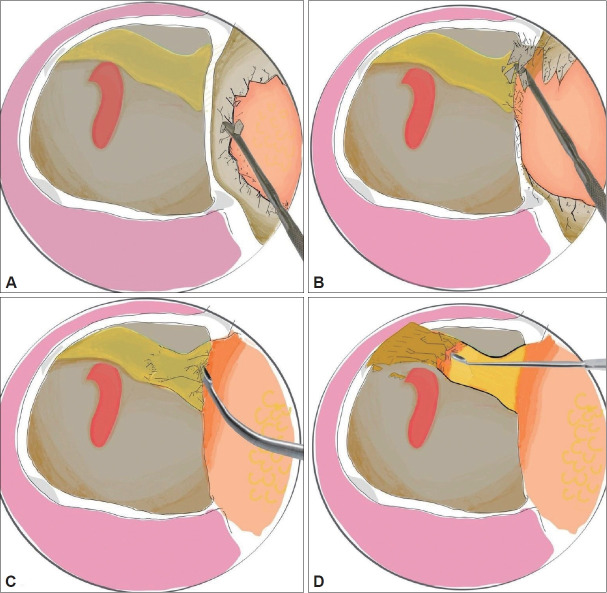
Methods: In a retrospective study, we examined the period from 2016 to 2022 at a tertiary care hospital in Mumbai, India. The analysis included 11 patients who presented with progressive diminution of vision. All patients received a regimen of broad-spectrum intravenous antibiotics and high-dose intravenous steroids. This was followed by either endoscopic optic nerve decompression or orbital decompression. Subsequent improvements in vision were documented, and any complications were evaluated.

Results: A total of 11 patients were treated with medical management followed by successful surgery, with 10 patients demonstrating significant vision improvement.

Conclusion: Identifying the etiology of vision loss and managing the condition can present challenges for otorhinolaryngologists. A thorough grasp of the underlying pathophysiology, combined with active surveillance of clinical and radiological indicators, can enable these clinicians to achieve effective and rewarding outcomes.

Keywords: Fibrous dysplasia; Mucocele; Non-traumatic; Optic nerve decompression; Sinusitis.


Conflict of interest statement

Conflicts of Interest The authors have no potential conflicts of interest to disclose.

Figures

Fig. 1.
Fig. 1.
Endoscopic anatomy of optic nerve. A: Schematic representation of intracanalicular part of optic nerve that can be assessed by transnasal endoscopy. B: Endoscopic anatomical relations of the right optic nerve showing carotid artery, opticocarotid recess and posterior ethmoidal artery in the Onodi cell in cadaveric dissection
Fig. 2.
Fig. 2.
Schematic diagram showing the steps of left trans-nasal endoscopic optic nerve decompression. A and B: The relationship of the left optic nerve with lamina papyracea and carotid. Removal of the lamina papyracea with a Freer’s elevator. C and D: Removal of the optic canal and annulus of Zinn with curette to decompress the optic nerve.
Fig. 3.
Fig. 3.
Contrast-enhanced computed tomography of the paranasal sinuses in a 62-year-old male patient presenting with headache and diminution of vision. A and B: Imaging revealed a sphenoid mucocele causing compression of the optic nerves. C: The patient underwent endoscopic marsupialization of the sphenoid mucocele and a postoperative scan for the same was done.
Fig. 4.
Fig. 4.
Coronal (A) and axial (B) computed tomography images of the paranasal sinuses showing ground glass opacity in the Onodi cell. A 25-year-old female patient presented to our outpatient department with left-sided diminution of vision and diplopia and left ophthalmoplegia of insidious onset for 15 days. On starting intravenous steroids, the ophthalmoplegia had improved. CT scan of paranasal sinuses revealed ground glass opacification of the Onodi cell suggestive of fibrous dysplasia with involvement of optic nerve on the left. Fundoscopy was done to check the viability of the optic disc plan endoscopic decompression. The patient required drilling of the dense fibrous dysplasia lesion in order to release the compression from the left optic nerve.
Fig. 5.
Fig. 5.
A rare case of osteopetrosis was identified in our study population (20-year-old male). The patient had presented with a recent onset, progressive diminution of vision on the left side. A: Axial CT scan revealed dense bone around the optic nerve causing optic canal stenosis. B: Endoscopic decompression of the left optic nerve was achievable only with the use of micro-drill. On follow-up, the progression of vision deterioration had halted and the patient was taken for endoscopic decompression of optic nerve on the right side after 3 months.

References

    1. Samardzic K, Samardzic J, Janjetovic Z, Samardzic I, Sekelj S, Latic-Hodzic L. Traumatic optic neuropathy - to treat or to observe? Acta Inform Med. 2012;20(2):131–2. - PMC - PubMed
    1. Rodriguez-Beato FY, De Jesus O. In: StatPearls [Internet] Treasure Island: StatPearls Publishing; 2023. Compressive optic neuropathy. [cited 2023 Aug 23]. Available from: https://www.ncbi.nlm.nih.gov/books/NBK560583. - PubMed
    1. Newton N, Jr, Baratham G, Sinniah R, Lim A. Bilateral compressive optic neuropathy secondary to bilateral sphenoethmoidal mucoceles. Ophthalmologica. 1989;198(1):13–9. - PubMed
    1. DeLano MC, Fun FY, Zinreich SJ. Relationship of the optic nerve to the posterior paranasal sinuses: a CT anatomic study. AJNR Am J Neuroradiol. 1996;17(4):669–75. - PMC - PubMed
    1. Lal D, Stankiewicz JA. Endoscopic optic nerve decompression. Oper Tech Otolayngol Head Neck Surg. 2009;20(2):96–100.

LinkOut - more resources