. 2024 Oct 28;20(15):5888-5909.
doi: 10.7150/ijbs.104351. eCollection 2024.

Development of Advanced Oral-on-a-Chip: Replicating the Intricate Human Oral Microenvironment

Affiliations

Development of Advanced Oral-on-a-Chip: Replicating the Intricate Human Oral Microenvironment

Soo-Rim Kim et al. Int J Biol Sci. .

Abstract

The interactions between various cellular populations in the oral cavity, including gingival keratinocytes, tonsil-resident stem cells, periodontal ligament fibroblasts, and vascular endothelial cells, are crucial for maintaining oral health. These interactions regulate essential functions like tooth support and pathogen defense. However, conventional 2D and 3D in vitro models often fail to capture the complexity of these interactions and the multicellular architecture of the oral environment. To address this limitation, we developed an advanced 3D oral-on-a-chip system that mimics the dynamic microenvironment of oral tissues. This system incorporates multiple oral cells into a 3D structure made from natural polymers such as collagen and hyaluronic acid, crosslinked by blood-coagulating factors. Our study revealed that tonsil-resident stem cells are more sensitive to toxic exposure compared to differentiated cells like fibroblasts and endothelial cells. SERPINB2 was identified as a key biomarker of oral toxicity, with significant upregulation observed in tonsil-resident stem cells after exposure to toxins. Based on this, we developed a fluorescence-linked toxicity detection system using SERPINB2, enabling sensitive and quantitative assessments of oral toxicity. This integrated system provides a valuable tool for evaluating the oral toxicity of drug candidates.

Keywords: Natural polymers; Oral on a chip; SERPINB2; Tonsil-resident stem cells; Toxicity screening.


Conflict of interest statement

Competing Interests: The authors have declared that no competing interest exists.

Figures
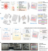
Figure 1
Figure 1
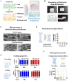
Fundamental design of the human oral-on-a-chip that effectively replicates the structural and multicellular complexities of oral tissue. The diagram illustrates the fundamental design of the multi-compartmentalized oral-on-a-chip platform. The dynamic interactions among cells across different compartments include the tonsil stem cell, gingival keratinocyte, and periodontal ligament chambers. This system combines diverse cellular components and 3D structural intricacies made from natural polymers. A thin porous barrier seamlessly connects the adjacent chambers, facilitating bidirectional communication among various embedded cells via the transfer of growth factors and cytokines. Moreover, to effectively replicate the complex multicellular interactions typical of oral tissue environments, these chambers are surrounded by media channels (A). The human oral-on-a-chip system utilizes a 3D casting mold to stably reproduce the structural characteristics of oral tissues, crafted using PLA-based 3D printing. PDMS was subsequently injected into the 3D printed mold, and the resulting chip platform was extracted following polymerization process (B). To accurately mimic the physiological features and multicellular complexity of oral tissue, the PDMS-based oral-on-a-chip platform was embedded with various human oral cell types that included gingival keratinocytes, tonsil-resident stem cells, periodontal ligament fibroblasts, and vascular endothelial cells. The cells were incorporated with a mixture of natural polymers, specifically hyaluronic acid and collagen, and combined with the thrombin and fibrinogen coagulating factors. This tonsil stem cell chamber is supported by adjacent chambers housing human gingival keratinocytes and periodontal ligament fibroblasts, facilitating a comprehensive representation of the oral tissue microenvironment (C). The PDMS-based oral-on-a-chip platform is rectangular (67 × 47 mm, with a height of 8 mm). This device simulates the microenvironment of oral tissues, promoting multicellular interactions between the tonsil stem cell chamber and neighboring compartments containing human gingival keratinocytes periodontal ligament fibroblasts (D).
Figure 2
Figure 2
Development of oral tissue model using natural polymers and analysis of structural characteristics. The various cell types found in oral tissue were combined with a mixture of natural polymers, such as hyaluronic acid and collagen, and supplemented with the non-toxic coagulants thrombin and fibrinogen (A). The fabricated tissue architecture has a refined surface, soft texture, and is white in color. (B) Cross-sectional and lateral views of the tissue matrix were analyzed using scanning electron microscopy (SEM) to explore their microstructural characteristics. SEM images revealed a uniformly distributed porous network within the tissue constructs, characterized by pores with diameters of 50 to 100 μm. This structure emerged from the interaction of natural polymers with blood coagulation factors (C). The mechanical properties of the fabricated tissue structures were assessed using a universal testing machine, which applied single-axis compression forces. Samples of the tissue, each 10 mm in diameter and height, were exposed to different levels of uniaxial compressive stress to analyze their mechanical response. To assess the exact breaking point of the fabricated samples, a uniaxial compression test was performed with a consistent load rate of 5 mm/min until failure occurred in the samples (D). To assess the absorption properties of the tissue models, the fabricated samples were submerged in distilled water and PBS (pH 7.4) at 37°C for 24 h. Following hydration, excess fluid was removed, and the samples were weighed to measure their water absorption capability (E). Viscosity measurements of the fabricated tissue structures were performed over a shear rate spectrum from 1 to 10/s. Viscosity markedly declined from about 1000 to 0 Pa-sec as the shear rate increased (F). All experiments were performed in triplicate. Significant differences are indicated as follows: *, p < 0.05; **, p < 0.005; and ***, p < 0.001 (two-sample t-test).
Figure 3
Figure 3
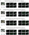
Analysis of sustained viability and metabolic functions of multiple cells embedded within each segment of the oral-on-a-chip model. The arrangement of various cell types within each segment of the chip was assessed. This involved culturing the tissue constructs in a cell-specific medium for 24 h, followed by staining with the DNA-specific dye 4′,6-diamidino-2-phenylindole (DAPI) to visualize the cells' spatial distribution. Fluorescent microscopy was utilized to examine the distribution patterns of cells throughout the tissue structures on the chip. The oral tissue cells in the chip compartments were tonsil-derived stem cells (A), periodontal ligament fibroblasts (B), vascular endothelial cells (C), and gingival keratinocytes (D). Subsequently, cells were placed in medium specialized for each cell type, and cultured for 1, 7, 14, 21, or 28 days after integration. Viability was evaluated with a commercial assay using fluorescent dyes to differentiate live (green) and dead (red) cells. The extended viability of the cells in each chamber was determined by fluorescent imaging. Over 60% of the cells embedded within each segment of the chip remained viable for 28 days. Furthermore, each segment was exposed to a serum-free environment with CCK-8 solution for 48 h. The metabolic activity of these cells was measured by assessing the optical density at 450 nm. All experiments were performed in triplicate. Significant differences are indicated as: *, p < 0.05; **, p < 0.005; and ***, p < 0.001 (two-sample t-test).
Figure 4
Figure 4
Preservation of distinct molecular properties within the natural polymer-based tissue matrix. Evaluations were conducted to verify whether various cell types maintained their molecular characteristics after being incorporated into specific tissue microenvironment of natural polymer-based tissue matrix. This process involved culturing the cells in specially formulated media for one week, followed by their evaluation using biomarkers unique to each cell type. Tonsil-derived stem cells were stained to identify CD105 and NANOG (A), while periodontal ligament fibroblasts were assessed for fibronectin and vimentin (B). Vascular endothelial cells were marked with labels for PECAM1 and vWF (C), and gingival keratinocytes were evaluated using markers for cytokeratin 5 and 8 (D). Each experiment was conducted three times. The nuclei in each field were stained with DAPI.
Figure 5
Figure 5
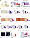
Treatment with the reference toxicant dioxin significantly inhibits various tonsil-derived stem cell functions in vitro. The effectiveness of dioxin was confirmed by its ability to trigger toxicity in tonsil-derived stem cells, measured at specific concentrations and durations of exposure (A). The effect of dioxin on tonsil-derived stem cell renewal was studied by administering various doses (1.25, 2.5, 5, 10, 20, and 40 ng/ml) and measuring the impact after 72 h using the (3-(4, 5-dimethylthiazolyl-2)-2, 5-diphenyltetrazolium bromide) (MTT assay. The impact of dioxin on cell survival was also assessed over 72 h using the same an MTT and fibroblasts, and vascular endothelial cells. Cell viability was quantified as a percentage compared to cells treated with a vehicle control (B). Tonsil-derived stem cells exposed to dioxin (5 ng/ml for 72 h exhibited reduced migratory abilities in a Transwell migration assay, compared to control cells (C). The effect of dioxin on the expression of the key cell migration regulators MMP-2 and MMP-9 was quantified using western blot analysis (D). Tonsil-derived stem cells were cultivated in medium designed to promote adipocyte or osteoblast differentiation; the cells were untreated or exposed to dioxin. The adverse effects of dioxin on the differentiation of the stem cells into adipocytes and osteoblasts were assessed using Oil Red O and Alizarin Red staining techniques. Quantitative analyses of calcium accumulation and lipid formation in the differentiated cells was performed by measuring the absorbance at 500 nm and 570 nm, respectively (E). The impact of exposure to dioxin on the expression of the pluripotency marker genes C-MYC, KLF4, NANOG, OCT4, and SOX2 in cells was assessed using real-time PCR (F). After a 72-h incubation with dioxin, the induced apoptotic DNA condensation and fragmentation were examined using DAPI staining (G). Concurrently, tonsil-derived stem cells that were untreated or cultured with dioxin were analyzed for increased levels of cleaved caspase-3 via western blotting to assess the apoptotic response to dioxin exposure (H). DAPI was used to label nuclei. β-Actin was used as the internal control. All experiments were performed in triplicate. The data are presented as the mean ± standard deviation. *, p < 0.05; **, p < 0.005; and ***, p < 0.001 (two-sample t-test).
Figure 6
Figure 6
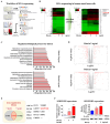
Identification and validation of a reliable marker predicting oral toxicity using human tonsil-derived stem cells. Illustration of the fundamental steps in the RNA-seq process, including the experimental setup, alignment of sequences, quantification, and visual depiction of data (A).The RNA-seq data was visualized in a heatmap, showing gene expression differences between control groups and those treated with low (5 ng/ml) and high (7 ng/ml) doses of athe reference toxicant, dioxin. This visualization displays upregulated genes in red and downregulated genes in green, compared to mRNA levels in the control samples (B). KEGG pathway analysis performed to explore the interrelated pathways and functions affected by exposure to dioxin (C). In the array of genes with altered expression, SERPINB2 expression in human tonsil-derived stem cells was significantly positively correlated with the response to dioxin concentrations of 5 ng/ml (D) and 9 ng/ml (E) dioxin exposure. Real-time PCR and western blot verification of the upregulation of SERPINB2 levels in response 5 and 9 ng/ml doses of dioxin (F). β-Actin was used as the internal protein control, and peptidyl-prolyl cis-trans isomerase (PPIA) was used as the housekeeping gene for real-time PCR. All experiments were performed in triplicate. The data are presented as the mean ± standard deviation. *, p < 0.05; **, p < 0.005; and ***, p < 0.001 (two-sample t-test).
Figure 7
Figure 7
Validating the reliability of SERPINB2 as a marker for oral toxicity in human tonsil-derived stem cells. Schematic illustration outlining the role of SERPINB2 in mediating the adverse effects triggered by dioxin in human tonsil-derived stem cells (A). Stem cells transfected with SERPINB2-specific shRNA were either untreated or exposed to dioxin (5 ng/ml) for 72 h. The effects on growth were analyzed using an MTT assay to assess the attenuating effects of SERPINB2 knockdown (B). Depleting SERPINB2 expression effectively counteracts the adverse effects of dioxin on the mobility of tonsil-derived stem cells, as validated by the Transwell migration/invasion assay (C) and western blot analysis of the production of MMP-2 and MMP-9 proteins (D). Human tonsil-derived stem cells were transfected with shRNA targeting SERPINB2 and exposed to 5 ng/ml dioxin. The cells were assessed for their differentiation into adipocytes and osteoblasts using Oil Red O and Alizarin Red staining (E). Changes in the gene expression of pluripotency markers C-MYC, KLF4, NANOG, OCT4, and SOX2 in cells assessed using real-time PCR (F). DNA fragmentation linked to apoptosis and caspase-3 related apoptotic processes were evaluated using nuclear staining techniques (G) and western blot analysis (H), respectively. β-actin was used as an internal control. All experiments were performed in triplicate. Data are presented as mean ± standard deviation. *, p < 0.05; **, p < 0.005; and ***, p < 0.001 (two-sample t-test).
Figure 8
Figure 8
Development of a fluorescence-based detection platform linked with a toxicity marker within the tonsil stem cell chamber of the oral-on-a-chip model. A fluorescence toxicity marker detection system equipped with SERPINB2-GFP reporter vector was successfully integrated into human tonsil-derived stem cells. The activation of SERPINB2 by exposure to dioxin was manifest as GFP fluorescence allowing the evaluation of oral toxicity in drug candidates using both qualitative and quantitative analyses by measuring the intensity of the fluorescent signal (A). Human tonsil-derived stem cells were stably transfected with GFP-labeled SERPINB2 reporter vector that produces green fluorescence. After these cells were exposed to 5 ng/ml dioxin, immunostaining revealed the notable increase in SERPINB2 activity, evident as green fluorescence (B). This fluorescence-based detection system was integrated into human tonsil-derived stem cells strategically placed within the tonsil stem cell chamber of the oral-on-a-chip. The spatial arrangement of these cells within the designated chamber was analyzed using the fluorescence imaging system (C). Exposure to 5 ng/ml dioxin significantly increased SERPINB2 activity, clearly evident as green fluorescence in the tonsil stem cell chamber of the chip (D). The data are presented as the mean ± standard deviation. *, p < 0.05; **, p < 0.005; and ***, p < 0.001 (two-sample t-test).
Figure 9
Figure 9
Exposure to various types of toxicants triggers the activation of SERPINB2, resulting in emission of green fluorescence signal within the tonsil stem cell chamber of the chip. A detection system utilizing GFP-tagged SERPINB2 reporter was successfully integrated within human tonsil-derived stem cells. These cells were then strategically placed within the tonsil stem cell chamber of the chip platform (A). To evaluate the accuracy and reliability of the SERPINB2-tagged fluorescence detection system, various toxic substances were added to the tonsil stem cell chamber of the chip. These substances included aristolochic acid I (10 μM), benzidine (10 μM), benzo[a]pyrene (2 μM), semustine (0.5 mM), TPA (5 nM), 1,2-dichloropropane (100 mM), 1,3-butadiene (10 mM), and 4,4'-methylenebis (5 μM). The evaluation of the SERPINB2 response to the toxic substances was performed by both qualitative observation and quantitative analysis, measuring the intensity of the emitted SERPINB2-linked green fluorescence signal (B). Significant differences are indicated as follows: *, p < 0.05; **, p < 0.005; and ***, p < 0.001 (two-sample t-test).

References

    1. Van Norman GA. Limitations of Animal Studies for Predicting Toxicity in Clinical Trials: Is it Time to Rethink Our Current Approach? JACC Basic Transl Sci. 2019;4:845–54. - PMC - PubMed
    1. Poornima K, Francis AP, Hoda M, Eladl MA, Subramanian S, Veeraraghavan VP. et al. Implications of Three-Dimensional Cell Culture in Cancer Therapeutic Research. Front Oncol. 2022;12:891673. - PMC - PubMed
    1. Huang C, Sanaei F, Verdurmen WPR, Yang F, Ji W, Walboomers XF. The Application of Organs-on-a-Chip in Dental, Oral, and Craniofacial Research. J Dent Res. 2023;102:364–75. - PMC - PubMed
    1. Pierfelice TV, D'Amico E, Petrini M, Romano M, D'Arcangelo C, Sbordone L. et al. A Systematic Review on Organ-on-a-Chip in PDMS or Hydrogel in Dentistry: An Update of the Literature. Gels. 2024;10:102. - PMC - PubMed
    1. Muniraj G, Tan RHS, Dai Y, Wu R, Alberti M, Sriram G. Microphysiological Modeling of Gingival Tissues and Host-Material Interactions Using Gingiva-on-Chip. Adv Healthc Mater. 2023;12:e2301472. - PMC - PubMed

LinkOut - more resources