. 2024 Jan-Mar;2(1):013016.
doi: 10.1103/prxlife.2.013016. Epub 2024 Mar 26.

Finding the last bits of positional information

Affiliations

Finding the last bits of positional information

Lauren McGough et al. PRX Life. 2024 Jan-Mar.

Abstract

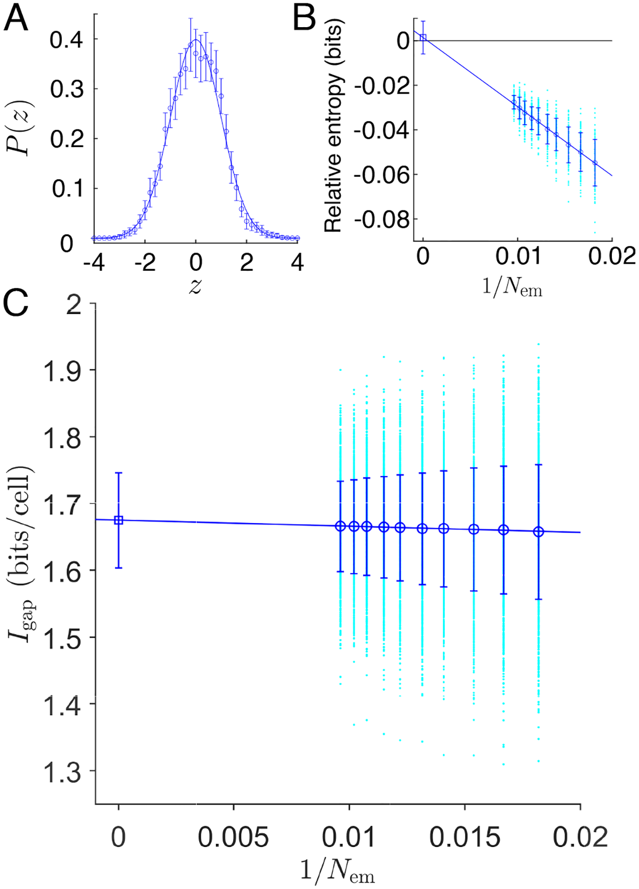
In a developing embryo, information about the position of cells is encoded in the concentrations of morphogen molecules. In the fruit fly, the local concentrations of just a handful of proteins encoded by the gap genes are sufficient to specify position with a precision comparable to the spacing between cells along the anterior-posterior axis. This matches the precision of downstream events such as the striped patterns of expression in the pair-rule genes, but is not quite sufficient to define unique identities for individual cells. We demonstrate theoretically that this information gap can be bridged if positional errors are spatially correlated, with correlation lengths ~ 20% of the embryo length. We then show experimentally that these correlations are present, with the required strength, in the fluctuating positions of the pair-rule stripes, and this can be traced back to the gap genes. Taking account of these correlations, the available information matches the information needed for unique cellular specification, within error bars of ~ 2%. These observation support a precisionist view of information flow through the underlying genetic networks, in which accurate signals are available from the start and preserved as they are transformed into the final spatial patterns.


Figures

FIG. A1:
FIG. A1:
pair-rule stripe positions. (A) Concentration of Eve protein in a single embryo. Colored circles indicate regions which were fitted with a Gaussian function to calculate the stripe position. Each stripe is fitted individually, with fits shown in red. Red triangles indicate centers of each fitted peak. (B) Stripe positions as a function of time in the nuclear cycle 14. Linear fits from Eq. (A1) are shown as black lines. (C) Peak positions xnt0 corrected to t0=45min. (D) Positional error of the pair-rule stripes. Magnitude of the error σx(n) is plotted against the mean position xn for each of the eve, prd, and rnt stripes. Errors in xn are standard errors of the mean; errors in σx are standard deviations across random halves of the data. Dashed line marks the rough estimate σx/L~0.01.
FIG. A2:
FIG. A2:
(A) Positional errors are well approximated as Gaussian. An estimate of the distribution of normalized errors, Eq. (A4). Open circles are means pooled across all stripes and embryos; error bars are standard deviations across random halves of the embryos; and the line is the Gaussian with zero mean and unit variance. (B) The entropy difference between this estimated distribution and the Gaussian, as a function of the (inverse) number of embryos we include in our analysis. Points (cyan) are examples from random choices out of the full ensemble of embryos; open circles with error bars are the mean and standard deviations of these points; and the line is a linear extrapolation [, –33]. (C) Estimates of the information gap, Eq. (A5). Points (cyan) are examples from random choices out of the full ensemble of embryos; open circles (blue) with error bars are the mean and standard deviations of these points; and the line is a linear extrapolation to Igap=1.68±0.07bits/cell.
FIG. A3:
FIG. A3:
Counting nuclei in nuclear cycle 14. (A) Fluorescence image of an embryo with labeled histones highlighting the nuclei underlaid with a brightfield image of the same embryo. Focus is in the midsagittal plane. (B) Zoom in to central 80% on the dorsal side, showing that we can count nuclei by hand. (C) Results from n=26 embryos. Histogram has mean ± std of 72 ± 3.
FIG. A4:
FIG. A4:
Entropy reduction by correlations among the pair-rule stripe fluctuations, estimated from different numbers of embryos Nem;N=10 stripes at left and N=20 stripes at right. Points (cyan) are examples from random choices out of the full ensemble of embryos; open circles (blue) with error bars are the mean and standard deviations of these points; and the line is a linear extrapolation to the square.
FIG. A5:
FIG. A5:
Decoding gap gene expression levels in a single embryo and correlations in the resulting pattern of positional errors. (A) Expression of Hb (blue), Kr (green), Gt (red), and Kni (cyan). Thin solid lines are means across Nem=38 embryos in a small window 40t44min in nuclear cycle 14; dense points are data from a single embryo [13]. (B) Positional errors computed from Eq. (D9). (C) Correlations in the positional noise inferred from gap gene expression. For each embryo α we compute the correlation function in Eq. (D12) and then normalize to give C˜(Δx)=C(Δx)/C(0). Blue circles with error bars are mean and standard error across Nem=38 embryos; solid red line is a smooth curve to guide the eye.
FIG. A6:
FIG. A6:
Correlations between noise in peak positions of the eve, run, and prd stripe patterns, as in Fig. 4, but with stripe positions measured along the ventral side of the embryo. Error bars estimated from the standard deviation across random halves of the data. With three genes, each having seven stripes, we observe (21 × 20)/2 = 210 distinct elements of the correlation matrix Cnm. Solid red line is a smooth curve to guide the eye.
FIG. 1:
FIG. 1:
Segmented Drosophila body plan. (A) Brightfield color image of a 5 mm long 3rd instar larva of the fruit fly Drosophila melanogaster [10] with clearly visible segments. (B) An optical section through an embryo stained for three of the pair-rule proteins, 50 min into nuclear cycle 14 (~ 3 h after oviposition), showing striped patterns that align with the body segments; data from Ref [13]. (C) As in (B), from multiple embryos, illustrating the pattern reproducibility. Time in nuclear cycle 14 indicated at bottom right of each profile. Asterisk marks the image in (B).
FIG. 2:
FIG. 2:
Probability of “crossed signals” between two neighboring cells as a function of the positional error, assuming that noise is independent in each cell, from Eq. (5). Dashed vertical line marks the experimental value of positional noise, σx~0.01L, which corresponds to less than the mean distance between neighboring cells L/N [11].
FIG. 3:
FIG. 3:
Extra information from correlations, as a function of the correlation length. (A) Numerical results for N=50 and N=100 from Eq. (9) with the correlation matrix in Eq. (10); analytic results for N from Eq. (15). Compare with the information gap from Appendix A (solid black line bracketed by dashed error bars). Intersection at ξ=(19.5±1.9)(L/N) marked by vertical line and arrow. (B) Probability Perror of at least two signals being “crossed,” xˆn+1<xˆn, in a line of N=90 cells, with σx/L=0.01.
FIG. 4:
FIG. 4:
Correlations between noise in peak positions of the eve, run, and prd stripe patterns, from Eq. (17), as a function of the mean separation between stripes. Error bars estimated from the standard deviation across random halves of the data. With three genes, each having seven stripes, we observe (21×20)/2 = 210 distinct elements of the correlation matrix Cnm. Solid red line is a smooth curve to guide the eye.
FIG. 5:
FIG. 5:
Extra information from correlations, ΔS/N, computed from the observed correlations in pair-rule stripe fluctuations Cnm through Eq. (9), including different numbers of contiguous stripes. Circles and error bars (blue) are the extrapolated estimates from Appendix C. Red dashed lines are ± one s.e.m. around the best estimate of the information gap Igap=1.68±0.07bits/cell from Appendix A.

Similar articles

Cited by

References

    1. Turing AM, The chemical basis of morphogenesis. Philos Trans R Soc Lond B, Biol Sci 237, 37–71 (1952). - PMC - PubMed
    1. Wolpert L, Positional information and the spatial pattern of cellular differentiation. J Theor Biol 25, 1–47 (1969). - PubMed
    1. Tkačik G and Gregor T, The many bits of positional information. Development 148, dev176065 (2021). - PMC - PubMed
    1. Nusslein-Vollhard C and Wieschaus E, Mutations affecting segment number and polarity in Drosophila. Nature 287, 795–801 (1980). - PubMed
    1. Lawrence PA. The Making of a Fly: The Genetics of Animal Design (Blackwell Scientific, Oxford, 1992).

LinkOut - more resources