

A super-giant basal cell carcinoma of the scalp
- PMID: 39664916
- PMCID: PMC11631210
- DOI: 10.1093/omcr/omae149
A super-giant basal cell carcinoma of the scalp
Abstract
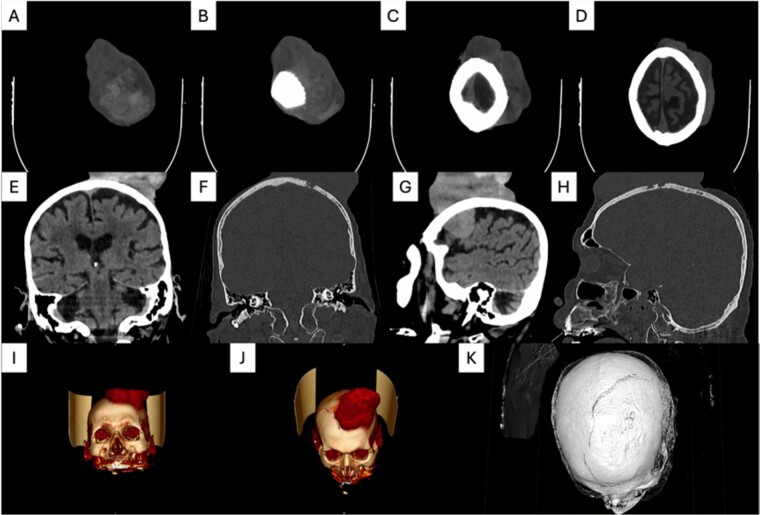
Basal cell carcinoma is a malignant skin cancer, originating from basal cells. However, it is regarded more benign than other skin cancers, in the majority of the cases. If left untreated, it can lead to various complications, degradation of quality of life and even mortality to the patient. A basal cell carcinoma with one dimension more than 20 cm, is defined as super-giant. In this report, we present a case of a super-giant basal cell carcinoma occupying most of the scalp in an elderly patient, causing him severe anemia and general malaise.
Keywords: basal cell carcinoma; cancer; skin; super-giant.
© The Author(s) 2024. Published by Oxford University Press.
Conflict of interest statement
No conflicts of interest.
Figures


References
-
- Cameron MC, Lee E, Hibler BP. et al. Basal cell carcinoma: epidemiology; pathophysiology; clinical and histological subtypes; and disease associations. J Am Acad Dermatol 2019;80:303–17. - PubMed
-
- Erba P, Farhadi J, Wettstein R. et al. Morphoeic basal cell carcinoma of the face. Scand J Plast Reconstr Surg Hand Surg 2007;41:184–8. - PubMed
-
- Vaca-Aguilera MR, Guevara-Gutiérrez E, Barrientos-García JG. et al. Giant basal cell carcinoma: clinical-histological characteristics of 115 cases. Int J Dermatol 2019;58:1430–4. - PubMed
Publication types
LinkOut - more resources
Full Text Sources
