. 2024 Nov 22;6(1):tzae041.
doi: 10.1093/bjro/tzae041. eCollection 2024 Jan.

Detection, measurement, and diagnosis of lung nodules by ultra-low-dose CT in lung cancer screening: a systematic review

Affiliations

Detection, measurement, and diagnosis of lung nodules by ultra-low-dose CT in lung cancer screening: a systematic review

Zhijie Pan et al. BJR Open. .

Abstract

Objective: There is a lack of recent meta-analyses and systematic reviews on the use of ultra-low-dose CT (ULDCT) for the detection, measurement, and diagnosis of lung nodules. This review aims to summarize the latest advances of ULDCT in these areas.

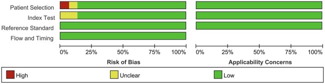
Methods: A systematic review of studies in PubMed and Web of Science was conducted, using search terms specific to ULDCT and lung nodules. The included studies were published in the last 5 years (January 2019-August 2024). Two reviewers independently selected articles, extracted data, and assessed the risk of bias and concerns using the Quality Assessment of Diagnostic Accuracy Studies-II (QUADAS-II) tool. The standard-dose, low-dose, or contrast-enhanced CT served as the reference-standard CT to evaluate ULDCT.

Results: The literature search yielded 15 high-quality articles on a total of 1889 patients, of which 10, 3, and 2 dealt with the detection, measurement, and diagnosis of lung nodules. QUADAS-II showed a generally low risk of bias. The mean radiation dose for ULDCT was 0.22 ± 0.10 mSv (7.7%) against 2.84 ± 1.80 mSv for reference-standard CT. Nodule detection rates ranged from 86.1% to 100%. The variability of diameter measurements ranged from 2.1% to 14.4% against contrast-enhanced CT and from 3.1% to 8.29% against standard CT. The diagnosis rate of malignant nodules ranged from 75% to 91%.

Conclusions: ULDCT proves effective in detecting lung nodules while substantially reducing radiation exposure. However, the use of ULDCT for the measurement and diagnosis of lung nodules remains challenging and requires further research.

Advances in knowledge: When ULDCT reduces radiation exposure to 7.7%, it detects lung nodules at a rate of 86.1%-100%, with a measurement variance of 2.1%-14.4% and a diagnostic accuracy for malignancy of 75%-91%, suggesting the potential for safe and effective lung cancer screening.

Keywords: diagnostic test; lung nodule; systematic review; ultra-low-dose CT.


Conflict of interest statement

The authors of this manuscript declare no relationships with any companies whose products or services may be related to the subject matter of the article.

Figures

Figure 1.
Figure 1.
Literature review and selection flowchart.
Figure 2.
Figure 2.
Assessment of individual risk of bias domains and concerns regarding applicability.
Figure 3.
Figure 3.
Assessment of risk of bias and concerns regarding applicability according to the QUADAS-II tool.
Figure 4.
Figure 4.
Representative cases. Comparison of ULDCT images (left) and diagnostic contrast-enhanced CT images (right). The first two ULDCT scans (A and B) had a radiation dose of 0.07 mSv (70 kV/20 mA). The third ULDCT scan (C) had a radiation dose of 0.14 mSv (70 kV/40 mA). The mean radiation dose of the contrast-enhanced CT scans (A, B, and C) is 2.24 mSv (120 kV/100-200 mA). The dose of ULDCT is only 3%-6% of that of contrast-enhanced CT. (A) A 64-year-old male patient with a 30-mm lobulated nodule in the basal segment of the lower lobe of the right lung, as detected and measured by ULDCT and contrast-enhanced CT, which was pathologically confirmed as invasive squamous carcinoma. (B) A 56-year-old male patient with chronic inflammation of the bronchial mucosa detected by biopsy, had a part-solid nodule in the apical posterior segment of the upper lobe of the left lung measuring 17.5 mm by ULDCT and 18.6 mm by contrast-enhanced CT. The morphology of this nodule is irregular with bronchogram. (C) A 57-year-old man with a lobulated nodule in the basal segment of the lower lobe of the right lung, measuring 26.2 mm by ULDCT and 26.7 mm by contrast-enhanced CT, was pathologically confirmed as invasive adenocarcinoma. Although the malignant nodules detected by ULDCT appear to be ill-defined compared with diagnostic contrast-enhanced CT, the shape of these nodules could be recognized and their size could be measured. (These cases are from the author's institute.)
Figure 5.
Figure 5.
Forest plot of sensitivity, specificity, positive likelihood ratio (LR+), negative likelihood ratio (LR-), and DOR for included studies.

Similar articles

Cited by

  • Lung Cancers Associated with Cystic Airspaces.
    Valsecchi C, Petrella F, Freguia S, Frattini M, Argentieri G, Puligheddu C, Treglia G, Rizzo S. Valsecchi C, et al. Cancers (Basel). 2025 Jan 18;17(2):307. doi: 10.3390/cancers17020307. Cancers (Basel). 2025. PMID: 39858089 Free PMC article. Review.

References

    1. Wood DE, Kazerooni EA, Aberle D, et al. NCCN Guidelines® Insights: Lung Cancer Screening, Version 1.2024. 2023. Accessed August 4, 2023. https://www.nccn.org/professionals/physician_gls/pdf/lung_screening.pdf
    1. de Koning HJ, van der Aalst CM, de Jong PA, et al.Reduced lung-cancer mortality with volume CT screening in a randomized trial. N Engl J Med. 2020;382(6):503-513. 10.1056/NEJMoa1911793 - DOI - PubMed
    1. Oliver AL. Lung cancer: epidemiology and screening. Surg Clin North Am. 2022;102(3):335-344. 10.1016/j.suc.2021.12.001 - DOI - PubMed
    1. Demb J, Chu P, Yu S, et al.Analysis of computed tomography radiation doses used for lung cancer screening scans. JAMA Intern Med. 2019;179(12):1650-1657. 10.1001/jamainternmed.2019.3893 - DOI - PMC - PubMed
    1. Perisinakis K, Seimenis I, Tzedakis A, et al.Radiation burden and associated cancer risk for a typical population to be screened for lung cancer with low-dose CT: a phantom study. Eur Radiol. 2018;28(10):4370-4378. 10.1007/s00330-018-5373-7 - DOI - PubMed

LinkOut - more resources