

Sonographic Characterization of Red-Blue Neurofibromas in Patients With Neurofibromatosis Type 1: An Observational Prospective Study
- PMID: 39665148
- PMCID: PMC11892087
- DOI: 10.1002/jum.16632
Sonographic Characterization of Red-Blue Neurofibromas in Patients With Neurofibromatosis Type 1: An Observational Prospective Study
Abstract
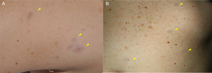
Objectives: Red-blue neurofibromas (RBNs), found in up to 29% of adult neurofibromatosis type 1 (NF1) patients, present as red-blue macules measuring 1-2 cm in diameter, primarily on the trunk. Despite their prevalence, RBNs often go unnoticed due to their subtle appearance. Ultrasound characterization serves as a diagnostic clue yet lacks comprehensive studies in both adult and pediatric populations. This study aims to define and compare RBNs' prevalence, characteristics, and ultrasound features in adult and pediatric patients with NF1.
Methods: This prospective study involved 118 patients (92 pediatric patients and 26 adults) diagnosed with NF1. Clinical examinations combined with cutaneous ultrasound scans using linear multifrequency probes (L4-12t, L10-22, ML6-15, or L8-18 MHz) were performed in order to determine the prevalence, and clinical and sonographic characteristics of RBN in both populations. Statistical analyses were performed using t tests and chi-square tests.
Results: RBNs were found in 26.3% (31) of the patients after clinical examination, including 179 lesions. RBN prevalence differed significantly between pediatric (10.9%) and adult (66.7%) patients. Lesions were primarily on the trunk and exhibited similar clinical characteristics. Ultrasound reveals RBNs as hypoechoic, oval lesions with irregular borders. Our results show that pediatric RBNs are typically more superficial and hypoechogenic, while adult RBNs are deeper and more heterogeneous.
Conclusion: Ultrasound findings showed subtle differences in lesion depth, morphology, and echogenicity between these 2 age-related groups. These changes highlight ultrasound's role in identifying RBNs in patients with NF1 and monitoring their evolution.
Keywords: childhood; cutaneous; neurofibroma; neurofibromatosis 1; red blue macules; ultrasound.
© 2024 The Author(s). Journal of Ultrasound in Medicine published by Wiley Periodicals LLC on behalf of American Institute of Ultrasound in Medicine.
Figures


References
-
- Vabres P, de Lonlay P, Amiel J, Lyonnet S, Munnich A, de Prost Y. Angiomatous and cerulodermic macules: early cutaneous signs of neurofibromatosis type I. Ann Dermatol Venereol 1998; 125:593–594. - PubMed
Publication types
MeSH terms
LinkOut - more resources
Full Text Sources
Research Materials
Miscellaneous
