. 2025 Jan;45(1):e16207.
doi: 10.1111/liv.16207.

Alpha-1 Antitrypsin Inclusions Sequester GRP78 in a Bile Acid-Inducible Manner

Affiliations

Alpha-1 Antitrypsin Inclusions Sequester GRP78 in a Bile Acid-Inducible Manner

Igor Spivak et al. Liver Int. 2025 Jan.

Abstract

Background and aims: The homozygous PiZ mutation (PIZZ genotype) constitutes the predominant cause of severe alpha-1 antitrypsin (AAT) deficiency and leads to liver disease via hepatocellular AAT aggregation. We systematically analysed the composition of AAT aggregates and studied the impact of bile acids.

Methods: AAT inclusions were isolated from livers of PiZ overexpressing mice and PIZZ humans via fluorescence-activated and immunomagnetic sorting (FACS/MACS), while insoluble proteins were obtained via Triton-X extraction. Inclusion composition was evaluated through mass-spectrometry (MS), immunoblotting and immunostaining. Hepatocytes with versus without AAT aggregates were obtained via microdissection. Serum bile acids were assessed in 57 PIZZ subjects and 19 controls. Mice were administered 2% cholic acid (CA)-supplemented chow for 7 days.

Results: MS identified the key endoplasmic reticulum chaperone 78 kDa glucose-regulated protein (GRP78) in FACS/MACS pulldowns. GRP78 was also enriched in insoluble fractions from PiZ mice versus wild types and detected in insoluble fractions/MACS isolates from PIZZ liver explants. In cultured cells/primary hepatocytes, PiZ overexpression was associated with increased GRP78 mRNA/protein levels. In human livers, hepatocytes with AAT aggregates had higher GRP78 levels than hepatocytes without. PIZZ subjects displayed higher serum bile acid levels than controls and the highest levels were seen in individuals with liver injury/fibrosis. In PiZ mice, CA-mediated bile acid challenge resulted in increased liver injury and translocation of GRP78 into the aggregates.

Conclusions: Our results demonstrate that GRP78 is sequestered within AAT inclusions. Bile acid accumulation, as seen in PIZZ subjects with liver disease, may promote GRP78 segregation and thereby augment liver damage.

Trial registration: NCT02929940.

Keywords: ER chaperone; cholestatic liver injury; genetic liver disease; rare liver disease; α‐1 antitrypsin deficiency.


Conflict of interest statement

P.S. reports receiving grants and honoraria from Arrowhead Pharmaceuticals, CSL Behring, Grifols Inc., consulting fees or honoraria from Alnylam Pharmaceuticals, Arrowhead Pharmaceuticals, BioMarin Pharmaceutical, BridgeBio, Dicerna Pharmaceuticals, GSK, Intellia Pharmaceuticals, Takeda Pharmaceuticals, Novo Nordisk and Ono Pharmaceuticals, participating in leadership or fiduciary roles in Alpha1‐Deutschland, Alpha1 Global and material transfer support for Vertex Pharmaceuticals and Dicerna Pharmaceuticals.

Figures

FIGURE 1
FIGURE 1
Purification of aggregated proteins from PiZ mouse livers. (A) Total liver lysates, Triton‐X soluble and insoluble fractions from PiZ transgenic and nontransgenic littermates (WT) were separated by SDS‐PAGE and stained with Coomassie blue. Immunoblot analysis was performed with an antibody against AAT. GAPDH and K8 were used as controls for loading and fractionation. (B) Fractionation of proteins from PiZ and WT livers, followed by separation via SDS‐PAGE and Coomassie‐staining or immunoblotting with an antibody against AAT and the nuclear protein HDAC2. (C) The nuclear isolates underwent FACS. A unique fraction of particles seen in PiZ but not WT samples is highlighted with a rectangle. SDS‐PAGE and Coomassie staining was performed to visualise the protein composition, while immunoblotting quantified the AAT content. (D) Nuclear fractions from PiZ and WT mouse livers underwent labelling with AAT antibody and a pulldown with secondary antibodies‐coupled magnetic beads (AAT‐MACS). The flowthrough (FT) contained the unbound proteins. The different fractions underwent SDS‐PAGE separation, Coomassie staining as well as immunoblotting with an antibody against AAT and the cytoplasmic protein GAPDH. AAT, alpha‐1 antitrypsin; FACS, fluorescence‐activated cell sorting; FSC, forward scatter; FT, flowthrough; GAPDH, glyceraldehyde‐3‐phosphate dehydrogenase; HDAC2, histone deacetylase 2; K8, keratin 8; MACS, magnetic activated cell separation; SSC, side scatter.
FIGURE 2
FIGURE 2
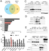
Identification of GRP78 as a component of mouse and human AAT aggregates. (A) Venn diagram illustrating the number of proteins identified by mass spectrometry to be enriched in the insoluble fractions of livers from PiZ transgenic mice versus nontransgenic littermates (WT) (blue circle) and present in the magnetic beads–mediated pulldown of AAT‐containing particles (MACS) from PiZ mouse livers (yellow circle). Out of the overlapping proteins, (B) illustrates the log2 intensity‐based absolute quantification (iBAQ) values of the proteins most abundant in the MACS pulldowns from PiZ mouse livers (upper panel) and shows log2 label‐free quantification (LFQ) values of these proteins in insoluble fractions from PiZ and WT mouse livers (lower panel). (C–E) Immunoblot analysis was performed with antibodies against GRP78 and other endoplasmic reticulum chaperones in the insoluble and soluble pool of PiZ and WT mouse livers (C), FACS isolates from nuclear fractions of PiZ mouse livers (D) and MACS pulldowns from nuclear fractions of PiZ and WT mouse livers (E). (F) Human liver explants from PIZZ individuals and subjects without the PiZ variant (PIMM genotype) were subdivided into insoluble and soluble fractions (upper panel) or subjected to MACS pulldown of nuclear fractions (lower panel) followed by immunoblotting with antibodies against AAT and GRP78. GAPDH and K8 were used as controls for loading and fractionation. AAT, alpha‐1 antitrypsin; FACS, fluorescence‐activated cell sorting; GAPDH, glyceraldehyde‐3‐phosphate dehydrogenase; GRP78, 78 kDa glucose‐regulated protein; GRP94, 94 kDa glucose‐regulated protein; iBAQ, intensity‐based absolute quantification; K8, keratin 8; LFQ, label‐free quantification; MACS, magnetic‐activated cell separation.
FIGURE 3
FIGURE 3
GRP78 and GRP94 levels in a human‐derived bronchial epithelial cell line (IB3) expressing wild‐type (WT) alpha‐1 antitrypsin (AAT) or its PiZ variant and in microdissected human PIZZ liver specimen. (A) GRP78 (n = 4 per group) and GRP94 mRNA (n = 3 per group) levels were quantified by RT‐qPCR in WT‐ and PiZ‐IB3 cells, 18s ribosomal RNA was used as an internal reference. (B) Immunoblotting against GRP78 and GRP94 (n = 3 per group), with corresponding stain‐free gels for normalisation, was used for band intensity quantification. (C) A human liver explant from a subject with PIZZ genotype was PAS‐D stained and laser microdissection was carried out to collect hepatocytes with and without Z‐AAT inclusions. GRP78 and GRP94 abundance ratios were quantified with mass‐spectrometry analysis. Levels of GRP78 and GRP94 in controls were arbitrarily set as 1 and levels in Z‐AAT groups expressed as ratio. Results are shown as mean ± SD (A–B) or median ± range (C). *p < 0.05; **p < 0.01. AAT, alpha‐1 antitrypsin; GRP78, 78 kDa glucose‐regulated protein; GRP94, 94 kDa glucose‐regulated protein; MW, molecular weight marker lane.
FIGURE 4
FIGURE 4
Bile acids in PIZZ adults and cholic acid challenge in PiZ mice. (A) Total serum bile acid concentrations in subjects without the PiZ variant (PIMM genotype) and PIZZ individuals without liver disease (no LD), with elevated liver enzymes but no fibrosis (LInj) or with significant liver fibrosis (LFib). Boxes display median ± IQR and whiskers indicate the 10–90 percentile. Outliers are depicted by circles. (B) Serum AST, ALT and AP levels were measured in PiZ transgenic mice and nontransgenic littermates (WT) fed with normal (control) or 2% cholic acid (CA)–supplemented chow. TUNEL‐staining of liver sections (C) with corresponding quantification. Scale bar = 200 μm. AST and AP are displayed with boxes indicating median ± IQR and whiskers showing the range of the data, ALT and TUNEL‐positive cells as mean ± SD. *p < 0.05; **p < 0.01. ALT, alanine aminotransferase; AP, alkaline phosphatase; AST, aspartate aminotransferase, CA, cholic acid; TUNEL, terminal deoxynucleotidyl transferase dUTP nick end labelling.
FIGURE 5
FIGURE 5
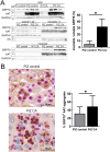
Effect of bile acid challenge on composition of AAT aggregates. (A) Immunoblotting against AAT and GRP78 was performed in the total liver lysates, soluble and insoluble fractions from untreated PiZ transgenic mice (control) and transgenic animals fed with 2% cholic acid (CA)–supplemented chow. GAPDH and K8 were used as controls for loading and fractionation. Band intensity quantification demonstrates the ratio of GRP78 in the insoluble versus soluble pool of PiZ mice. (B) PAS‐D staining combined with GRP78 immunohistochemistry was used to demonstrate colocalisation of GRP78 with the AAT aggregates in PiZ control and CA‐fed animals. Scale bar = 25 μm. Quantification of total AAT aggregates and those with positive GRP78 staining shows the ratio of GRP78+ aggregates in PiZ mice. Results are displayed as mean ± SD. *p < 0.05. AAT, alpha‐1 antitrypsin; CA, cholic acid; GAPDH, glyceraldehyde‐3‐phosphate dehydrogenase; GRP78, 78 kDa glucose‐regulated protein; K8, keratin 8; PAS‐D, periodic acid–Schiff‐diastase.

References

    1. Chiti F. and Dobson C. M., “Protein Misfolding, Amyloid Formation, and Human Disease: A Summary of Progress Over the Last Decade,” Annual Review of Biochemistry 86 (2017): 27–68. - PubMed
    1. Soto C. and Pritzkow S., “Protein Misfolding, Aggregation, and Conformational Strains in Neurodegenerative Diseases,” Nature Neuroscience 21 (2018): 1332–1340. - PMC - PubMed
    1. Lee W.‐C. M., Yoshihara M., and Littleton J. T., “Cytoplasmic Aggregates Trap Polyglutamine‐Containing Proteins and Block Axonal Transport in a Drosophila Model of Huntington's Disease,” Proceedings of the National Academy of Sciences of the United States of America 101 (2004): 3224–3229. - PMC - PubMed
    1. Strnad P., McElvaney N. G., and Lomas D. A., “Alpha1‐Antitrypsin Deficiency,” New England Journal of Medicine 382 (2020): 1443–1455. - PubMed
    1. Lomas D. A., Evans D. L., Finch J. T., and Carrell R. W., “The Mechanism of Z Alpha 1‐Antitrypsin Accumulation in the Liver,” Nature 357 (1992): 605–607. - PubMed

MeSH terms

Associated data