

Radiomics-driven spectral profiling of six kidney stone types with monoenergetic CT reconstructions in photon-counting CT
- PMID: 39665989
- PMCID: PMC12081576
- DOI: 10.1007/s00330-024-11262-w
Radiomics-driven spectral profiling of six kidney stone types with monoenergetic CT reconstructions in photon-counting CT
Abstract
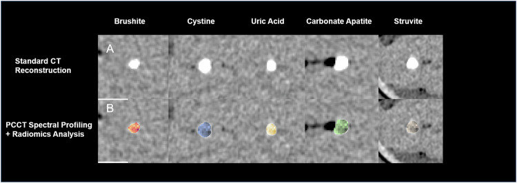
Objectives: Urolithiasis, a common and painful urological condition, is influenced by factors such as lifestyle, genetics, and medication. Differentiating between different types of kidney stones is crucial for personalized therapy. The purpose of this study is to investigate the use of photon-counting computed tomography (PCCT) in combination with radiomics and machine learning to develop a method for automated and detailed characterization of kidney stones. This approach aims to enhance the accuracy and detail of stone classification beyond what is achievable with conventional computed tomography (CT) and dual-energy CT (DECT).
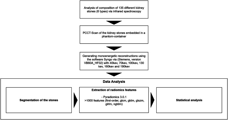
Materials and methods: In this ex vivo study, 135 kidney stones were first classified using infrared spectroscopy. All stones were then scanned in a PCCT embedded in a phantom. Various monoenergetic reconstructions were generated, and radiomics features were extracted. Statistical analysis was performed using Random Forest (RF) classifiers for both individual reconstructions and a combined model.
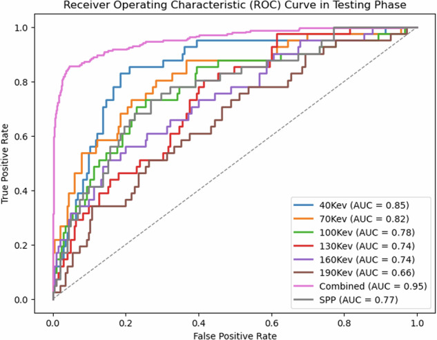
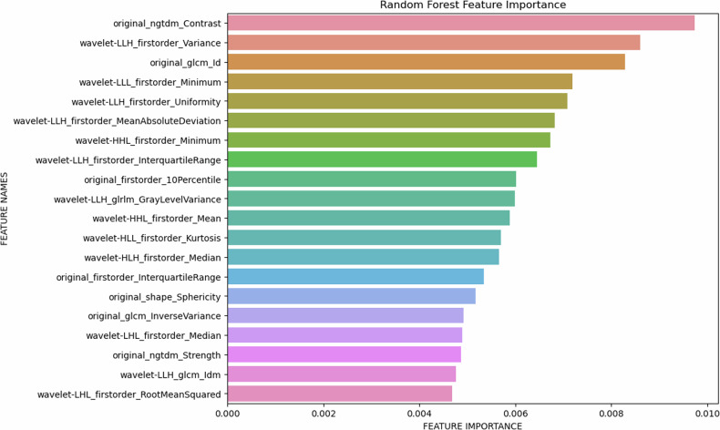
Results: The combined model, using radiomics features from all monoenergetic reconstructions, significantly outperformed individual reconstructions and SPP parameters, with an AUC of 0.95 and test accuracy of 0.81 for differentiating all six stone types. Feature importance analysis identified key parameters, including NGTDM_Strength and wavelet-LLH_firstorder_Variance.
Conclusion: This ex vivo study demonstrates that radiomics-driven PCCT analysis can improve differentiation between kidney stone subtypes. The combined model outperformed individual monoenergetic levels, highlighting the potential of spectral profiling in PCCT to optimize treatment through image-based strategies.
Key points: Question How can photon-counting computed tomography (PCCT) combined with radiomics improve the differentiation of kidney stone types beyond conventional CT and dual-energy CT, enhancing personalized therapy? Findings Our ex vivo study demonstrates that a combined spectral-driven radiomics model achieved 95% AUC and 81% test accuracy in differentiating six kidney stone types. Clinical relevance Implementing PCCT-based spectral-driven radiomics allows for precise non-invasive differentiation of kidney stone types, leading to improved diagnostic accuracy and more personalized, effective treatment strategies, potentially reducing the need for invasive procedures and recurrence.
Keywords: Kidney stones; Machine learning; Photon-counting CT; Radiomics; Spectral profiling.
© 2024. The Author(s).
Conflict of interest statement
Compliance with ethical standards. Guarantor: The scientific guarantor of this publication is Alexander Hertel. Conflict of interest: The authors, S.F., M.J., and B.S. are employed by Siemens Healthineers. The remaining authors declare no conflicts of interest. Statistics and biometry: One of the authors (A.V.) has significant statistical expertise. Informed consent: Since this is an ex vivo phantom study, no written consent was needed. Ethical approval: Not applicable. Study subjects or cohorts overlap: The results of the infrared spectroscopy and photon-counting CT scans have partly been published in previous publications, and the dataset has been evaluated regarding conventional automated stone detection and differentiation of uric vs non-uric acid stones (not published) (Siener et al [32]; Nestler et al [33]). Methodology: Diagnostic or prognostic study The urinary stone analysis was performed at the urinary stone analysis center in Bonn The phantom scans were performed at the Federal Armed Services Hospital in Koblenz
Figures


References
MeSH terms
LinkOut - more resources
Full Text Sources
Medical
Miscellaneous
