. 2024 Dec 12;15(1):299.
doi: 10.1186/s13244-024-01876-5.

Preoperative detection of extraprostatic tumor extension in patients with primary prostate cancer utilizing [68Ga]Ga-PSMA-11 PET/MRI

Affiliations

Preoperative detection of extraprostatic tumor extension in patients with primary prostate cancer utilizing [68Ga]Ga-PSMA-11 PET/MRI

Clemens P Spielvogel et al. Insights Imaging. .

Abstract

Objectives: Radical prostatectomy (RP) is a common intervention in patients with localized prostate cancer (PCa), with nerve-sparing RP recommended to reduce adverse effects on patient quality of life. Accurate pre-operative detection of extraprostatic extension (EPE) remains challenging, often leading to the application of suboptimal treatment. The aim of this study was to enhance pre-operative EPE detection through multimodal data integration using explainable machine learning (ML).

Methods: Patients with newly diagnosed PCa who underwent [68Ga]Ga-PSMA-11 PET/MRI and subsequent RP were recruited retrospectively from two time ranges for training, cross-validation, and independent validation. The presence of EPE was measured from post-surgical histopathology and predicted using ML and pre-operative parameters, including PET/MRI-derived features, blood-based markers, histology-derived parameters, and demographic parameters. ML models were subsequently compared with conventional PET/MRI-based image readings.

Results: The study involved 107 patients, 59 (55%) of whom were affected by EPE according to postoperative findings for the initial training and cross-validation. The ML models demonstrated superior diagnostic performance over conventional PET/MRI image readings, with the explainable boosting machine model achieving an AUC of 0.88 (95% CI 0.87-0.89) during cross-validation and an AUC of 0.88 (95% CI 0.75-0.97) during independent validation. The ML approach integrating invasive features demonstrated better predictive capabilities for EPE compared to visual clinical read-outs (Cross-validation AUC 0.88 versus 0.71, p = 0.02).

Conclusion: ML based on routinely acquired clinical data can significantly improve the pre-operative detection of EPE in PCa patients, potentially enabling more accurate clinical staging and decision-making, thereby improving patient outcomes.

Critical relevance statement: This study demonstrates that integrating multimodal data with machine learning significantly improves the pre-operative detection of extraprostatic extension in prostate cancer patients, outperforming conventional imaging methods and potentially leading to more accurate clinical staging and better treatment decisions.

Key points: Extraprostatic extension is an important indicator guiding treatment approaches. Current assessment of extraprostatic extension is difficult and lacks accuracy. Machine learning improves detection of extraprostatic extension using PSMA-PET/MRI and histopathology.

Keywords: Extraprostatic extension; Machine learning; PET/MRI; PSMA; Prostate cancer.


Conflict of interest statement

Declarations. Ethics approval and consent to participate: This study was approved by the institutional review board with ethics ID EK 1985/2014 at the General Hospital of Vienna and was performed in line with the principles of the Declaration of Helsinki. Informed Consent was obtained from all patients participating in the study. Consent for publication: Not applicable. Competing interests: M. Hacker has received lecture fees from Siemens Healthineers and GE Healthcare. M. Hacker has received consulting fees from Evomics. P.C. has received lecture fees from Siemens Healthineers and is the upcoming Editor-in-Chief of Insights into Imaging. As such, they did not participate in the selection nor review processes for this article. The remaining authors have nothing to disclose.

Figures

Fig. 1
Fig. 1
Cohort flow and validation diagram. A total of 107 patients were included in the overall analysis, 77 of whom were in the development cohort (training and cross-validation) and 30 who were part of the independent validation cohort. EPE, extraprostatic extension
Fig. 2
Fig. 2
Study workflow. A total of 77 patients with localized prostate cancer (PCa) underwent PET/CT examination and radical prostatectomy. Three approaches for the assessment of extraprostatic extension were compared: (1) Visual qualitative assessment as performed in clinical routine, (2) Machine learning modeling using non-invasive parameters, and (3) Machine learning modeling using invasive and non-invasive parameters. Comparative diagnostic performance was assessed, and explainability methods for the machine learning models were integrated. The model using invasive and non-invasive parameters was then validated on an independently collected cohort of additional 30 PCa patients
Fig. 3
Fig. 3
Comparison of the three investigated approaches for pre-operative EPE detection. a Diagnostic performance metrics of the three approaches. b Confusion matrix of the visual clinical PET/MRI read-out. c Confusion matrix for the machine learning model based on non-invasive features only. d Confusion matrix for the machine learning model based on non-invasive and invasive features. e Confusion matrix for the machine learning model with non-invasive and invasive features on the independent test cohort. For c and d, predictions are shown cumulatively over all 100 folds. ACC, accuracy; SNS, sensitivity; SPC, specificity; PPV, positive predictive value; NPV, negative predictive; BAC, balanced accuracy; AUC, area under the receiver operating characteristic curve; EPE, extraprostatic extension
Fig. 4
Fig. 4
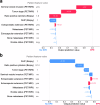
Global SHAP feature importance for the machine learning models with (a) non-invasive features only and (b) for all features. Features are ranked in order of their importance with the most important feature at the top. Each dot represents a single patient’s prediction. Colors indicate the magnitude of the feature value in each row for the given patient. The x-axis indicates the model’s tendency for the prediction of EPE or no-EPE due to the given feature value for the given patient. ISUP, International Society of Urological Pathology (grade); EPE, extraprostatic extension
Fig. 5
Fig. 5
SHAP importance for two example patients correctly predicted by the non-invasive model. Blue arrows indicate a shift of the model prediction toward non-EPE due to the given feature while pink arrows indicate a shift toward a prediction of EPE. In a, an example patient with EPE is shown. Despite the feature with the highest importance (ISUP grade) being 1, therefore indicating a tendency toward the prediction of non-EPE, the final prediction of the model was EPE-positive since features including seminal vesicle invasion, tumor stage, and ratio of positive biopsy cylinders were in favor for the presence of EPE. In b, the model predicted the patient correctly as EPE-negative even though the visual PET/MRI-based assessment by the imaging physician indicated positivity for both, extraprostatic and extracapsular extension. ISUP, International Society of Urological Pathology (grade); EPE, extraprostatic extension

References

    1. Mottet N, van den Bergh RCN, Briers E et al (2021) EAU-EANM-ESTRO-ESUR-SIOG guidelines on prostate cancer—2020 update. Part 1: Screening, diagnosis, and local treatment with curative intent. Eur Urol 79:243–262 - PubMed
    1. Costello AJ (2020) Considering the role of radical prostatectomy in 21st century prostate cancer care. Nat Rev Urol 17:177–188 - PubMed
    1. Reeves F, Preece P, Kapoor J et al (2015) Preservation of the neurovascular bundles is associated with improved time to continence after radical prostatectomy but not long-term continence rates: results of a systematic review and meta-analysis. Eur Urol 68:692–704 - PubMed
    1. Potter SR, Epstein JI, Partin AW (2000) Seminal vesicle invasion by prostate cancer: prognostic significance and therapeutic implications. Rev Urol 2:190–195 - PMC - PubMed
    1. de Rooij M, Hamoen EHJ, Witjes JA, Barentsz JO, Rovers MM (2016) Accuracy of magnetic resonance imaging for local staging of prostate cancer: a diagnostic meta-analysis. Eur Urol 70:233–245 - PubMed

LinkOut - more resources