. 2025 Feb;12(5):e2406565.
doi: 10.1002/advs.202406565. Epub 2024 Dec 12.

Utilizing AI for the Identification and Validation of Novel Therapeutic Targets and Repurposed Drugs for Endometriosis

Affiliations

Utilizing AI for the Identification and Validation of Novel Therapeutic Targets and Repurposed Drugs for Endometriosis

Bonnie Hei Man Liu et al. Adv Sci (Weinh). 2025 Feb.

Abstract

Endometriosis affects over 190 million women globally, and effective therapies are urgently needed to address the burden of endometriosis on women's health. Using an artificial intelligence (AI)-driven target discovery platform, two unreported therapeutic targets, guanylate-binding protein 2 (GBP2) and hematopoietic cell kinase (HCK) are identified, along with a drug repurposing target, integrin beta 2 (ITGB2) for the treatment of endometriosis. GBP2, HCK, and ITGB2 are upregulated in human endometriotic specimens. siRNA-mediated knockdown of GBP2 and HCK significantly reduced cell viability and proliferation while stimulating apoptosis in endometrial stromal cells. In subcutaneous and intraperitoneal endometriosis mouse models, siRNAs targeting GBP2 and HCK notably reduced lesion volume and weight, with decreased proliferation and increased apoptosis within lesions. Both subcutaneous and intraperitoneal administration of Lifitegrast, an approved ITGB2 antagonist, effectively suppresses lesion growth. Collectively, these data present Lifitegrast as a previously unappreciated intervention for endometriosis treatment and identify GBP2 and HCK as novel druggable targets in endometriosis treatment. This study underscores AI's potential to accelerate the discovery of novel drug targets and facilitate the repurposing of treatment modalities for endometriosis.

Keywords: GBP2; HCK; ITGB2; artificial intelligence; drug repurposing; endometriosis; lifitegrast; target discovery.


Conflict of interest statement

B.H.M.L., X.L., A.G., F.R., A.Z., and F.W.P. are affiliated with Insilico Medicine, a commercial company developing AI solutions for aging research, drug discovery, and longevity medicine. Y.L., S.W.H., and C.C.W. declare no competing interests.

Figures
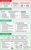
Figure 1
Figure 1
Workflow for identifying therapeutic targets and repurposing drugs for endometriosis using artificial intelligence techniques. 1) Dataset selection: With the input of 11 ectopic endometrium transcriptomic datasets, 11 case‐control comparisons were generated using 437 cases collected from patients with endometriosis and 216 healthy control samples for therapeutic target identification. 2) Target prioritization: With its proprietary AI and bioinformatic models, PandaOmics is a generative AI system that determines target‐disease association and prioritizes targets using the listed sources and characteristics. This prioritization step also integrates a druggability assessment of candidate targets. 3) Target selection: Following the prioritization of targets based on the listed characteristics, GBP2 and HCK were selected as novel targets. In parallel, ITGB2 was nominated as a high‐confidence target for drug repurposing. 4) Target validation: Proposed targets were validated by histology in patient samples and functional validation in preclinical endometriosis models. GEO: Gene Expression Omnibus; LINCS: Library of Integrated Network‐Based Cellular Signatures; OMIM: Online Mendelian Inheritance in Man; OGEE: Online Gene Essentiality; TTD: Therapeutic Target Database.
Figure 2
Figure 2
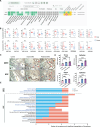
GBP2 and HCK were identified as potential therapeutic targets for endometriosis. A) Screenshot of the Target ID page of PandaOmics for endometriosis meta‐analysis. GBP2 and HCK were revealed as novel druggable targets for endometriosis. B) GBP2 and HCK transcript levels in the eleven ectopic endometrium‐related comparisons were displayed in box plots. *FDR < 0.05, **FDR < 0.01, ***FDR < 0.001. FDR < 0.05 indicates a significant differential expression. C) Representative images of the paraffin‐embedded human endometriosis and normal endometrium tissues stained with anti‐GBP2 and anti‐HCK antibodies. G, endometrium glands; g: endometriotic glands; blue arrows, positive staining in endometrial or endometriotic stromal cells; red arrows, positive staining in endometrial or endometriotic epithelial cells. Magnifications, 40x. D) GBP2 and HCK protein expression in the epithelial and stromal cells was assessed by H‐score. Control (n = 10), ovarian EMS (n = 7), and peritoneal EMS (n = 10). Data are represented as mean ± SEM. *P < 0.05, **P < 0.01, ***P < 0.001. P < 0.05 is considered as statistically significant. E) Dysregulated pathways associated with GBP2 (upper) and HCK (lower) in endometriosis comparisons were displayed. Pathways were annotated by the Reactome database, and the iPANDA algorithm determined the degree of pathway dysregulation. Blue and red bar colors indicate the number of comparisons with significant activation or inactivation of the corresponding pathway, respectively. Pathways labeled in blue are immune‐associated.
Figure 3
Figure 3
GBP2 and HCK ablation inhibited proliferation and promoted apoptosis of endometriotic stromal cells. Endometriotic stromal cells were transiently transfected with non‐target siRNA control (Control), siRNA targeting HCK (siHCK), and siRNA target GBP2 (siGBP2). mRNA and protein expressions of A,B) GBP2 and C,D) HCK in the transfected cells were examined by qPCR and immunofluorescence staining with anti‐GBP2 and anti‐HCK antibodies, respectively. Representative images of the transfected cells stained with anti‐HCK and anti‐GBP2 antibodies were shown. E) CCK8 assay to measure relative proliferation in the indicated groups. F) Ki67 immunofluorescence staining of the indicated groups. G) TUNEL assay to measure apoptosis in transfected cells. Mean apoptotic cell percentages are plotted. Data are represented as mean ± SEM. *P < 0.05, **P < 0.01. P < 0.05 is considered as statistically significant. Each experimental group contained 6 samples. Differences between group comparisons were evaluated using Kruskal‐Wallis Test. Adjustment by Bonferroni correction was used for multiple comparisons.
Figure 4
Figure 4
GBP2 and HCK knockdown independently suppressed endometriosis in vivo. The effect of knocking down GBP2 and HCK in endometriosis was assessed by both subcutaneous A–G) and intraperitoneal H–N) endometriosis mouse models (n = 5). A,H) The volume and weight of the excised lesions were recorded and compared. Representative images of the paraffin‐embedded murine ectopic endometrium xenografts stained with hematoxylin and eosin were shown. mRNA and protein expressions of (B‐C, I‐J) GBP2 and (D‐E, K‐L) HCK in murine endometriosis xenografts were analyzed by qPCR and IHC. Representative images of the paraffin‐embedded xenografts stained with anti‐GBP2 and anti‐HCK antibodies were shown. (F, M) Cell proliferation and (G, N) apoptosis status of the xenograft treated with siRNA against GBP2 and HCK were analyzed by Ki‐67 IHC staining and TUNEL assay, respectively. Representative images of each experimental group were displayed. Data are represented as mean ± SEM. *P < 0.05, **P < 0.01. P < 0.05 is considered as statistically significant. Differences between group comparisons were evaluated using Kruskal‐Wallis Test. Adjustment by Bonferroni correction was used for multiple comparisons.
Figure 5
Figure 5
Identification of ITGB2 as a potential high‐confidence target for endometriosis. A) Screenshot of the Target ID page of PandaOmics for endometriosis meta‐analysis. ITGB2 was revealed as a druggable target with high confidence for endometriosis. B) The expressions of ITGB2 in the eleven endometriosis‐related comparisons were displayed in box plots. *FDR < 0.05, **FDR < 0.01, ***FDR < 0.001. FDR < 0.05 indicates a significant differential expression. C) ITGB2 protein expression in the paraffin‐embedded human endometriosis and normal endometrium tissues were assessed by H‐score. Control (n = 10), ovarian EMS (n = 7), and peritoneal EMS (n = 10). Representative images of the human tissues stained with anti‐ITGB2 antibody were displayed. G) endometrium glands; g: endometriotic glands; blue arrows, positive staining in endometrial or endometriotic stromal cells; red arrows, positive staining in endometrial or endometriotic epithelial cells. Magnifications, 40x. Data are represented as mean ± SEM. *P < 0.05, **P < 0.01, ***P < 0.001. P < 0.05 is considered as statistically significant. D) Dysregulated pathways associated with ITGB2 in endometriosis comparisons were displayed. Pathways labeled in blue are immune‐associated. Differences between group comparisons were evaluated using Kruskal‐Wallis Test. Adjustment by Bonferroni correction was used for multiple comparisons.
Figure 6
Figure 6
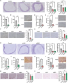
Lifitegrast inhibited murine endometriotic growth. The efficacy of lifitegrast in treating endometriosis was determined by subcutaneous A–F) and intraperitoneal G–L) administration of lifitegrast in endometriosis mouse models (n = 4). A,G) The volume and weight of the excised lesions treated with DMSO or Lifitegrast at the indicated doses were recorded and compared. B,H) mRNA and C,I) protein expressions of ITGB2 in the murine ectopic endometrium xenograft were analyzed by qPCR and IHC. D,J) Cell proliferation and E,K) apoptosis status of the xenograft were analyzed by Ki‐67 IHC staining and TUNEL assay, respectively. F,L) Representative images of paraffin‐embedded murine ectopic endometrium xenografts stained with hematoxylin and eosin, anti‐ITGB2 antibody, Ki‐67 IHC staining, and TUNEL assay in each experimental group were displayed. Data are represented as mean ± SEM. *P < 0.05, **P < 0.01, ***P < 0.001. P < 0.05 is considered as statistically significant. Differences between group comparisons were evaluated using Kruskal‐Wallis Test. Adjustment by Bonferroni correction was used for multiple comparisons.

References

    1. Moingeon P., Ann. Pharm. Fr. 2021, 79, 566. - PubMed
    1. Vamathevan J., Clark D., Czodrowski P., Dunham I., Ferran E., Lee G., Li B., Madabhushi A., Shah P., Spitzer M., Zhao S., Nat. Rev. Drug Discovery 2019, 18, 463. - PMC - PubMed
    1. Swinney D. C., Anthony J., Nat. Rev. Drug Discovery 2011, 10, 507. - PubMed
    1. Plenge R. M., Scolnick E. M., Altshuler D., Nat. Rev. Drug Discovery 2013, 12, 581. - PubMed
    1. Zhavoronkov A., Ivanenkov Y. A., Aliper A., Veselov M. S., Aladinskiy V. A., Aladinskaya A. V., Terentiev V. A., Polykovskiy D. A., Kuznetsov M. D., Asadulaev A., Volkov Y., Zholus A., Shayakhmetov R. R., Zhebrak A., Minaeva L. I., Zagribelnyy B. A., Lee L. H., Soll R., Madge D., Xing L., Guo T., Aspuru‐Guzik A., Nat. Biotechnol. 2019, 37, 1038. - PubMed

LinkOut - more resources