. 2024 Dec 13;386(6727):1243-1250.
doi: 10.1126/science.add8468. Epub 2024 Dec 12.

A synthetic protein-level neural network in mammalian cells

Affiliations

A synthetic protein-level neural network in mammalian cells

Zibo Chen et al. Science. .

Abstract

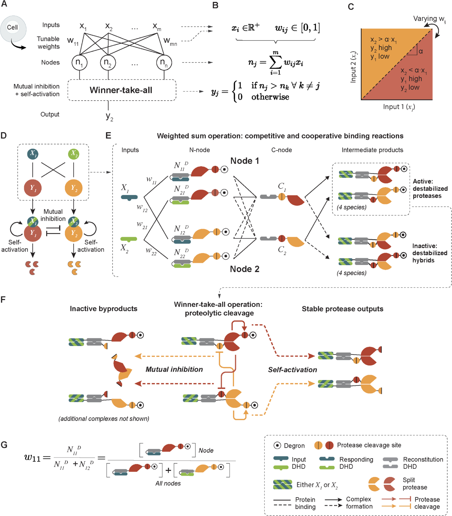
Artificial neural networks provide a powerful paradigm for nonbiological information processing. To understand whether similar principles could enable computation within living cells, we combined de novo-designed protein heterodimers and engineered viral proteases to implement a synthetic protein circuit that performs winner-take-all neural network classification. This "perceptein" circuit combines weighted input summation through reversible binding interactions with self-activation and mutual inhibition through irreversible proteolytic cleavage. These interactions collectively generate a large repertoire of distinct protein species stemming from up to eight coexpressed starting protein species. The complete system achieves multi-output signal classification with tunable decision boundaries in mammalian cells and can be used to conditionally control cell death. These results demonstrate how engineered protein-based networks can enable programmable signal classification in living cells.


Conflict of interest statement

Competing interests: Z.C. and M.B.E. have filed a provisional patent application based on this work. M.B.E. is a co-founder, scientific advisory board member, or consultant at TeraCyte, Primordium Labs (now Plasmidsaurus), and Spatial Genomics.

Figures

Figure 1.
Figure 1.. Winner-take-all neural network computation can be implemented using engineered proteins.
(A) A winner-take-all neural network, operating inside cells, would use a set of interacting proteins (Nj) to activate exactly one of its outputs (colored proteases, yj) depending on the relative values of its inputs (Xi). (B) Formal description of the system in (A). The network consists of m inputs (Xi), each taking positive real values. The inputs interact with the n nodes (Nj) with weights wij (1 ≤ i ≤ m, 1 ≤ j ≤ n, 0 ≤ wij ≤ 1) connecting input Xi to node Nj. Each node performs weighted sum operations to integrate the input signals it receives, and winner-take-all is achieved through self-activation and mutual inhibition. The output yj from a node Nj is active only if its weighted sum is greater than that from any other node. (C) The decision boundary, α, for a 2-input, 2-node network can be tuned by varying the weights wij. (D) In a 2-input, 2-node circuit, each input protein (X1, X2) activates either node protein (Y1, Y2) by forming input-node complexes. Such complexes then undergo self-activation and mutual inhibition to perform the winner-take-all computation. The final state of the system is defined by the abundance of the active node. (E) The weighted sum operation is carried out through competitive and cooperative binding. The two inputs are de novo designed orthogonal DHDs (X1 and X2). Each node consists of two groups of proteins: the N-nodes, where the cognate binding partners of X1 and X2 are caged by a genetically fused DHD caging domain, and further linked to the N-terminal half of a protease, its cleavage site, and a DHFR degron; the C-nodes, made from the cognate binding partner of the DHD caging domain in primary half-nodes, fused to the cleavage sequence of the other protease, and the C-terminal half of a protease. The inputs, N-node, and C-node bind cooperatively, such that neither of the two proteins can bind with high affinity without the third protein. They also interact competitively, such that the N-nodes compete to bind to the input protein, and the C-nodes. Two types of reconstituted proteases result from these 3-way binding events: the active but destabilized proteases where the two protease halves reconstitute a functional protease, or the inactive and destabilized hybrids where the two protease halves do not match. The blue and green stripes indicate that the DHD domains can be either blue (X1) or green (X2). (F) Winner-take-all operation is achieved through two types of reactions: mutual inhibition, where each protease can inactivate the opposite protease type by cleaving its C-terminal half proteases off the C-nodes; self-activation, where the intermediate DHD-protease complexes cleave off DHFR degrons from their N-nodes, converting them to stable proteases, which can in turn activate and inhibit other protease complexes. (G) The weights connecting inputs to nodes are set based on the abundance of each primary half-node complex. For example, w11, the weight that connects input X1 to Node 1, is defined as the concentration of the N11 N-node divided by the sum of the concentrations of all N-nodes that can potentially bind to X1, in this case N11 + N12.
Figure 2.
Figure 2.. Simulated two-input circuits perform winner-take-all classification.
(A) Simulations of circuit variants (left) reveal circuit dynamics (middle) and classification ability (right). Input values, weights, and weighted sums (denoted Σ) are indicated on the circuit diagrams, with larger weights represented by thicker lines. Each cell in the heatmap represents the difference between active Y1 and Y2 proteases at steady state. Both the full circuit and the comparator are able to classify across the full range of input levels, while circuits lacking self-activation or mutual inhibition only classify within a limited input range. (B) The decision boundary (gray lines) of the comparator circuit can be tuned by varying the relative levels of the two node proteins, N11D and N22D. (C) Stochastic simulations of the comparator. Twenty simulations were performed for each condition (light traces), and their average traces are plotted in dark lines. Colors indicate the input levels (legend). See supplementary materials for simulation methods. (D) Percentage of 50 equivalent simulations that correctly classify inputs as a function of the concentration difference between the two inputs. Input X1 is fixed at 0.05 molecules/s. Even with stochasticity, input differences of at least 20% classify correctly ~95% of the time.
Figure 3.
Figure 3.. The winner-take-all neural network circuit classifies inputs in mammalian cells.
Unless otherwise noted, transfections were conducted using DNA. (A) The stable reporter cell line constitutively co-expresses Citrine and mCherry fluorescent proteins that can be cleaved at the N-terminus by TEVP and TVMVP, respectively, to reveal N-terminal degrons that destabilize the fluorescent proteins. PGK, 3-phosphoglycerate kinase promoter. (B) Engineered protease can respond to inputs, self-activate, and mutually inhibit. Normalized protease activities under different experimental setups indicate expected functions. (C) Testing the weight multiplication module by fixing input X1 and varying nodes N11D and N12D. Ideal behaviors are shown in solid lines, experimental data points from mRNA transfections are mean ± s.d from three biological repeats. (D) A fully connected 2-input, 2-node circuit that compares relative input levels (left). (E) A fully connected 2-input, 2-node circuit that, by construction, should always result in Y1 being the winner. (F) The decision boundaries of a two-input comparator can be tuned by varying the ratios of Y1 to Y2 protein concentrations. Data in D-F are averages of two biological replicates. (G) A cell death circuit is placed downstream of the perceptein classifier, where activation of Node 2 results in cell death. (H) HEK293 cells were transfected with the perceptein-apoptosis circuit (two inputs, two nodes, and TEVP-activatable caspase-3) and an mCherry co-transfection marker. The percentage of apoptotic (Annexin-positive) cells were calculated using flow cytometry, after gating on transfected cells based on an mCherry co-transfection marker. Data points are averages of four biological replicates. (I) Red fluorescence images of cells treated with the perceptein-apoptosis circuit along with the mCherry co-transfection marker. Dead cells show reduced or no expression of mCherry.
Figure 4.
Figure 4.. Scaling of the winner-take-all circuit.
(A) A thresholded two-input comparator where the third node (Y3) participates in self-activation and mutual inhibition, but does not directly respond to inputs (X1 and X2). (B) Simulation results predict three distinct classification outcomes (red, green, and blue) depending on input levels, and a fourth unclassified state (black). (C) The thresholded two-input comparator was tested in a three-color reporter cell line transiently transfected with perceptein components. For each cell, normalized fluorescent levels from the Citrine, mCherry, and miRFP680 channels were converted to a three-element RGB vector that represents the color of the cell. Data points represent averages of three biological replicates. (D) The number of reactions in a comparator circuit increases roughly linearly with its size, as the circuit dynamic range decreases. (E) A 2-input classifier can generate distinct responses to all 4 input states. (F) A 3-input winner-take-all circuit performs the (X1 OR X3) AND NOT X2 calculation. Node 1 wins if the condition is met. (G) A 3-input winner-take-all circuit performs “Any 2 out of 3” logic. A fourth “hidden unit” input was added to set the threshold and make the circuit more compact. Node 1 wins if the condition is met.

Comment in

  • Bringing neural networks to life.
    Galloway K, Johnstone C. Galloway K, et al. Science. 2024 Dec 13;386(6727):1225-1226. doi: 10.1126/science.adu1327. Epub 2024 Dec 12. Science. 2024. PMID: 39666818

References

    1. Hafner A, Bulyk ML, Jambhekar A, Lahav G, The multiple mechanisms that regulate p53 activity and cell fate. Nat. Rev. Mol. Cell Biol 20, 199–210 (2019). - PubMed
    1. Zagorski M, Tabata Y, Brandenberg N, Lutolf MP, Tkačik G, Bollenbach T, Briscoe J, Kicheva A, Decoding of position in the developing neural tube from antiparallel morphogen gradients. Science 356, 1379–1383 (2017). - PMC - PubMed
    1. Antebi YE, Reich-Zeliger S, Hart Y, Mayo A, Eizenberg I, Rimer J, Putheti P, Pe’er D, Friedman N, Mapping differentiation under mixed culture conditions reveals a tunable continuum of T cell fates. PLoS Biol 11, e1001616 (2013). - PMC - PubMed
    1. Hong M, Clubb JD, Chen YY, Engineering CAR-T Cells for Next-Generation Cancer Therapy. Cancer Cell, doi: 10.1016/j.ccell.2020.07.005 (2020). - DOI - PubMed
    1. Benenson Y, Biomolecular computing systems: principles, progress and potential. Nat. Rev. Genet 13, 455–468 (2012). - PubMed

Publication types

Substances

LinkOut - more resources