Review
. 2024 Dec 12;13(1):61.
doi: 10.1186/s40035-024-00457-2.

Cellular senescence as a key contributor to secondary neurodegeneration in traumatic brain injury and stroke

Affiliations
Review

Cellular senescence as a key contributor to secondary neurodegeneration in traumatic brain injury and stroke

Zhihai Huang et al. Transl Neurodegener. .

Abstract

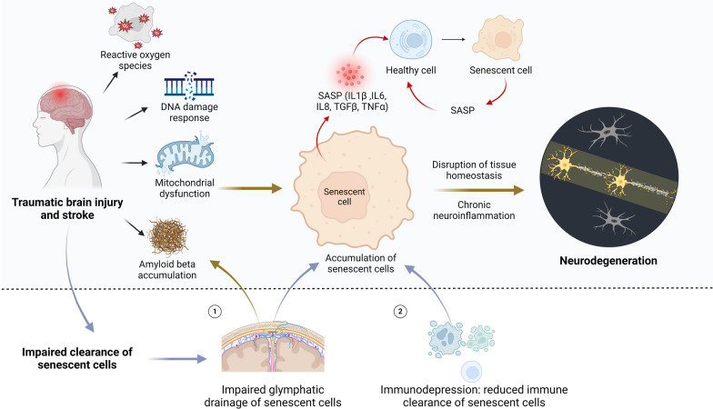
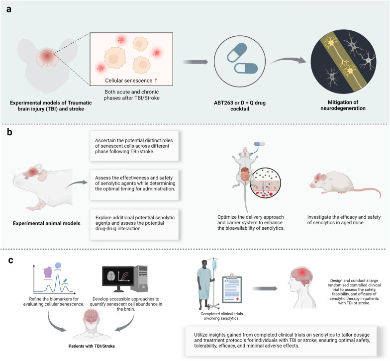
Traumatic brain injury (TBI) and stroke pose major health challenges, impacting millions of individuals globally. Once considered solely acute events, these neurological conditions are now recognized as enduring pathological processes with long-term consequences, including an increased susceptibility to neurodegeneration. However, effective strategies to counteract their devastating consequences are still lacking. Cellular senescence, marked by irreversible cell-cycle arrest, is emerging as a crucial factor in various neurodegenerative diseases. Recent research further reveals that cellular senescence may be a potential driver for secondary neurodegeneration following brain injury. Herein, we synthesize emerging evidence that TBI and stroke drive the accumulation of senescent cells in the brain. The rationale for targeting senescent cells as a therapeutic approach to combat neurodegeneration following TBI/stroke is outlined. From a translational perspective, we emphasize current knowledge and future directions of senolytic therapy for these neurological conditions.

Keywords: Cellular senescence; Neurodegeneration; Senolytic therapy; Stroke; Traumatic brain injury.


Conflict of interest statement

Declarations. Ethics approval and consent to participate: Not applicable. Consent for publication: All authors have read and approved the final manuscript for publication. Competing interests: The authors declare that they have no competing interests.

Figures

Fig. 1
Fig. 1
Proposed mechanisms underlying cellular senescence following TBI and stroke. DNA damage response, ROS, and mitochondrial dysfunction resulting from TBI may initiate cellular senescence in the brain. These senescent cells subsequently induce SASP, potentially transforming healthy cells into senescent ones. Furthermore, TBI/stroke may impair glymphatic drainage function and induce immunodepression, exacerbating the accumulation of senescent cells in the brain. The accumulation of senescent cells disrupts tissue homeostasis, leading to chronic neuroinflammation, ultimately culminating in neurodegeneration. Abbreviations: SASP, Senescence-associated secretory phenotype; TBI, Traumatic brain injury; ROS, Reactive oxygen species
Fig. 2
Fig. 2
Consequences of senescence in different cell types and their potential interplay. Breakdown of the BBB following brain injury can lead to the infiltration of cytotoxic mediators and immune cells into the brain parenchyma, creating a proinflammatory environment that perpetuates cellular senescence. Senescence in endothelial cells can further compromise the BBB, creating a negative feedback loop that exacerbates the damage. Prolonged microglial proliferation may promote senescence within themselves, reducing their ability to phagocytose cellular debris and beta-amyloid, thereby hindering remyelination after brain injury. The compromised phagocytosis by microglia may also induce astrocytes to phagocytose myelin, potentially worsening demyelination. Senescent astrocytes may have diminished glutamate transport capacity, resulting in reduced glutamate uptake. This leads to the accumulation of extracellular glutamate, causing excessive calcium influx and, ultimately, neuronal damage. Oligodendrocyte senescence, triggered by proinflammatory signals, can inhibit their maturation into myelinating oligodendrocytes, leading to insufficient myelination and possible axonal degeneration. Inflammation may be a mediator underlying the communications between different cells. Senescent cells not only exacerbate inflammation but may also be induced by a proinflammatory microenvironment. Abbreviations: BBB, Blood–brain barrier
Fig. 3
Fig. 3
Summary of potential approaches to enhance the therapeutic targeting and efficacy of senolytic agents. a A diagram showing the mechanism of action of a β-galactosidase-targeted senolytic prodrug. After administration, the prodrug is cleaved into active molecules by β-galactosidase expressed in senescent cells. This allows the senolytic to specifically target these cells and induce their apoptosis. b Summary of the potential application of photodynamic therapy with senolytics. After administration, the photoactivatable senolytics are activated by light, leading to the release of active molecules in the targeted tissue. c Summary of the potential application of combining nanoparticles with senolytics. Utilizing desired nanoparticles, the drug molecules may more effectively cross the blood–brain barrier, resulting in higher bioavailability
Fig. 4
Fig. 4
Summary of experimental evidence supporting the therapeutic value of senolytic therapy for TBI/stroke and directions for future research. a Experimental evidence indicates that cellular senescence occurs during both acute and chronic phases following TBI/stroke. Senolytics such as ABT263 and the D + Q drug cocktail, administered during the subacute or chronic phases post-TBI/stroke, demonstrate efficacy in mitigating secondary neurodegeneration. b Summary of future directions for research on senolytic therapy for TBI/stroke. c Clinical translation of senolytic therapy for TBI/stroke. Abbreviations: TBI, Traumatic brain injury; D, Dasatinib; Q, Quercetin

References

    1. Feigin VL, Vos T, Nichols E, Owolabi MO, Carroll WM, Dichgans M, et al. The global burden of neurological disorders: translating evidence into policy. Lancet Neurol. 2020;19(3):255–65. - PMC - PubMed
    1. Capizzi A, Woo J, Verduzco-Gutierrez M. Traumatic brain injury: an overview of epidemiology, pathophysiology, and medical management. Med Clin North Am. 2020;104(2):213–38. - PubMed
    1. Saini V, Guada L, Yavagal DR. Global epidemiology of stroke and access to acute ischemic stroke interventions. Neurology. 2021;97(20 Suppl 2):S6–16. - PubMed
    1. Bramlett HM, Dietrich WD. Pathophysiology of cerebral ischemia and brain trauma: similarities and differences. J Cereb Blood Flow Metab. 2004;24(2):133–50. - PubMed
    1. Kunz A, Dirnagl U, Mergenthaler P. Acute pathophysiological processes after ischaemic and traumatic brain injury. Best Pract Res Clin Anaesthesiol. 2010;24(4):495–509. - PubMed

Publication types

MeSH terms

LinkOut - more resources