. 2024;20(12):1989-1996.
doi: 10.1038/s41567-024-02652-4. Epub 2024 Oct 8.

Emergent actin flows explain distinct modes of gliding motility

Affiliations

Emergent actin flows explain distinct modes of gliding motility

Christina L Hueschen et al. Nat Phys. 2024.

Abstract

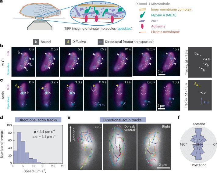
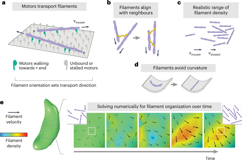
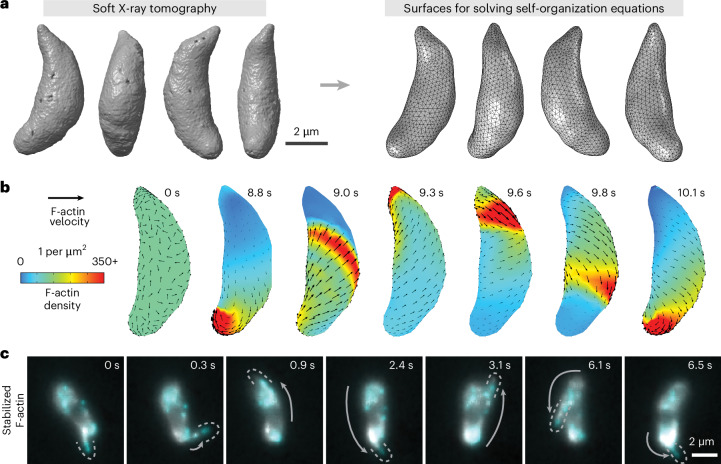
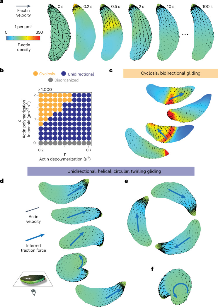
During host infection, Toxoplasma gondii and related unicellular parasites move using gliding, which differs fundamentally from other known mechanisms of eukaryotic cell motility. Gliding is thought to be powered by a thin layer of flowing filamentous (F)-actin sandwiched between the plasma membrane and a myosin-covered inner membrane complex. How this surface actin layer drives the various gliding modes observed in experiments-helical, circular, twirling and patch, pendulum or rolling-is unclear. Here we suggest that F-actin flows arise through self-organization and develop a continuum model of emergent F-actin flow within the confines provided by Toxoplasma geometry. In the presence of F-actin turnover, our model predicts the emergence of a steady-state mode in which actin transport is largely directed rearward. Removing F-actin turnover leads to actin patches that recirculate up and down the cell, which we observe experimentally for drug-stabilized actin bundles in live Toxoplasma gondii parasites. These distinct self-organized actin states can account for observed gliding modes, illustrating how different forms of gliding motility can emerge as an intrinsic consequence of the self-organizing properties of F-actin flow in a confined geometry.

Keywords: Biological physics; Cellular motility.


Conflict of interest statement

Competing interestsThe authors declare no competing interests.

Figures

Fig. 1
Fig. 1. Toxoplasma gondii actin transport direction is heterogeneous, not uniformly rearward.
a, Schematic of the intermembrane actomyosin layer that drives apicomplexan gliding and of TIRF imaging of speckle-labelled actin or MLC1. Inset: actin is speckle-labelled with single Janelia Fluor dyes (cyan). b,c, Examples of MLC1 (b) and actin (c) movement (speckles; cyan) over time in extracellular parasites, with higher-density labelling (bulk; magenta) to show cell position. Arrowheads highlight examples of specific protein behaviours. Images denoised with noise2void. Protein trajectories (far right) are shown for an equal time interval (1.3 s) to allow comparison of bound (white), diffusive (yellow) and directional (purple) movements. Experiment repeated in n = 7 (MLC1) and n = 18 (actin) independent cells. d, Histogram of speeds of directional actin tracks from 18 cells. μ, mean; s.d., standard deviation. e, Directional actin tracks (n = 54 tracks in 18 overlaid cells from three experiments). Cells were grouped by the cell side visible, aligned with anterior end up and superimposed. Cell polarity was determined by microtubule labelling or tracking of the posterior end following posterior-down cell twirling (Supplementary Information Section 5), and directional actin tracks were aligned with respect to the parasite long axis. f, Polar histogram of the orientation of directional actin displacements with respect to cell polarity (n = 231 displacements, 54 tracks, 18 cells).
Fig. 2
Fig. 2. Toxoplasma actin self-organization: theoretical model.
Rules of local actin filament behaviour, implemented in equation (2): a, Actin filaments (purple) are transported with their minus ends leading at speed vmyosin, as indicated by arrows. b, Neighbouring filaments align by steric effects or crosslinking proteins (yellow). c, Filament density remains within a realistic range, with filament speed slowing if entering a pile-up. d, Filament orientation is biased towards lower curvature. e, Example of numerically solving for filament self-organization using the finite element method, predicting filament density and velocity over time. Black arrow size reflects velocity magnitude. To provide intuition, for each white box, the corresponding inset shows schematized F-actin (darker purple represents minus ends) whose density and orientation are consistent with the simulation density (colour) and velocity (black arrow).
Fig. 3
Fig. 3. Stable actin filaments circle the Toxoplasma cell.
a, Soft X-ray tomograms of cryo-fixed extracellular Toxoplasma gondii tachyzoites were used to generate triangle-meshed surfaces on which to solve our actin self-organization theoretical model. b, For stable filaments, solving the model constrained to Toxoplasma’s surface geometry predicts recirculating actin patches. c, In experiments, actin filaments briefly stabilized with jasplakinolide can circle around the cell. Cyan and grey both show actin, labelled at different dye densities. Images denoised with noise2void. Dotted lines outline protruding actin filaments, and grey arrows highlight the movement of the protrusion since the previous frame. Representative of n = 10 cells whose protrusion velocities are characterized in Supplementary Fig. 4.
Fig. 4
Fig. 4. Polarized actin turnover governs a transition between actin recirculation and unidirectional transport.
a, Incorporating F-actin depolymerization and anterior polymerization into the model enables the emergence of a unidirectional, stable velocity pattern. b, Tuning rates of F-actin polymerization and depolymerization move the cell between distinct self-organized states: bidirectional cyclosis, unidirectional and disorganized. c, Model prediction: recirculating F-actin cyclosis generates bidirectional traction force (blue arrows) to drive ‘patch’ gliding. df, Model prediction: the unidirectional self-organized F-actin state drives helical gliding (d), circular gliding (e) and twirling (f). In the images in cf, cells are viewed from below; cell-substrate contact occurs at the position of the inferred traction force.

References

    1. Tenter, A. M., Heckeroth, A. R. & Weiss, L. M. Toxoplasma gondii: from animals to humans. Int. J. Parasitol.30, 1217–1258 (2000). - PMC - PubMed
    1. Checkley, W. et al. A review of the global burden, novel diagnostics, therapeutics, and vaccine targets for cryptosporidium. Lancet Infect. Dis.15, 85–94 (2015). - PMC - PubMed
    1. White, N. J. et al. Malaria. Lancet383, 723–735 (2014). - PubMed
    1. Jensen, J. B. & Edgar, S. A. Effects of antiphagocytic agents on penetration of Eimeria magna sporozoites into cultured cells. J. Parasitol.62, 203–206 (1976). - PubMed
    1. Wetzel, D. M., Håkansson, S., Hu, K., Roos, D. & Sibley, L. D. Actin filament polymerization regulates gliding motility by apicomplexan parasites. Mol. Biol. Cell14, 396–406 (2003). - PMC - PubMed