. 2024 Nov 28:15:1506638.
doi: 10.3389/fimmu.2024.1506638. eCollection 2024.

Mucosal vaccination with outer membrane vesicles derived from Bordetella pertussis reduces nasal bacterial colonization after experimental infection

Affiliations

Mucosal vaccination with outer membrane vesicles derived from Bordetella pertussis reduces nasal bacterial colonization after experimental infection

E Rudi et al. Front Immunol. .

Abstract

Introduction: We previously identified Bordetella pertussis-derived outer membrane vesicles (OMVs) as a promising immunogen for improving pertussis vaccines. In this study, we evaluated the efficacy of our vaccine prototype in immunization strategies aimed at reducing disease transmission by targeting colonization in the upper airways while maintaining protection against severe disease by reducing colonization in the lower respiratory tract.

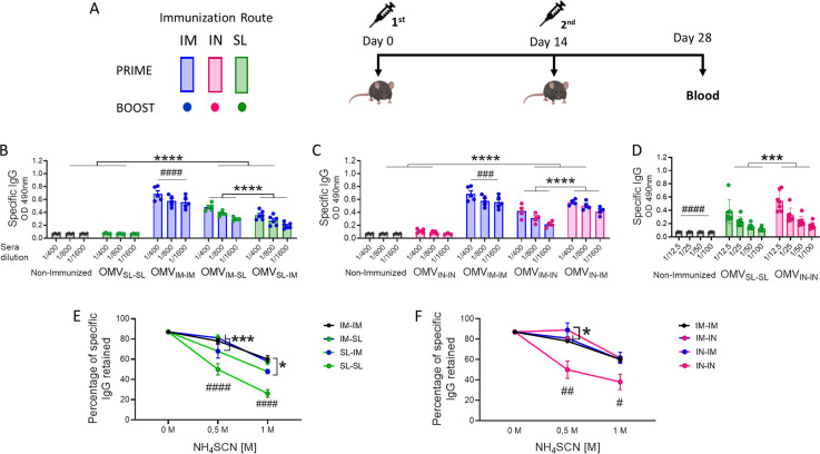
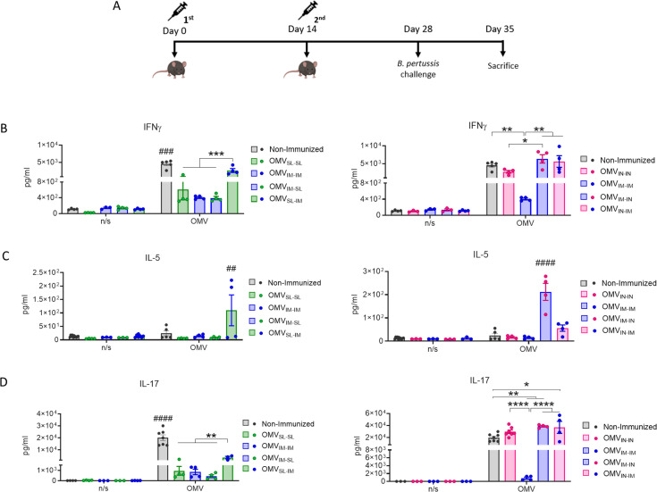
Methods: We assessed different mucosal administration strategies in a murine model, including homologous mucosal 2-dose prime-boost schedules and heterologous prime-boost strategies combining intramuscular (IM) systemic immunization with mucosal routes (intranasal, IN; or sublingual, SL). We utilized alum and c-di-AMP as adjuvants for the systemic and mucosal formulations of the OMV vaccine prototype, respectively. A homologous prime/boost IM immunization schedule and commercial vaccines were used for comparisons.

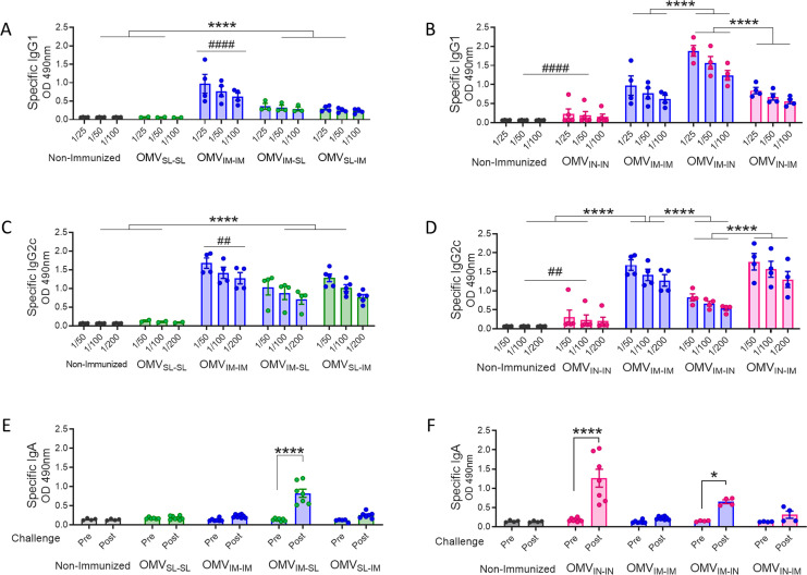
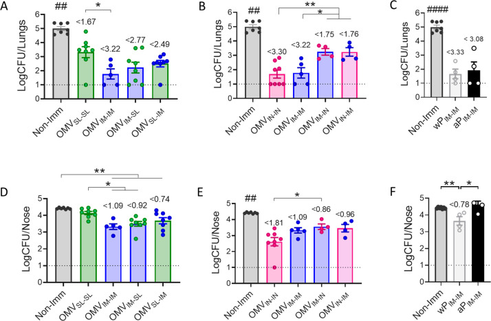
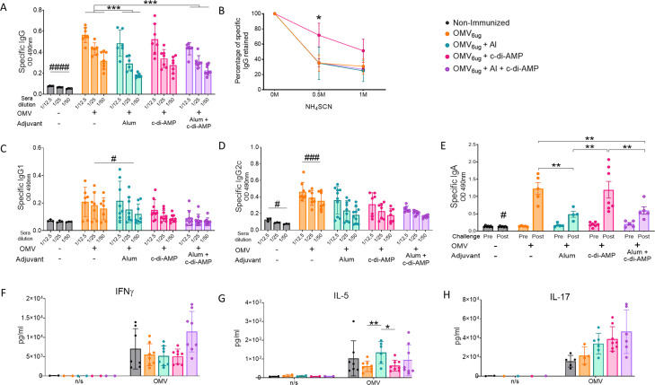
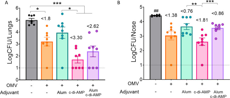
Results: All tested heterologous schemes induced higher levels of specific IgG with significant avidity, as well as higher levels of IgG1 and IgG2c, compared to the corresponding homologous prime-boost 2-dose schemes via mucosal routes (OMVIN-IN or OMVSL-SL). High IgA levels were observed post-B. pertussis challenge following OMVIN-IN treatments and heterologous treatments where the second dose was administered via a mucosal route (prime-pull scheme). Furthermore, schemes involving the intranasal route, whether in a homologous or heterologous scheme, induced the highest levels of IL-17 and IFN-γ. Accordingly, these schemes showed superior efficacy against nasal colonization than the commercial vaccines. Homologous intranasal immunization exhibited the highest protective capacity against nasal colonization while maintaining an excellent level of protection in the lower respiratory tract. To further enhance protection against nasal colonization, we performed a comparative analysis of formulations containing either single or combined adjuvants, administered via homologous intranasal route. These assays revealed that the use of alum combined with c-di-AMP, did not enhance the immune protective capacity in comparison with that observed for the formulation containing c-di-AMP alone.

Conclusions: All the experiments presented here demonstrate that the use of OMVs, regardless of the scheme applied (except for OMVSL-SL), significantly outperformed acellular pertussis (aP) vaccines, achieving a higher reduction in bacterial colonization in the upper respiratory tract (p<0.01).

Keywords: Bordetella pertussis; IgA; intranasal; mucosal; outer-membrane vesicles.


Conflict of interest statement

The authors declare that the research was conducted in the absence of any commercial or financial relationships that could be construed as a potential conflict of interest. The author(s) declared that they were an editorial board member of Frontiers, at the time of submission. This had no impact on the peer review process and the final decision.

Figures

Figure 1
Figure 1
Characterization of the humoral immune response induced by OMVs administered through heterologous and homologous immunization schemes. C57BL/6 mice (n=7/group) were immunized on days 0 and 14, with a 2-dose scheme administered with a formulation containing OMVs using mucosal routes (sublingual OMVSL or intranasal OMVIN) in homologous schemes (OMVSL-SL or OMVIN-IN) or heterologous schemes (OMVIM-SL OMVSL-IM, OMVIM-IN or OMVIN-IM),. A homologous 2-dose scheme using the intramuscular route (OMVIM-IM) was also incorporated for comparative analysis. Different bars colors are used to discriminate the route used for the first dose and the colors of circles denote the route used for the booster dose (A). Specific IgG levels induced by schedules that include SL administration (B), IN administration (C) or homologous mucosal administration (D) were determined in sera collected on day 14 after the last dose (absorbance values at 490 nm for 2 sera dilutions). The avidity of IgG antibodies was also measured 14 days post the second dose and the results are presented as percentages of B. pertussis-specific IgG antibodies retained after exposure with 0.5 M and 1M of ammonium thiocyanate (NH4SCN) (E, F). *p<0.05, ***p<0.001, ****p<0.0001 by two-way ANOVA using Bonferroni for multiple comparisons. The # symbol indicate significant differences between treatment and the other groups. #p<0.05, ##p<0.01, ###p<0.001, #### p <0.0001.
Figure 2
Figure 2
Specific B. pertussis IgG isotypes and IgA levels induced by OMVs administered through different heterologous and homologous immunization schemes. Groups of C57BL6 mice were immunized with a 2-dose scheme as described in panel A of Figure 1 . A homologous 2-dose scheme using the intramuscular route was incorporated for comparative analysis (OMVIM-IM) and a group of non-immunized mice was employed as a control. Specific IgG1 (A, B) and IgG2a (C, D) levels were determined in sera of immunized mice two weeks after last dose (absorbance values at 490 nm for 3 sera dilutions). (A, C): levels of IgG isotypes induced by schedules that include SL immunization. (B, D): levels of IgG isotypes induced by schedules that include IN administration. IgA antibody responses in serum were evaluated before and after intranasal B. pertussis challenge (absorbance values at 490 nm) in the groups of immunized mice with schedules that include SL (E) and IN (F) administration. *p<0.05, ****p<0.0001 by two-way ANOVA using Bonferroni for multiple comparisons. The # symbol indicate significant differences between OMVIM-IM and the other groups. # p<0.05, #### p <0.0001.
Figure 3
Figure 3
Characterization of cellular immune response induced by OMVs administered through heterologous and homologous immunization schemes. Mice were immunized on day 0 and 14, with a 2-dose scheme administered with a formulation containing OMVs using mucosal routes in homologous schemes or heterologous schemes, as described in Figure 1 . Seven days after challenge with a sublethal dose (1x10⁷-5x10⁷ CFU/40 ul), mice were sacrificed, and their spleen cells were restimulated ex-vivo with 2μg/ml of OMV derived from B. pertussis or non-stimulated as controls (n/s). (A) Levels of secreted IFN-γ (B), IL-5 (C) and IL-7 (D) following splenocytes stimulation with medium or OMVs were determined by ELISA. Bars are means ± SEM of pg/ml. *p<0.05, **p<0.01, ***p<0.001, ****p<0.0001 by two-way ANOVA using Bonferroni for multiple comparisons. The # symbol indicate significant differences between the treatment and the other groups. ##p<0.01, ###p<0.001, #### p <0.0001.
Figure 4
Figure 4
Effect of OMV mucosal homologous and heterologous immunization on protection against B. pertussis infection. C57BL/6 mice were immunized on days 0 and 14 with a 2-dose scheme OMV administered as described in panel A of Figure 1 . Groups of animals immunized with commercial aP, or commercial wP vaccines or non-immunized were included as positive and negative protection control. Mice from all groups were challenged with a sublethal dose (1x10⁷ – 5 x10⁷/40 μl) B. pertussis Tohama phase I 14 days after the second dose followed by sacrifice 7 days after challenge. The number of bacteria recovered from mouse lungs (A-C) or nose (D-F) expressed as the log10 of CFUs per lungs or nose, is plotted on the ordinate, the different treatments here tested are indicated on the abscissa, with the data representing the means ± the SD. The dotted horizontal line indicates the lower limit of detection. The reduction detected in protection levels induced by different formulations in comparison with non-immunized animals is indicated at the top of the figures. *p<0.05, **p<0.01 by one way ANOVA using Bonferroni for multiple comparisons. The # symbol indicate significant differences between the treatment and the other tested groups. ## p<0.01, #### p <0.0001.
Figure 5
Figure 5
Characterization of immunogenicity and the protection capacity induce by OMVs administered by intranasal route using different adjuvants. Groups of C57BL6 mice were immunized with a 2-dose scheme administered IN with a formulation containing OMVs formulated with c-di AMP, Alum, or c-di AMP plus Alum. As a control, a group of non-immunized were employed. Specific IgG levels induced by different schedules were determined in sera collected on day 14 after the last dose (absorbance values at 490 nm) (A). The avidity of IgG antibodies was also measured 14 days after the second dose and the results are presented as percentages of B. pertussis-specific IgG antibodies retained after exposure with 0.5 M and 1 M of ammonium thiocyanate (NH4SCN) (B). Specific IgG1 (C) and IgG2c levels (D) were determined in sera of immunized mice two weeks after last dose (absorbance values at 490 nm). Mice from all groups were challenged with a sublethal dose (1x10⁷ – 5 x10⁷/40 μl) B. pertussis Tohama phase I 14 days after the second dose followed by sacrifice 7 days after challenge. IgA antibody responses in serum were detected before and after intranasal B. pertussis challenge (absorbance values at 490 nm) (E). Levels of secreted IFN-γ (F), IL-5 (G) and IL-7 (H) following splenocytes stimulation with medium or OMVs were determined by ELISA. Bars are means ± SEM of pg/ml. *p<0.05, **p<0.01, ***p<0.001 by two-way ANOVA using Bonferroni for multiple comparisons. The # symbol indicate significant differences between the treatment and the other groups. # p<0.05, ### p <0.001.
Figure 6
Figure 6
Effect of OMV intranasal homologous immunization on protection against B. pertussis infection. The number of bacteria recovered from mouse lungs (left panel A) or nose (right panel B) expressed as the log10 of CFUs per lungs or nose, is plotted on the ordinate, the different treatments here tested are indicated on the abscissa, with the data representing the means ± SEM. The reduction detected in protection levels induced by different formulations in comparison with non-immunized animals is indicated at the top of the figures. The dotted horizontal line indicates the lower limit of detection. p<0.05, **p<0.01, ***p<0.001, by one way ANOVA using Bonforroni for multiple comparisons. The # symbol indicate significant differences between the treatment and the other tested groups. ## p<0.01.

Similar articles

References

    1. Tozzi AE, Gesualdo F, Rizzo C, Carloni E, Russo L, Campagna I, et al. . A data driven clinical algorithm for differential diagnosis of pertussis and other respiratory infections in infants. PloS One. (2020) 15:e0236041. doi: 10.1371/journal.pone.0236041 - DOI - PMC - PubMed
    1. Barbic J, Leef MF, Burns DL, Shahin RD. Role of gamma interferon in natural clearance of Bordetella pertussis infection. Infect Immun. (1997) 65:4904–8. doi: 10.1128/IAI.65.12.4904-4908.1997 - DOI - PMC - PubMed
    1. Mills KHG, Redhead K. Cellular immunity in pertussis. J Med Microbiol. (1993) 39:163–4. doi: 10.1099/00222615-39-3-163 - DOI - PubMed
    1. Higgs R, Higgins SC, Ross PJ, Mills KHG. Immunity to the respiratory pathogen Bordetella pertussis. Mucosal Immunol. (2012) 5:485–500. doi: 10.1038/mi.2012.54 - DOI - PubMed
    1. Mills KHG, Ryan M, McGuirk P, Griffin F, Murphy G, Mahon B. The immunology of Bordetella pertussis infection. Biologicals. (1999) 27:77–89. doi: 10.1006/biol.1999.0183 - DOI - PubMed

MeSH terms

LinkOut - more resources