. 2025 Jan 31;93(1):e0033024.
doi: 10.1128/iai.00330-24. Epub 2024 Dec 13.

Novel broadly reactive monoclonal antibody protects against Pseudomonas aeruginosa infection

Affiliations

Novel broadly reactive monoclonal antibody protects against Pseudomonas aeruginosa infection

Margalida Mateu-Borrás et al. Infect Immun. .

Abstract

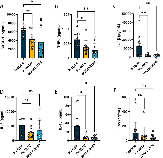
The incidence of infections attributed to antimicrobial-resistant (AMR) pathogens has increased exponentially over the recent decades reaching 1.27 million deaths worldwide in 2019. Without intervention, these infections are predicted to cause up to 10 million deaths a year and incur costs of up to 100 trillion US dollars globally by 2050. The emergence of AMR bacteria such as the ESKAPEE pathogens, and in particular Pseudomonas aeruginosa and species from the genus Burkholderia, underscores an urgent need for new therapeutic strategies. Monoclonal antibody (mAb) therapy offers a promising alternative to treat and prevent bacterial infections. In this study, we used peptides from highly conserved areas of the bacterial flagellin to generate monoclonal antibodies capable of broad binding to flagellated Gram-negative bacteria. We generated a broadly reactive IgG2bĸ mAb (WVDC-2109) that recognizes P. aeruginosa, Burkholderia sp., and other Gram-negative pathogens of interest. Characterization of the therapeutic potential of this antibody was determined using P. aeruginosa as model. In vitro characterization of WVDC-2109 demonstrated complement-mediated bactericidal activity and enhanced opsonophagocytosis of P. aeruginosa. Prophylactic administration of WVDC-2109 markedly improved survival and outcome in a lethal sepsis model and a sub-lethal murine pneumonia model of P. aeruginosa infection, reducing bacterial burden and inflammation. These findings suggest that WVDC-2109 and similar FliC-targeting antibodies could be valuable in preventing or treating diseases caused by P. aeruginosa as well as other life-threatening diseases of concern.IMPORTANCEAntimicrobial resistance (AMR) costs hundreds of thousands of lives and billions of dollars annually. To protect the population against these infections, it is imperative to develop new medical countermeasures targeting AMR pathogens like P. aeruginosa and Burkholderia sp. The administration of broadly reactive monoclonal antibodies can represent an alternative to treat and prevent infections caused by multi-drug-resistant bacteria. Unlike vaccines, antibodies can provide protection regardless of the immune status of the infected host. In this study, we generated an antibody capable of recognizing flagellin from P. aeruginosa and B. pseudomallei along with other Gram-negative pathogens of concern. Our findings demonstrate that the administration of the monoclonal antibody WVDC-2109 enhances survival rates and outcomes in different murine models of P. aeruginosa infection. These results carry significant implications in the field given that there are no available vaccines for P. aeruginosa.

Keywords: Burkholderia; Pseudomonas aeruginosa; anti-microbial resistance; monoclonal antibodies.


Conflict of interest statement

M.M.-B. and M.B. are inventors on a pending patent application related to the sequence of WVDC-2109. The remaining authors declare no competing interests.

Figures

Fig 1
Fig 1
B. pseudomallei and P. aeruginosa flagellin variability map. 3D representation of B. pseudomallei (A) and P. aeruginosa (B) FliC proteins. Residues colored in dark green represent amino acids 100% conserved across all sequences compared, light green highlights residues with one polymorphism, and yellow and white residues with two or more polymorphisms. (C) Sequence of B. pseudomallei FliC with the peptide selected for immunization in bold letter. In addition, residues highly conserved across species (one polymorphism/position or less) are highlighted in green.
Fig 2
Fig 2
WVDC-2109 binds to P. aeruginosa and B. pseudomallei FliC. (A) Whole-cell lysates of P. aeruginosa PAO1 were treated with proteinase K (PK) or sodium periodate (NaIO4) and immunoblotted with WVDC-2109. (B) Immunoblot against 10 µg of P. aeruginosa PAO1 and B. pseudomallei Bp82 cell lysates using WVDC-2109 antibody. (C) Immunoblot using WVDC-2109 against 3 µg of recombinant FliC purified from P. aeruginosa or B. pseudomallei Bp82. (D) Transmission electron microscopy with immunogold labeling of P. aeruginosa using PBS, an isotype control or WVDC-2109 followed by a gold-conjugated secondary antibody. All blots in this panel were performed in parallel. Mw, Molecular weight size.
Fig 3
Fig 3
WVDC-2109 interacts with FliC without blocking TLR5 activation. Model of the interaction between B. pseudomallei FliC (A) and P. aeruginosa FliC (B) and the predicted structure of WVDC-2109. In both panels A and B, the structure of FliC is depicted in gray. The WVDC-2109 antibody is illustrated in light blue, with residues unique to the WVDC-2109 heavy chain highlighted in dark blue. The peptide sequence is shown in green, indicating the sequence and its corresponding similar peptide in P. aeruginosa used for generating hybridomas. The amino acid residues in the heavy chain variable region involved in binding to FliC are shown in a box. (C) HEK-Blue hTLR5 cells stimulated with B. pseudomallei flagellin (0–100 ng/mL) were previously incubated with increasing concentrations of WVDC-2109 (0–100 µg/mL). (D) HEK-Blue hTLR5 cells stimulated with P. aeruginosa flagellin (0–100 ng/mL) were previously incubated with increasing concentrations of WVDC-2109 (0–100 µg/mL). SEAP activity was determined with HEK-Blue detection media at 655 nm. Bars graphs represent mean ± SD.
Fig 4
Fig 4
WVDC-2109 increases bacterial killing mediated by the complement system and opsonophagocytic killing by macrophages. (A) Complement bactericidal assay. Percentage of survival of P. aeruginosa PAO1 after 90 min of incubation with Heat-Inactivated (HI) Guinea Pig Complement, Guinea Pig Complement alone, HI-Guinea Pig Complement + IgG2 b Isotype control, HI-Guinea Pig Complement + WVDC-2109, Guinea Pig Complement + IgG2 b Isotype control, or Guinea Pig Complement + WVDC-2109. **** Denotes a comparison to all other groups. (B) Opsonophagocytic killing assay. Percentage of survival of P. aeruginosa PAO1 after 2 h of incubation with macrophage J774A.1 cells previously opsonized with or without WVDC-2109 or IgG2b isotype control. **** Denotes a comparison to all other groups. (C) Opsonophagocytic killing assay with the addition of HI-Guinea Pig Complement or Guinea Pig Complement. Percentage of survival of P. aeruginosa PAO1 after 2 h of incubation with macrophage J774A.1 cells previously opsonized with or without WVDC-2109 or IgG2b isotype control, with or without added HI-Guinea Pig Complement or Guinea Pig Complement. **** Denotes a comparison to all other groups, unless otherwise displayed. Statistical significance was determined by ordinary one-way ANOVA: *P < 0.05, **P < 0.01, ****P < 0.0001. The dotted line represents the above growth threshold. Each dot represents one replicate. Error bars represent the standard error of the mean.
Fig 5
Fig 5
WVDC-2109 protects mice against P. aeruginosa sepsis. Mice survival (A) and rectal temperatures (B) over time of mice challenged with P. aeruginosa in a murine lethal model of sepsis. Mice received WVDC-2109 (light blue) or an isotype IgG control (dark blue) 12 h before P. aeruginosa PAO1 IP infection and were monitored for morbidity and mortality over 96 h. Bacterial burden in the blood (C), kidneys, (D) and spleen (E) 6 and 12 h after P. aeruginosa PAO1 IP infection in mice that were prophylactically administered WVDC-2109 or an isotype control antibody. Statistical significance in A was determined using a Mantel–Cox test. Differences in bacterial burden were determined using the Mann-Whitney test. *P < 0.05.
Fig 6
Fig 6
WVDC-2109 reduces bacterial burden and lung edema during acute murine pneumonia. Bacterial burden in the respiratory tract 16 h after P. aeruginosa challenge in CD-1 mice. Mice received WVDC-2109, a non-specific isotype IgG control or Pa-WCV serum 12 h before P. aeruginosa PAO1 infection. Viable bacteria were quantified in the lung (A) and nasal lavage (B) using serial dilutions. Rectal temperature (C) and lung mass (D) were also determined at the time of euthanasia. (E) Pharmacokinetics of WVDC-2109. Mice were injected with IP WVDC-2109 (blue) or Pa-WCV serum (orange). The level of anti-P. aeruginosa PAO1 IgG in the mice sera was determined by ELISA. Each dot represents an individual animal. Results from statistical analysis of comparisons between animals treated with Pa-WCV serum and animals administered WVDC-2109 are represented with *, and Pa-WCV and WVDC-2109 to the limit of detection are represented with #. Statistical significance was determined by one-way ANOVA in A-D (*P < 0.05, **P < 0.005, ***P < 0.0005), and one-sample t-test and Mann-Whitney test in E (#P < 0.05, ##P < 0.01; *P < 0.05). The dotted line indicates the lowest limit of detection.
Fig 7
Fig 7
WVDC-2109 reduces lung inflammation during acute murine pneumonia. CXCL-1 (A), TNFα (B), IL-1β (C), IL-6 (D), IL-10, (E) and IFN-γ (F) in the lung homogenate of CD-1 mice after 16 h of P. aeruginosa PAO1 intranasal infection. Mice were passively immunized with WVDC-2109, with a non-specific isotype IgG as a negative control or serum from mice previously vaccinated with inactivated P. aeruginosa (Pa WCV) 12 h before P. aeruginosa PAO1 infection. Each dot represents an individual animal. Statistical significance was determined by one-way ANOVA. *P < 0.05, **P < 0.005. The dotted line indicates the lowest limit of detection.
Fig 8
Fig 8
Representative hematoxylin and eosin and Gram-stained sections obtained from the right lobe of the murine lungs. (A) Non-treated non-challenged (NTNC) mouse lung depicting (A.1) unremarkable pulmonary parenchyma and vasculature without evidence of inflammation (1×), (A.2) typical lung architecture consisting of alveolar spaces, bronchiole, and vessels with absence of inflammatory cells (20×) and (A.3, A.4) Gram stain revealing no bacilli in alveolar spaces, interstitium, or bronchial lumen (60×). (B) Isotype control mouse shows widespread acute inflammation (1×) (B.1), acute inflammation characterized by neutrophilic invasion of alveolar spaces and interstitium (20×). (B.3) Gram stain shows viable Gram-negative rods in alveolar spaces in a background of diffuse neutrophilic infiltration (60×). (C) Pa-WCV serum group mouse lung showing decreased net inflammation largely localized to peribronchial zones (1×) (C.1), a pair of bronchioles circumscribed by neutrophils characteristic of peribronchial inflammation (20×). (C.3, C.4) Gram stain depicting neutrophils infiltrating alveolar spaces without evidence of viable rods (60×). (D) WVDC-2109 inoculated mouse lung showing diffuse, acute inflammation (D.1) wish sheets of neutrophils infiltrating the alveoli (20×) (D.2). (D.3, D.4) Gram stain depicting scattered neutrophils with absence of viable bacilli (60×). (E) Percentage of the lung area affected by inflammation across all groups. Each dot represents an individual animal. Differences were determined using a one-way ANOVA with Dunnett´s multiple comparison test. *P < 0.05.
Fig 9
Fig 9
Cross-reactivity of WVDC-2109. (A) Western Blot using WVDC-2109 against 10 µg of WCL from P. aeruginosa PAO1, P. aeruginosa PAK and P. aeruginosa clinical isolates (CF8, CF9, CF11, CF21, CF40, CF51, CF112, CF121, CF130, CF147, CF172, and CF178). (B) Western blot against 10 µg of B. pseudomallei, B. thailandensis, B. cepacia, and B. cenocepacia WCL using WVDC-2109. All blots in this panel were performed in parallel. (C) ELISA using WVDC-2109 against whole S. marcescens, E. clocae, S. enterica, E. coli, and P. aeruginosa.

References

    1. Murray CJL, Ikuta KS, Sharara F, Swetschinski L, Robles Aguilar G, Gray A, Han C, Bisignano C, Rao P, Wool E, et al. . 2022. Global burden of bacterial antimicrobial resistance in 2019: a systematic analysis. Lancet 399:629–655. doi:10.1016/S0140-6736(21)02724-0 - DOI - PMC - PubMed
    1. Pachori P, Gothalwal R, Gandhi P. 2019. Emergence of antibiotic resistance Pseudomonas aeruginosa in intensive care unit; a critical review. Genes Dis 6:109–119. doi:10.1016/j.gendis.2019.04.001 - DOI - PMC - PubMed
    1. Reynolds D, Kollef M. 2021. The epidemiology and pathogenesis and treatment of Pseudomonas aeruginosa infections: an update. Drugs (Abingdon Engl) 81:2117–2131. doi:10.1007/s40265-021-01635-6 - DOI - PMC - PubMed
    1. Santajit S, Indrawattana N. 2016. Mechanisms of antimicrobial resistance in ESKAPE pathogens. Biomed Res Int 2016:2475067. doi:10.1155/2016/2475067 - DOI - PMC - PubMed
    1. Raman G, Avendano EE, Chan J, Merchant S, Puzniak L. 2018. Risk factors for hospitalized patients with resistant or multidrug-resistant Pseudomonas aeruginosa infections: a systematic review and meta-analysis. Antimicrob Resist Infect Control 7:79. doi:10.1186/s13756-018-0370-9 - DOI - PMC - PubMed

Publication types

MeSH terms

LinkOut - more resources