. 2024 Dec 12;16(22):13563-13587.
doi: 10.18632/aging.206173. Epub 2024 Dec 12.

Arginase-II gene deficiency reduces skeletal muscle aging in mice

Affiliations

Arginase-II gene deficiency reduces skeletal muscle aging in mice

Matteo Caretti et al. Aging (Albany NY). .

Abstract

Age-associated sarcopenia decreases mobility and is promoted by cell senescence, inflammation, and fibrosis. The mitochondrial enzyme arginase-II (Arg-II) plays a causal role in aging and age-associated diseases. Therefore, we aim to explore the role of Arg-II in age-associated decline of physical activity and skeletal muscle aging in a mouse model. Young (4-6 months) and old (20-24 months) wild-type (wt) mice and mice deficient in arg-ii (arg-ii-/-) of both sexes are investigated. We demonstrate a decreased physical performance of old wt mice, which is partially prevented in arg-ii-/- animals, particularly in males. The improved phenotype of arg-ii-/- mice in aging is associated with reduced sarcopenia, cellular senescence, inflammation, and fibrosis, whereas age-associated decline of microvascular endothelial cell density, satellite cell numbers, and muscle fiber types in skeletal muscle is prevented in arg-ii-/- mice. Finally, we demonstrate an increased arg-ii gene expression level in aging skeletal muscle and found Arg-II protein expression in endothelial cells and fibroblasts, but not in skeletal muscle fibers, macrophages, and satellite cells. Our results suggest that increased Arg-II in non-skeletal muscle cells promotes age-associated sarcopenia, particularly in male mice.

Keywords: aging; arginase-II; cellular senescence; fibrosis; physical activity; skeletal muscle.


Conflict of interest statement

CONFLICTS OF INTEREST: The authors declare there are no conflicts of interest related to this study.

Figures

Figure 1
Figure 1
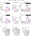
Arg-ii-/- mice reveal improved physical activity in aging. Diurnal voluntary physical activities (as revolution/hour on an average of 7 days in the wheel running cages under 12:12 light-dark conditions: 0-12, light-on (resting phase, white bar); 12–24, light-off (active phase, black bar) of young and old wild type (wt) and arg-ii-/- (ko) male (AC) and female mice (DF). (A, D) General activities of wheel running in males and females respectively; (B, E) Quantification of the general or total wheel-running activities during light-on and light-off in males and females, respectively. Graphs show the counting of total wheel revolutions during 24 hours in the young and old wt and ko mice. (C, F) Graphs show wheel running activities during light-on (white bar) and light-off (black bar) in males and females, respectively. Data are reported as means ± SEM. Combined one-way ANOVA and unpaired t-test with Welch’s correction were applied. n = 4 to 5 mice for each group. (G) Non-voluntary physical activities (treadmill fatigue test) in old male and female wt and arg-ii-/- mice. A parametric unpaired t-test with Welch’s correction was applied. n = 5 animals for each group. *p < 0.05. (H) Body weight in old male and female wt and arg-ii-/- mice. One-way ANOVA was applied with n = 5 animals for each group. ***p < 0.001. (I) Traction test measuring the success rate of lifting their weight to lean all the paws on a horizontally suspended bar. A contingency test was performed with n = 11–12 mice for each group. *p < 0.05, **p < 0.01, ***p < 0.001, and ****p < 0.0001 between the indicated groups.
Figure 2
Figure 2
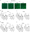
Arg-ii-/- mice reveal protection against age-associated sarcopenia (muscle fiber cross-sectional area, CSA). (A) Representative images of soleus sections stained for nuclei with DAPI (blue) and skeletal sarcolemma with WGA (green) in young and old wild type (wt) and arg-ii-/- (ko) male mice. Scale bar: 100 µm. (BE) left panels present quantification of CSA in µm2 of soleus and tibialis, in male and female mice, while the right panels present the fold change in CSA based on the ratio of old/young reflecting the age-dependent reduction of CSA. Combined parametric one-way ANOVA and unpaired t-test with Welch’s correction were applied; n = 4–7 mice for each group. *p < 0.05, **p < 0.01, ***p < 0.001 between the indicated groups.
Figure 3
Figure 3
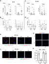
Arg-ii-/- mice reveal reduced senescent cells in aging skeletal muscles. (A) Representative confocal immunofluorescence staining of DAPI (blue) and anti-p16 (red) in tibialis from young and old wild type (wt) and arg-ii-/- (ko) male mice. Scale bar: 50 µm. (B) Representative image of old male mouse tibialis stained with DAPI (blue), anti-p16 (red), and anti-dystrophin (yellow, marker of sarcolemma). Negative control staining without the two primary antibodies. Scale bar: 20 µm. (C, D) Quantification of p16+ nuclei (senescent cells) expressed as the percentage of p16+ nuclei/total number of nuclei in the mm2 area. One-way ANOVA test was applied. n = 3–4 mice per group. *p < 0.05, **p < 0.01, ***p < 0.001, ****p < 0.0001 between the indicated groups.
Figure 4
Figure 4
Arg-ii-/- mice reveal reduced accumulation of centrally located nuclei in aging skeletal muscles. (A) Representative confocal immunofluorescence staining of DAPI (white) and WGA (red) for nuclei and skeletal muscle fiber sarcolemma, respectively, in the old soleus muscle from young and old wild type (wt) and arg-ii-/- (ko) male mice. Scale bar: 50 µm. (BE) The left graphics in each figure present the percentage of centrally nucleated fibers/total of fibers in the mm2 area for both soleus and tibialis in male and female mice as indicated. The fold changes based on the ratio of old/young (dotted line: young as 1) measuring the aging-dependent increase in centrally nucleated fibers were presented in the right bar graph in each panel. Combined one-way ANOVA and unpaired t-test with Welch’s correction were applied. n = 4–7 mice per group. *p < 0.05, **p < 0.01, ***p < 0.001 between the groups.
Figure 5
Figure 5
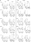
Arg-ii-/- mice reveal decreased inflammatory markers in aging skeletal muscles. mRNA expression levels of inflammatory markers and cytokines analyzed by qRT-PCR in soleus and tibialis from male (A) and female (B) young and old wild type (wt) and arg-ii-/- (ko) mice. One-way ANOVA was applied. n = 4–7 mice per group. *p < 0.05, **p < 0.01, ***p < 0.001 between the groups.
Figure 6
Figure 6
Arg-ii-/- mice reveal preserved microvascular density in aging skeletal muscle. (A) Representative confocal images showing CD31 staining (red; endothelial cells marker) in male tibialis of young and old male wild type (wt) and arg-ii-/- (ko) mice. DAPI (blue) is used to stain the nuclei. The graph reports the quantification of CD31 signal in the area (mm2). Scale bar: 50 µm. (B to D) Representative confocal images and relative quantification for CD31 (red) and DAPI (blue) in tibialis from young and old female wt and ko mice (B), and in soleus of young and old male (C) and female (D) wt and ko mice. Scale bar: 50 µm. One-way ANOVA was applied with n = 3 mice per group. *p < 0.05, **p < 0.01, ***p < 0.001, ****p < 0.0001 between the groups.
Figure 7
Figure 7
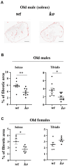
Arg-ii-/- mice reveal reduced skeletal muscle fibrosis in aging. (A) Representative masking images showing collagen signals (red) in soleus from old male wild type (wt) and arg-ii-/- (ko) mice. (B, C) Quantification of fibrosis area in both soleus and tibialis from old males and females. A parametric unpaired t-test with Welch’s correction was applied. n = 6–9 mice per group. *p < 0.05, **p < 0.01 between the groups. Scale bar: 250 µm.
Figure 8
Figure 8
Arg-ii deficiency prevents age-associated decrease in type-I and type-II muscle fibers. Representative confocal images showing Type-I (A) and Type-IIA (B) fibers in the soleus of young and old wt and arg-ii-/- (ko) male mice. DAPI (blue) is used to stain nuclei. Scale bar = 100 µm. The graphs present the percentage of Type-I (C) and Type-II (D) fibers over the total number of fibers in the image. (E) the graph shows the ratio of Type-I and Type-II fibers expressed as fold change (wt young group was taken as reference). One-way ANOVA test was applied. n = 3 mice per group. *p < 0.05, **p < 0.01, ***p < 0.001 between the indicated groups.
Figure 9
Figure 9
Arg-ii expression and cellular localization in aging skeletal muscles. Arg-ii mRNA levels were analyzed by qRT-PCR in skeletal muscles of wild-type (wt) male (A) and female (C) mice. (B, D) arg-i expression in soleus and tibialis from young and old wt and ko mice in both sexes. A parametric unpaired t-test with Welch’s correction was applied. n = 3–9 mice per group. *p < 0.05 between the indicated groups. (E) Representative confocal images of old wt and ko skeletal muscle showing co-localization of Arg-II (green) with CD31 (red, endothelial marker), and with Vimentin (red, fibroblasts marker). (F) Representative confocal images of old wt and ko muscle showing lack of co-localization of Arg-II (green) with F4-80 (red, macrophage marker), and with PAX-7 (red, satellite cells marker). DAPI (blue) stains cell nuclei. Scale bar: 10 µm (Arg-II/CD31); 25 µm (Arg-II/Vimentin); 10 µm (Arg-II/F4/80); 20 µm (Arg-II/PAX-7). Each experiment was repeated with 3 animals. (G) Representative confocal images of PAX-7 (red) in skeletal muscle of young and old wt and arg-ii-/- mice. Scale bar: 25 µm. (H) Quantification of PAX-7+ cells (satellite cells) in skeletal muscle expressed as the percentage of PAX-7+/total number of cells in the mm2 area. One-way ANOVA test was applied. n = 3 mice per group. ***p < 0.001 between the indicated groups.

References

    1. Khan SS, Singer BD, Vaughan DE. Molecular and physiological manifestations and measurement of aging in humans. Aging Cell. 2017; 16:624–33. 10.1111/acel.12601 - DOI - PMC - PubMed
    1. Campisi J, Kapahi P, Lithgow GJ, Melov S, Newman JC, Verdin E. From discoveries in ageing research to therapeutics for healthy ageing. Nature. 2019; 571:183–92. 10.1038/s41586-019-1365-2 - DOI - PMC - PubMed
    1. Clegg A, Young J, Iliffe S, Rikkert MO, Rockwood K. Frailty in elderly people. Lancet. 2013; 381:752–62. 10.1016/S0140-6736(12)62167-9 - DOI - PMC - PubMed
    1. Englund DA, Zhang X, Aversa Z, LeBrasseur NK. Skeletal muscle aging, cellular senescence, and senotherapeutics: Current knowledge and future directions. Mech Ageing Dev. 2021; 200:111595. 10.1016/j.mad.2021.111595 - DOI - PMC - PubMed
    1. Kunz HE, Lanza IR. Age-associated inflammation and implications for skeletal muscle responses to exercise. Exp Gerontol. 2023; 177:112177. 10.1016/j.exger.2023.112177 - DOI - PubMed