. 2025 Apr;52(5):1890-1900.
doi: 10.1007/s00259-024-07015-y. Epub 2024 Dec 13.

Evaluation of fibroinflammatory activity in thyroid eye disease using [18F]AlF-NOTA-FAPI-04 PET/CT: A prospective study

Affiliations

Evaluation of fibroinflammatory activity in thyroid eye disease using [18F]AlF-NOTA-FAPI-04 PET/CT: A prospective study

Hui Li et al. Eur J Nucl Med Mol Imaging. 2025 Apr.

Abstract

Purpose: This study evaluates the value of [18F]AlF-NOTA-FAPI-04PET/CT in assessing the disease activity of thyroid eye disease (TED).

Methods: Twenty-three TED patients and ten healthy controls were prospectively recruited and underwent [18F]AlF-NOTA-FAPI-04 PET/CT scans. TED eyes were classified by Clinical Activity Score (CAS) as active (CAS +) or inactive (CAS-). PET analysis included qualitative and quantitative evaluations. Qualitative analysis categorized TED eyes as positive (FAPI +) or negative (FAPI-), while quantitative analysis including SUVmax and target-to-backgroud ratio (TBR) of each extraocular muscle (EOM). Pathological assessment of 16 EOM samples from 15 TED eyes was used as a gold standard to evaluate the diagnostic accuracy of CAS-driven and FAPI-driven assessments.

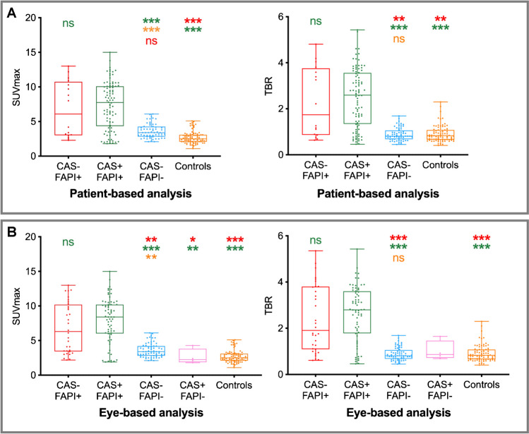
Results: Eye-based analysis included 42 TED eyes and 20 control eyes. SUVmax did not differ significantly between CAS-/FAPI + (n = 8) and CAS + /FAPI + (n = 17) eyes; though both groups had significantly higher SUVmax than CAS-/FAPI- (n = 16), CAS + /FAPI- (n = 1), and control eyes (n = 20) (all P < .05). Diffuse high FAPI uptake with excellent TBR was observed in pathologically active EOMs and had higher SUVmax and TBR than inactive EOMs (SUVmax: 9.57 ± 2.79 vs. 4.04 ± 1.02, TBR: 2.99 ± 1.00 vs. 1.00 ± 0.35, both P < .001). An SUVmax cutoff value of 5.95 yielded an area under the receiver operating characteristic curve of 0.98 for predicting active disease. Sensitivity and specificity of CAS-driven and FAPI-driven assessments for TED activity were 72.7% and 100%, 100% and 100%, respectively. SUVmax positively correlated with pathological fibroinflammatory score (r = 0.68, P = .004).

Conclusion: The diffuse high FAPI uptake with excellent TBR in the EOMs could illustrate the activity of TED, positively correlated with pathological fibroinflammatory scores. These findings suggest that [18F]AlF-NOTA-FAPI-04 PET/CT may serve as a promising reliable non-invasive approach for assessing disease activity in TED, potentially superior to the CAS.

Keywords: Clinical activity score; Extraocular muscles; Fibroblast activation protein inhibitor; Thyroid eye disease.


Conflict of interest statement

Declarations. Ethics approval: This study was performed in line with the principles of the Declaration of Helsinki. Approval was granted by the Ethics Committee of Peking University Third Hospital (M2023762 and M2024362). Consent to participate: Informed consent was obtained from all individual participants included in the study. Consent to publish: Informed consent for publication was obtained from the individual participant for whom identifying information is included in this article. Competing interests: The authors declare that they have no competing interests.

Figures

Fig. 1
Fig. 1
Flowchart of participant enrollment and study profile. CAS = Clinical Activity Score, FAPI = fibroblast activation protein inhibitor, TED = thyroid eye disease
Fig. 2
Fig. 2
Maximum-intensity-projection (MIP) images of [18F]AlF-NOTA-FAPI-04 PET/CT from all participants and heatmap for eye-based analysis. A MIP images of FAPI-positive (FAPI +) patients, showing partial or all extraocular muscles (EOMs) with high FAPI uptake. Patients were assigned as #1-#15. Clinical Activity Score (CAS)-driven assessment results were assigned to each eye. B MIP images of FAPI-negative (FAPI-) patients, showing all EOMs with mild to moderate FAPI uptake. Patients were assigned as #16-#23. CAS-driven assessment results were assigned to each eye. C MIP images of control participants, assigned as Control #1-#10, demonstrating faint to mild FAPI uptake in the EOMs of control eyes. D Heatmap illustrating the CAS-driven and FAPI-driven assessment results, along with the maximum standardized uptake value (SUVmax) for each eye (L = left eye, R = right eye). NA = not applicable, postoperative eyes were excluded
Fig. 3
Fig. 3
Quantitative analyses across all groups. A Patient-based analysis. B Eye-based analysis. The differences in maximum standardized uptake value (SUVmax) and target-to-background ratio (TBR) of extraocular muscles were significant among all groups in patient-based and eye-based analyses (all P < .001). CAS = Clinical Activity Score, CAS- = patients or eyes with inactive CAS scores. CAS +  = patients or eyes with active CAS scores. FAPI = fibroblast activation protein inhibitor, FAPI- = patients or eyes classified as negative on [18F]AlF-NOTA-FAPI-04 PET/CT. FAPI +  = patients or eyes classified as positive on [18F]AlF-NOTA-FAPI-04 PET/CT. All statistically significant differences and important non-statistically significant differences among multiple comparisons were summarized in the figure. Statistical significance is indicated as follows: *P < .05, **P < .01, ***P < .001, and ns = no significant difference
Fig. 4
Fig. 4
Images of patients #22, #3, and #5 with consistent Clinical Activity Score (CAS)-driven and Fibroblast Activation Protein Inhibitor (FAPI)-driven assessment results. (A-C) Left panel: external photograph. Middle panel: [18F]AlF-NOTA-FAPI-04 PET/CT images. Right panel: representative histopathological figures (low-power microscopic examination with scale bar of 200 μm, high-power microscopic examination with scale bar of 50 μm) using Hematoxylin–eosin (H&E) staining (left), Masson trichrome staining (middle), and fibroblast activation protein (FAP) immunochemistry staining (right) of the extraocular muscles (EOMs). A Patient #22, mild to moderate FAPI uptake in EOMs was observed. Histopathological figures from the left eye inferior rectus muscle (arrows, SUVmax = 4.3). IG = I0-I1, FG = F0-F1. B Patient #3, high FAPI uptake in EOMs was observed. Histopathological figures from the right eye medial rectus muscle (arrows, SUVmax = 6.4) showed high expression of FAP. IG = I3, FG = F2. C Patient #5, intense FAPI uptake in EOMs was observed. Histopathological figures from the left eye inferior rectus muscle (arrows, SUVmax = 13.8) showed high expression of FAP. IG = I3, FG = F3. IG = Inflammation grade, FG = fibrosis grade
Fig. 5
Fig. 5
Images of patients #10 and #6 with inconsistent Clinical Activity Score (CAS)-driven and Fibroblast Activation Protein Inhibitor (FAPI)-driven assessment results. (A-C) Left panel: external photograph. Middle panel: [18F]AlF-NOTA-FAPI-04 PET/CT images. Right panel: representative histopathological figures (low-power microscopic examination with scale bar of 200 μm, high-power microscopic examination with scale bar of 50 μm) using Hematoxylin–eosin (H&E) staining (left), Masson trichrome staining (middle), and fibroblast activation protein (FAP) immunochemistry staining (right) of the extraocular muscles (EOMs). (A) Patient #10, intense FAPI uptake in EOMs of right eye was observed. Histopathological figures from the right eye medial rectus muscle (arrows, SUVmax = 12.2) showed high expression of FAP. IG = I4, FG = F4. (B) Patient #6, intense FAPI uptake in EOMs was observed. Histopathological figures from the left eye medial rectus muscle (arrows, SUVmax = 10.9) showed high expression of FAP. IG = I3, FG = F3. IG = Inflammation grade, FG = fibrosis grade

References

    1. Bahn RS. Graves’ ophthalmopathy. N Engl J Med. 2010;362:726–38. 10.1056/NEJMra0905750. - PMC - PubMed
    1. Perros P, Neoh C, Dickinson J. Thyroid eye disease. Bmj. 2009;338: b560. 10.1136/bmj.b560. - PubMed
    1. Bartalena L, Tanda ML. Current concepts regarding Graves’ orbitopathy. J Intern Med. 2022;292:692–716. 10.1111/joim.13524. - PMC - PubMed
    1. Mourits MP, Koornneef L, Wiersinga WM, Prummel MF, Berghout A, van der Gaag R. Clinical criteria for the assessment of disease activity in Graves’ ophthalmopathy: a novel approach. Br J Ophthalmol. 1989;73:639–44. 10.1136/bjo.73.8.639. - PMC - PubMed
    1. Bartalena L, Kahaly GJ, Baldeschi L, Dayan CM, Eckstein A, Marcocci C, et al. The 2021 European Group on Graves’ orbitopathy (EUGOGO) clinical practice guidelines for the medical management of Graves’ orbitopathy. Eur J Endocrinol. 2021;185:G43-g67. 10.1530/eje-21-0479. - PubMed

LinkOut - more resources