. 2024 Dec 17;121(51):e2412327121.
doi: 10.1073/pnas.2412327121. Epub 2024 Dec 13.

Temperature-dependent fold-switching mechanism of the circadian clock protein KaiB

Affiliations

Temperature-dependent fold-switching mechanism of the circadian clock protein KaiB

Ning Zhang et al. Proc Natl Acad Sci U S A. .

Abstract

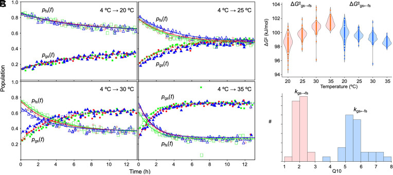
The oscillator of the cyanobacterial circadian clock relies on the ability of the KaiB protein to switch reversibly between a stable ground-state fold (gsKaiB) and an unstable fold-switched fold (fsKaiB). Rare fold-switching events by KaiB provide a critical delay in the negative feedback loop of this posttranslational oscillator. In this study, we experimentally and computationally investigate the temperature dependence of fold switching and its mechanism. We demonstrate that the stability of gsKaiB increases with temperature compared to fsKaiB and that the Q10 value for the gsKaiB → fsKaiB transition is nearly three times smaller than that for the reverse transition in a construct optimized for NMR studies. Simulations and native-state hydrogen-deuterium exchange NMR experiments suggest that fold switching can involve both partially and completely unfolded intermediates. The simulations predict that the transition state for fold switching coincides with isomerization of conserved prolines in the most rapidly exchanging region, and we confirm experimentally that proline isomerization is a rate-limiting step for fold switching. We explore the implications of our results for temperature compensation, a hallmark of circadian clocks, through a kinetic model.

Keywords: NMR; circadian clock; molecular dynamics; protein folding; temperature compensation.


Conflict of interest statement

Competing interests statement:The authors declare no competing interest.

Figures

Fig. 1.
Fig. 1.
KaiB switches reversibly between two distinct folds. Three proline residues—P63, P70, and P72—are trans in the ground-state fold of KaiB (gsKaiB, Left) and cis in the fold-switched fold of KaiB (fsKaiB, Right). PDB IDs 1VGL and 5JYT were used with VMD (13) to generate the ribbon diagrams for gsKaiB and fsKaiB, respectively.
Fig. 2.
Fig. 2.
The gsKaiBD91R ⇌ fsKaiBD91R equilibrium is sensitive to temperature. (A) A sample of 15N-enriched KaiBD91R was incubated at 4 °C for 24 h before inserting it into a 14.1 T NMR spectrometer that was set to 35 °C. 15N,1H HSQC spectra were collected over time and some representative regions are shown here. (B) Residue-specific ΔG values for the gsKaiBD91R → fsKaiBD91R reaction are plotted as red, green, and blue points, one color for each replicate, and are superimposed on distribution diagrams for each temperature sampled. The blue line is a fit to mean values using the equation ΔG = ΔHTΔS. Fitting the data for individual residues yields ΔH = –82 ± 18 kJ mol−1 and ΔS = –276 ± 58 J mol−1 K−1, indicating that the gsKaiBD91R → fsKaiBD91R reaction is enthalpically driven.
Fig. 3.
Fig. 3.
The kinetics of gsKaiBD91R → fsKaiBD91R and gsKaiBD91R ← fsKaiBD91R fold switching differ in their dependence on temperature. (A) Kinetics of gsKaiBD91R ← fsKaiBD91R fold switching of residue G16 under different temperature jumps. A sample of 15N-enriched KaiBD91R was incubated at 4 °C for at least 24 h before inserting it into a 14.1 T NMR spectrometer that was set at either 20 °C, 25 °C, 30 °C, or 35 °C. 15N,1H HSQC spectra were collected at regular intervals after a dead time of approximately 4 min. Open and solid symbols represent fractional populations of residue G16 in the gsKaiBD91R and fsKaiBD91R folds, respectively. Red, green, and blue colors represent separate experiments, each of which used a freshly prepared sample. HSQC peak volumes were determined by nmrPipe and nmrDraw. (B) ΔGgs→fs and ΔGgs←fs as a function of temperature for all residues, and (C) histograms of residue-specific Q10 values for kgs→fs and kgs←fs. The forward and reverse reactions are rendered in pink and blue, respectively. In (B) and (C) the three replicates were pooled. Residue-specific plots of Q10 values are presented in SI Appendix, Fig. S5.
Fig. 4.
Fig. 4.
Fold switching in an Upside model of KaiBD91R involves partially and completely unfolded intermediates. (A) Potential of mean force as a function of the fraction of N-terminal contacts (QN) and the difference in the fractions of gsKaiB and fsKaiB C-terminal contacts (Qfs,CQgs,C). From Left to Right, the Upside temperature is increasing. Contour lines are drawn every 2 kBT. The approximate location of the transition state (q = 0.5) is marked as a dashed line. (B) Selected structures corresponding to points marked on the potentials of mean force in (A). The sequence is colored by the position of secondary structures in fsKaiB and P63, P70, P71, and P72 are shown as balls and sticks.
Fig. 5.
Fig. 5.
HDX rates for KaiBHDX indicate that β1-α1-β2 and α3 are more stable than α2-β3-β4. Observed exchange rates (log10 of kobs in h–1) at (A) pH 5.5 and (B) pH 6.5. In (A) and (B), blue circles, gold triangles, green diamonds, red squares, inverted purple triangles, and brown circles represent data in 0.0, 0.5, 1.0, 1.5, 2.0, and 2.5 M urea, respectively. Each data point represents the mean of two replicates, except for those at 2.0 M urea at pH 5.5 and 1.5 M urea at pH 6.5, for which there was only one measurement. The SE in the mean was 0.05 on average for datasets at both pH values. Residues with a measurable kobs value at only a single urea concentration were not included in these plots. The secondary structure cartoons along the Bottom of (B) represent the fold-switched structure.
Fig. 6.
Fig. 6.
Proline isomerization is rate-limiting for gsKaiB ← fsKaiB fold-switching of KaiBD91R. (A) An Upside simulation starting from fsKaiB with P63, P70, and P72 fixed in trans. (Top) Secondary structure assigned via DSSP (46) and (Bottom) RMSD to either the C-terminal half of fsKaiB, the C-terminal half of gsKaiB, or the N-terminal half (SI Appendix, Table S2) during a section of a successful fold-switching trajectory from fsKaiB to gsKaiB. RMSDs are plotted as a moving average over 20 frames. The dotted lines indicate the approximate start and end times of the fold-switching event. (B) Observed rates of fold switching, kobs, after a jump to 35 °C for KaiBD91R samples pre-equilibrated at 4 °C as a function of the concentration of human PPIA. 190 to 200 µM 15N-enriched KaiBD91R samples stored at –80 °C were incubated at 4 °C for 24 h before inserting them into the NMR spectrometer, which was set to a sample temperature of 35 °C. 15N,1H HSQC spectra were collected every 18 min over a span of 7 h. Residue-specific rates were determined from these spectra as described in SI Appendix. Circles represent kobs values from globally fitting data from residues with resolved and assigned gsKaiB and fsKaiB HSQC peaks (35 residues total). Replicates are depicted in different colors (blue and gold). Triangles represent kobs values where 55 µM cyclosporin A (CsA) was added to the samples. Replicates are depicted in different colors (green and pink). Error bars represent uncertainties from the global fits.
Fig. 7.
Fig. 7.
Kinetics of formation of the fsKaiBD91R–CI complex is partially temperature compensated. (A) Reaction scheme of KaiBD91R fold switching and binding the CI domain of KaiC. (B) Calculated kinetics of fsKaiBD91R–CI complex formation when ΔG and ΔG values are fixed at their experimentally determined 30 °C values. (C) Calculated kinetics of fsKaiBD91R–CI complex formation using ΔG and ΔG values experimentally determined at each temperature modeled.

Update of

References

    1. Kolodny R., Pereyaslavets L., Samson A. O., Levitt M., On the universe of protein folds. Annu. Rev. Biophys. 42, 559–582 (2013). - PubMed
    1. Porter L. L., Fluid protein fold space and its implications. Bioessays 45, 2300057 (2023). - PMC - PubMed
    1. Kim A. K., Porter L. L., Functional and regulatory roles of fold-switching proteins. Structure 29, 6–14 (2021). - PMC - PubMed
    1. Murzin A. G., Metamorphic proteins. Science 320, 1725–1726 (2008). - PubMed
    1. Chang Y.-G., et al. , A protein fold switch joins the circadian oscillator to clock output in cyanobacteria. Science 349, 324–328 (2015). - PMC - PubMed

MeSH terms

Substances

LinkOut - more resources