. 2024 Dec 13;20(12):e1011496.
doi: 10.1371/journal.pgen.1011496. eCollection 2024 Dec.

Integrin adhesome axis inhibits the RPM-1 ubiquitin ligase signaling hub to regulate growth cone and axon development

Affiliations

Integrin adhesome axis inhibits the RPM-1 ubiquitin ligase signaling hub to regulate growth cone and axon development

Jonathan Amezquita et al. PLoS Genet. .

Abstract

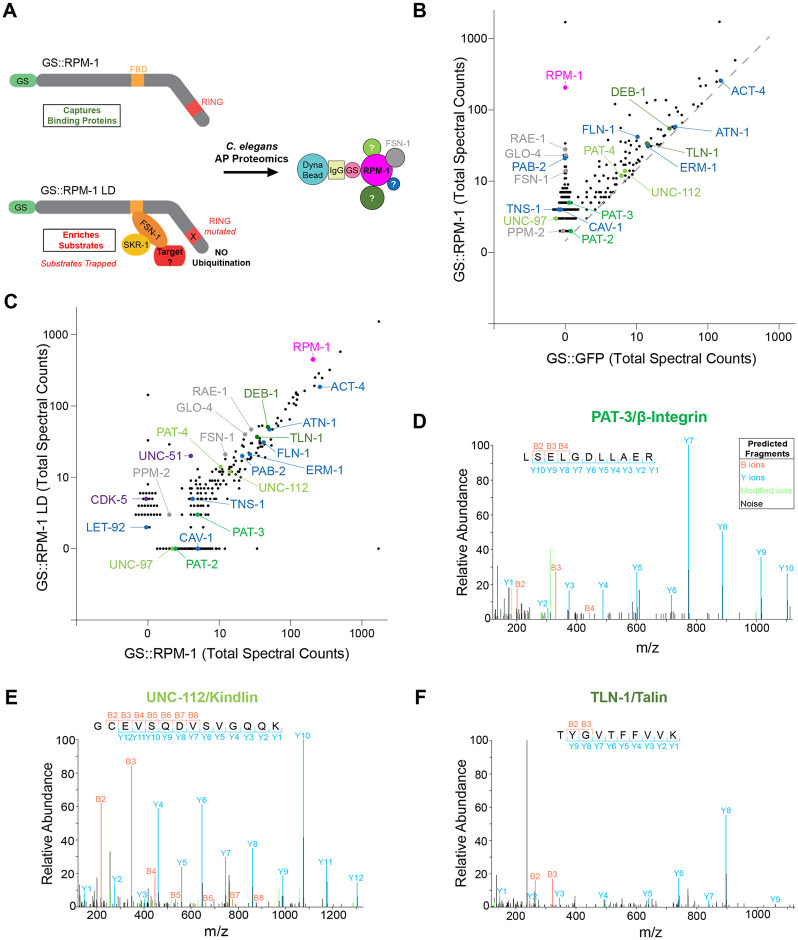
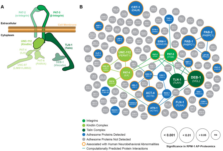
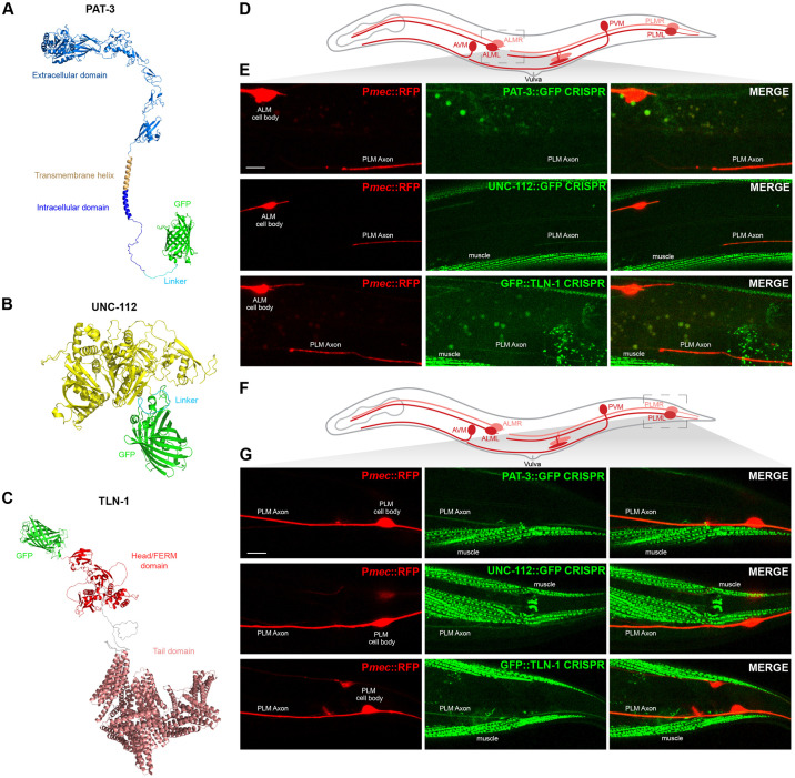
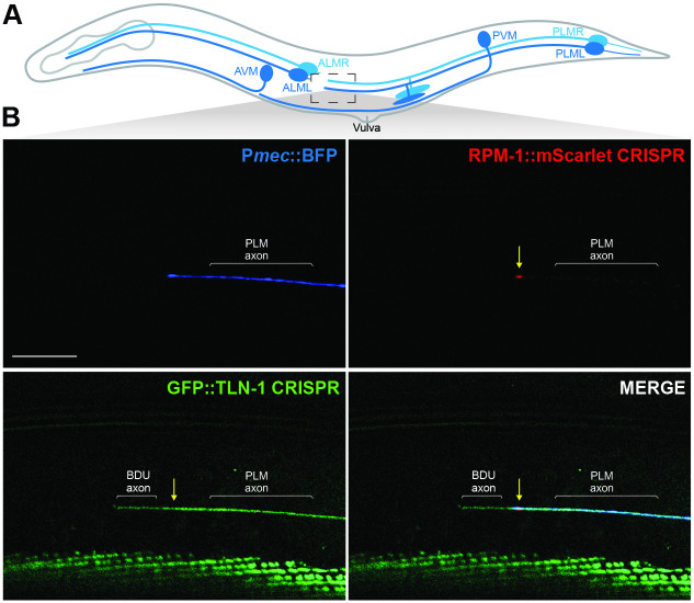
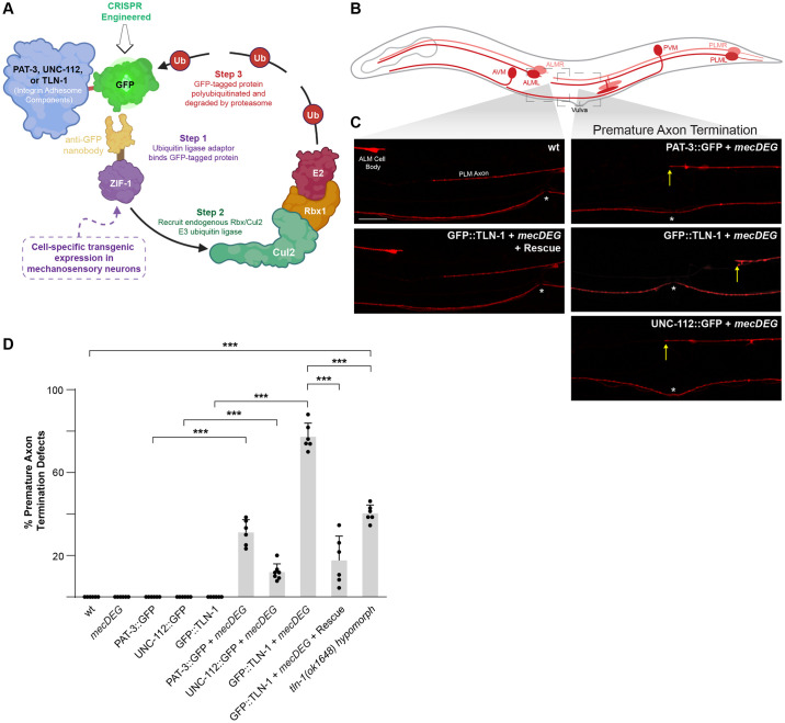
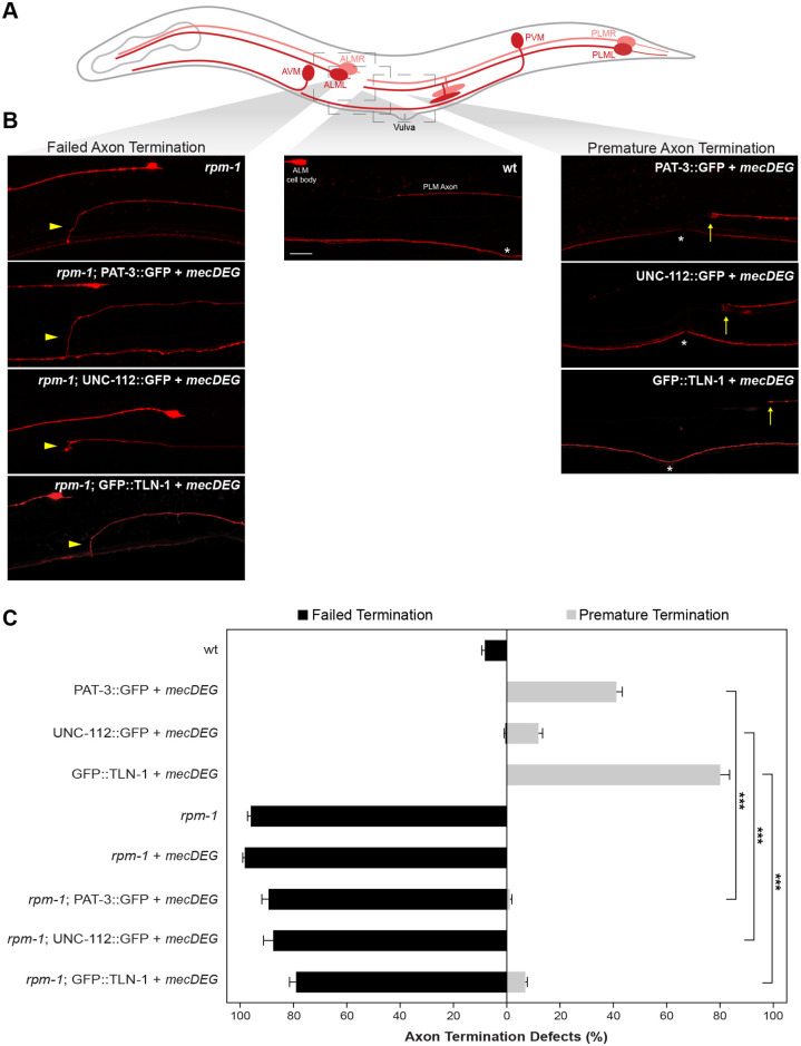
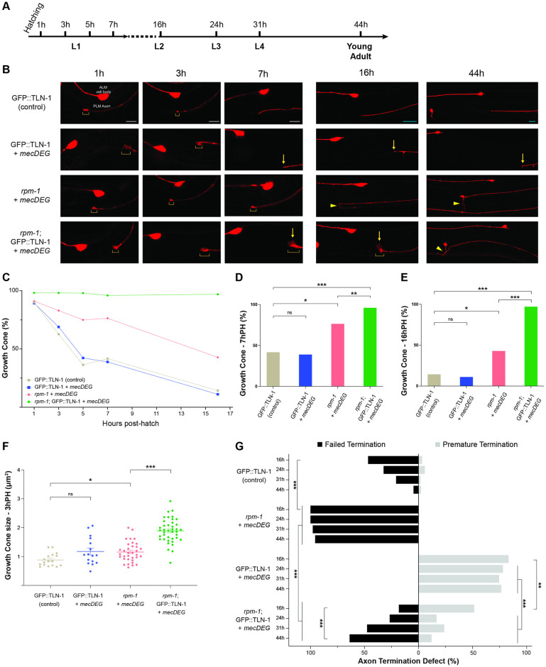
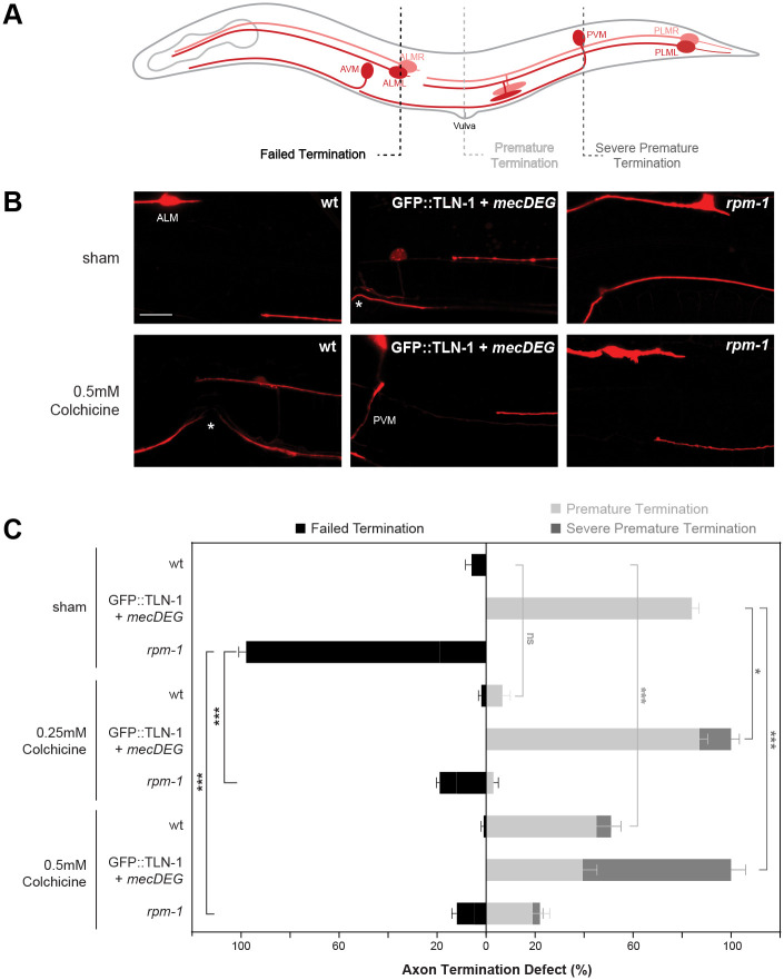
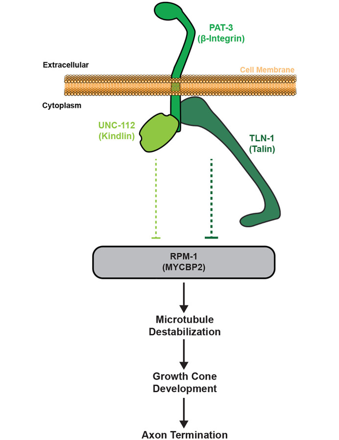
Integrin signaling plays important roles in development and disease. An adhesion signaling network called the integrin adhesome has been principally defined using bioinformatics and cell-based proteomics. To date, the adhesome has not been studied using integrated proteomic and genetic approaches. Here, proteomic studies in C. elegans identified physical associations between the RPM-1 ubiquitin ligase signaling hub and numerous adhesome components including Talin (TLN-1), Kindlin (UNC-112) and β-integrin (PAT-3). C. elegans RPM-1 is orthologous to human MYCBP2, a prominent player in nervous system development recently associated with a neurodevelopmental disorder. After curating and updating the conserved C. elegans adhesome, we identified an adhesome subnetwork physically associated with RPM-1 that has extensive links to human neurobehavioral abnormalities. Using neuron-specific, CRISPR loss-of-function strategies, we demonstrate that a PAT-3/UNC-112/TLN-1 adhesome axis regulates axon termination in mechanosensory neurons by inhibiting RPM-1. Developmental time-course studies and pharmacological results suggest TLN-1 inhibition of RPM-1 affects growth cone collapse and microtubule dynamics during axon outgrowth. These results indicate the PAT-3/UNC-112/TLN-1 adhesome axis restricts RPM-1 signaling to ensure axon outgrowth is terminated in a spatially and temporally accurate manner. Thus, our findings orthogonally validate the adhesome using an organismal setting, identify an adhesome axis that inhibits RPM-1 (MYCBP2), and highlight important new links between the adhesome and brain disorders.


Conflict of interest statement

The authors have declared that no competing interests exist.

Figures

Fig 1
Fig 1. RPM-1 AP-proteomics identifies numerous integrin adhesome components.
A) Schematic of RPM-1 ubiquitin ligase constructs used for AP-proteomics from C. elegans. Both GS::RPM-1 and GS::RPM-1 LD capture binding proteins (e.g. FSN-1), while GS::RPM-1 LD enriches ubiquitination substrates. B) Example of single AP-proteomics experiment showing individual proteins identified by LC-MS/MS. Shown are results from GS::RPM-1 sample compared with GS::GFP (negative control). Highlighted (shades of green) are integrin receptors, PAT-3/β-Integrin and PAT-2/α-Integrin, and two canonical integrin signaling complexes, UNC-112/Kindlin complex and TLN-1/Talin complex. Additional adhesome components (blue) were identified. Also highlighted are RPM-1 (magenta) and known RPM-1 binding proteins (gray). Grey dashed line represents 1.5x enrichment of spectra for proteomic hits in GS::RPM-1 sample compared to GS::GFP negative control. C) Results from single AP-proteomics experiment showing GS::RPM-1 compared to GS::RPM-1 LD ubiquitination substrate ‘trap’. Highlighted are previously validated substrates (CDK-5 and UNC-51, purple). D-F) Examples of LC-MS/MS peptide spectrum for D) PAT-3/β-Integrin, E) UNC-112/Kindlin and F) TLN-1/Talin.
Fig 2
Fig 2. PAT-3/UNC-112/TLN-1 adhesome axis and other adhesome components present in RPM-1 proteomics are linked to human neurobehavioral deficits.
A) Illustration of PAT-3/UNC-112/TLN-1 adhesome axis that includes two integrin receptors PAT-3 (β-Integrin, green) and PAT-2 (α-Integrin, green), the UNC-112 Kindlin complex (light green), and the TLN-1 Talin complex (dark green). B) Summary of RPM-1 AP-proteomics data from C. elegans integrated with a computationally predicted protein-protein interaction network (lines) and genetic links to human neurobehavioral abnormalities (orange halo). Adhesome components enriched in RPM-1 AP-proteomics are highlighted in color with increasing circle size denoting significance (GS::RPM-1 test samples versus GS::GFP negative controls). Highlighted (shades of green) are Integrins, UNC-112 Kindlin complex and TLN-1 Talin complex. Also shown are additional adhesome components detected (blue) or absent (gray) in RPM-1 AP-proteomics. Orthologous human protein annotated in brackets. Data is presented from 7 independent RPM-1 AP-proteomics experiments. Significance determined using Mann-Whitney test. ns = not significant.
Fig 3
Fig 3. PAT-3/UNC-112/TLN-1 adhesome axis is expressed in C. elegans mechanosensory neurons and localized to axons.
A-C) Alpha-fold predictions showing adhesome signaling components that were GFP-tagged using CRISPR engineering in C. elegans. Structural predictions indicate GFP is not likely to interfere with protein folding. A) C. elegans PAT-3::GFP, B) UNC-112::GFP, and C) GFP::TLN-1. D) Schematic of C. elegans mechanosensory neurons highlighting region imaged to visualize axon termination sites for PLM neurons (light gray box). E) Representative images showing that PAT-3::GFP, UNC-112::GFP and GFP::TLN-1 are expressed in PLM neurons and localized to axons. Pmec-7::mRFP (jsIs973) labels mechanosensory neurons. F) Schematic highlights cell body and initial axon segment (light gray box) of PLM neurons. G) Representative images showing PAT-3::GFP, UNC-112::GFP and GFP::TLN-1 primarily localized to PLM axon. For E and G, note that integrin components also show prominent expression in muscles. Scale bars 10μm.
Fig 4
Fig 4. TLN-1 and RPM-1 colocalize at axon termination sites in PLM mechanosensory neurons.
A) Schematic of C. elegans mechanosensory neurons highlighting region imaged to visualize axon termination sites for PLM neurons (light gray box). B) Representative super-resolution images demonstrating CRISPR engineered GFP::TLN-1 and RPM-1::mScarlet colocalize at terminated axon tips of PLM neurons. PLM axons were visualized using transgenic BFP expressed in mechanosensory neurons (Pmec17::mTagBFP, bggEx180). Note GFP::TLN-1 expression anterior to the PLM termination site is a neurite from the adjacent BDU neuron. Scale bar 10μm.
Fig 5
Fig 5. Impairing PAT-3/UNC-112/TLN-1 adhesome axis with a CRISPR-based, cell-specific protein degradation system results in premature axon termination.
A) Illustration of CRISPR-based cell-specific protein degradation system used to impair adhesome components in C. elegans mechanosensory neurons (mecDEG). Adapted from Wang et al. 2017. B) Schematic of mechanosensory neurons with imaged regions highlighted (light gray boxes). C) Representative images of PLM axons for indicated genotypes visualized using Pmec-7::mRFP (jsIs973). Premature axon termination defects (arrows) occur when mecDEG targets GFP that is CRISPR engineered onto adhesome components (PAT-3::GFP, UNC-112::GFP and GFP::TLN-1). Vulva (asterisks) used as anatomical reference point for premature termination. D) Quantitation of premature axon termination defects in PLM neurons for indicated genotypes. Note premature axon termination occurs with mecDEG targeting of adhesome components and in tln-1(ok1648) hypomorphic mutants. Means (bars) are shown for 5 or more counts (black dots) with 20 or more animals/count for each genotype. Error bars indicate SEM. Significance assessed using Student’s t-test with Bonferroni correction for multiple comparisons. ***p<0.001. Scale bar 10μm.
Fig 6
Fig 6. PAT-3/UNC-112/TLN-1 adhesome axis inhibits RPM-1 to regulate axon termination.
A) Schematic of C. elegans mechanosensory neurons with imaged regions highlighted (light gray boxes). B) Representative images of PLM axons for indicated genotypes visualized using Pmec-7::mRFP (jsIs973). rpm-1 mutants display failed axon termination defects (arrowhead) and mecDEG targeting of adhesome components (PAT-3::GFP, UNC-112::GFP and GFP::TLN-1 CRISPR) results in premature termination defects (arrows). rpm-1 double mutants with adhesome component degradation (e.g. rpm-1; PAT-3::GFP + mecDEG) predominantly display failed termination defects. Vulva (asterisks) used as anatomical reference point for premature termination. C) Quantitation of failed axon termination defects (black) and premature termination defects (light gray) in PLM neurons for indicated genotypes. Premature termination defects are strongly suppressed in double mutants lacking RPM-1 and PAT-3, UNC-112 or TLN-1 adhesome components. Mean (bars) are shown for 5 or more counts (20 or more animals/count) for each genotype. Error bars indicate SEM. Significance assessed using Student’s t-test with Bonferroni correction. ***p<0.001. Scale bar 10μm.
Fig 7
Fig 7. Time-course studies indicate TLN-1 inhibits RPM-1 to regulate growth cone collapse and axon termination during development.
A) Timeline of C. elegans larval development and key time points evaluated. B) Representative images of PLM axons for indicated genotypes visualized using Pmec-7::mRFP (jsIs973). Shown are axonal growth cones (brackets), premature termination sites (arrows) and failed termination defects (arrowheads). Double mutants (rpm-1; GFP::TLN-1 + mecDEG) display failed growth cone collapse with persistent, enlarged growth cones. In double mutants, growth cones are initially in locations corresponding to premature axon termination (1 to 16h PH) but failed axon termination becomes the primary phenotype as development progresses (44h PH). Note AVM cell body is only visible at 44h PH on one side of the animal. C) Summary of quantitative results for growth cone frequency during development for indicated genotypes. D-E) Quantitative analysis of growth cone frequency at D) 7h PH and E) 16h PH. F) Quantitation of growth cone size at 3h PH for indicated genotypes. G) Quantitation of failed axon termination defects (black) and premature termination defects (light gray) in PLM neurons for indicated genotypes at specified time points in development. For C-G, minimum of 21 PLM neurons scored for each time point and genotype. Mean (square, bar or line) are shown for each genotype. For F, dots represent a single animal. Error bars indicate SEM. For C-E and G, significance assessed using Fisher’s exact test. For F, significance assessed using Student’s t-test with Bonferroni correction for growth cone size. ns = not significant, *p<0.05, **p<0.01, ***p<0.001. Scale bars 5μm (1-7h PH, white bars) or 10μm (16-44h PH, teal bars).
Fig 8
Fig 8. TLN-1 and RPM-1 influence axon termination via effects on microtubule stability.
A) Schematic of C. elegans mechanosensory neurons. Dashed lines indicate different regions where axon termination defects were visualized. B) Representative images of PLM axons for indicated genotypes under sham conditions (DMSO) or treated with the microtubule destabilizing drug colchicine (0.5mM). Mechanosensory neurons visualized using Pmec-7::mRFP (jsIs973). C) Quantitation of failed termination (black), premature termination (light gray) and severe premature termination (dark gray) defects for indicated genotypes and treatment. rpm-1 mutants display failed axon termination defects that are suppressed by colchicine. GFP::TLN-1 degraded by mecDEG results in premature termination defects with colchicine treatment resulting in more severe premature termination defects. Vulva (asterisks) and PVM are anatomical reference points for premature and severe premature termination defects, respectively. Mean (bars) are shown for 5 counts (20 or more animals/count) for each genotype and treatment. Error bars indicate SEM. Significance assessed using Student’s t-test with Bonferroni correction. ns = not significant, *p<0.05, **p<0.01, ***p<0.001. Scale bar 10μm.
Fig 9
Fig 9. Summary of results indicating PAT-3/UNC-112/TLN-1 adhesome axis inhibits RPM-1 to influence microtubules and growth cone dynamics during axon termination.

Update of

References

    1. Myers JP, Santiago-Medina M, Gomez TM. Regulation of axonal outgrowth and pathfinding by integrin-ECM interactions. Dev Neurobiol. 2011;71(11):901–23. doi: 10.1002/dneu.20931 . - DOI - PMC - PubMed
    1. Park YK, Goda Y. Integrins in synapse regulation. Nat Rev Neurosci. 2016;17(12):745–56. Epub 2016/11/05. doi: 10.1038/nrn.2016.138 . - DOI - PubMed
    1. Lilja J, Ivaska J. Integrin activity in neuronal connectivity. J Cell Sci. 2018;131(12). Epub 20180615. doi: 10.1242/jcs.212803 . - DOI - PubMed
    1. Eva R, Fawcett J. Integrin signalling and traffic during axon growth and regeneration. Curr Opin Neurobiol. 2014;27:179–85. Epub 20140504. doi: 10.1016/j.conb.2014.03.018 . - DOI - PubMed
    1. Wickstrom SA, Radovanac K, Fassler R. Genetic analyses of integrin signaling. Cold Spring Harb Perspect Biol. 2011;3(2). Epub 20110201. doi: 10.1101/cshperspect.a005116 . - DOI - PMC - PubMed

MeSH terms

LinkOut - more resources