. 2024 Dec 13;10(50):eads5735.
doi: 10.1126/sciadv.ads5735. Epub 2024 Dec 13.

Effect of pandemic influenza A virus PB1 genes of avian origin on viral RNA polymerase activity and pathogenicity

Affiliations

Effect of pandemic influenza A virus PB1 genes of avian origin on viral RNA polymerase activity and pathogenicity

Stephanie L Williams et al. Sci Adv. .

Abstract

Zoonotic influenza A virus (IAV) infections pose a substantial threat to global health. The influenza RNA-dependent RNA polymerase (RdRp) comprises the PB2, PB1, and PA proteins. Of the last four pandemic IAVs, three featured avian-origin PB1 genes. Prior research linked these avian PB1 genes to increased viral fitness when reassorted with human IAV genes. This study evaluated chimeric RdRps with PB1 genes from the 1918, 1957, and 1968 pandemic IAVs in a low pathogenic avian influenza (LPAI) virus background to assess polymerase activity and pathogenicity. Substituting in the pandemic PB1 genes reduced polymerase activity, virulence, and altered lung pathology, while the native LPAI PB1 showed the highest pathogenicity and polymerase activity. The native LPAI PB1 virus caused severe pneumonia and high early viral RNA levels, correlating with elevated host cytokine signaling. Increased genetic distance from the LPAI PB1 sequence correlated with reduced polymerase activity, IFN-β expression, viral replication, and pathogenicity.


Figures

Fig. 1.
Fig. 1.. Chimeric LPAI polymerases with pandemic PB1 show reduced polymerase activity, RNA levels, and IFN-β expression.
(A) Minigenome assays used to assess the impact of the PB1 genes of the 1918, 1957, and 1968 pandemic IAVs on the LPAI polymerase. Created in BioRender.com. (B) Activity of chimeric IAV polymerases in a luciferase reporter assay. Chimeric polymerase activities were presented as a percent fold compared to the activity of the LPAI polymerase. Three biological replicate ratios with SEM are shown. A one sample t test was done to test each ratio = 1 with Bonferroni correction. (C) vRNA, mRNA, and cRNA levels generated by chimeric IAV polymerases using NA vRNA template. PB1 expression levels with α-β-actin as a loading control is shown. Data are a mean of four biological replicates showing SEM. Data were normalized to the 5S rRNA loading control and expressed as a percent fold compared to the LPAI polymerase. Statistical significance was determined by two-way ANOVA and Dunnett’s multiple comparison test compared to the LPAI PB1. (D) Measuring IFN-β expression using a minigenome luciferase reporter gene under the control of an IFN-β promoter. Created in BioRender.com. (E) IFN-β levels in an IAV minigenome assay. IFN-β expression levels induced by polymerase activity were presented as a percent fold compared to the activity of the LPAI polymerase. Three biological replicate ratios with SEM are shown. A one sample t test was done to test each ratio = 1 with Bonferroni correction.
Fig. 2.
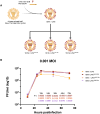
Fig. 2.. Chimeric virus rescue and corresponding growth kinetics in MDCK cells.
(A) Recombinant viruses were generated using a LPAI prototypical virus, A/green-winged teal/Ohio/175/1986 H2N1. As previously described, the LPAI HA segment was replaced with the 1918 HA gene segment in the backbone, along with the nonsynonymous PB2 E627K mutation in PB2. The LPAI PB1 was then systematically substituted with that of the early three IAV pandemics: 1918, 1957, and 1968. Image created with BioRender.com. (B) Viral replication kinetics as assessed across 72 hours at an MOI of 0.001. Bars represent the SD between three technical replicates. Statistical significance was determined by two-way ANOVA and Dunnett’s multiple comparison test compared to the LPAI PB1 virus.
Fig. 3.
Fig. 3.. Virulence and replication of chimeric 18HA + LPAI viruses in BALB/c mice.
(A) Weight loss of infected BALB/c mice across 13 days at the 103 PFU dose in three separate experiments where groups of five mice were infected per virus (n = 15). SEM of data shown. A dashed line represents the 75% initial body ethical endpoint for euthanasia. (B) Kaplan-Meier survival curve demonstrating percent survival of mice following infection with rescued viruses at the 103 PFU dose. (C) Viral titers of chimeric 18 HA + LPAI viruses postinfection from day 1 through day 4 (n = 3 per virus per day, except for 1957 day 4, which was n = 2 due to sample contamination). Bars represent the SD between three technical replicates. Statistics were completed via two-way ANOVA, mixed-effect analysis with Dunnett test for multiple comparisons compared to the LPAI PB1 virus. (D) Total viral RNA levels quantified across days 1 through 4 postinfection. Bars represent the SD between three technical replicates. Statistics were completed via two-way ANOVA, mixed-effect analysis with Dunnett test for multiple comparisons compared to the LPAI PB1 virus.
Fig. 4.
Fig. 4.. Pathology and IHC influenza A and MPO staining of mouse lung tissue.
IAV and MPO antibody staining of BALB/c mouse lung at day 7 postinfection of LPAI model virus with respective pandemic PB1 genes. (A) DMEM negative-control mock-infected mouse lung with H&E staining. (B to E) H&E staining for chimeric 18HA + LPAI viruses. (F to J) α-IAV staining of day 7 lung histopathology postinfection with chimeric PB1 genes and DMEM mock. (K to O) α-MPO staining of day 7 lung histopathology. (P) Lung pathology scores counted for two replicates of each infection group evaluated on a 0 to 3 scale. Zero indicated no pathology, and 3 represented severe pathology and the highest tissue involvement. (Q) Cell counting of MPO staining across 10 frames. Bars represent SD. Statistics were done in Prism 9 with repeated-measures, one-way ANOVA and Dunnett multiple comparison test to the LPAI PB1 virus.

References

    1. Webster R. G., Bean W. J., Gorman O. T., Chambers T. M., Kawaoka Y., Evolution and ecology of influenza A viruses. Microbiol. Rev. 56, 152–179 (1992). - PMC - PubMed
    1. Taubenberger J. K., Reid A. H., Lourens R. M., Wang R., Jin G., Fanning T. G., Characterization of the 1918 influenza virus polymerase genes. Nature 437, 889–893 (2005). - PubMed
    1. Taubenberger J. K., Kash J. C., Morens D. M., The 1918 influenza pandemic: 100 years of questions answered and unanswered. Sci. Transl. Med. 11, eaau5485 (2019). - PMC - PubMed
    1. Patrono L. V., Vrancken B., Budt M., Düx A., Lequime S., Boral S., Gilbert M. T. P., Gogarten J. F., Hoffmann L., Horst D., Merkel K., Morens D., Prepoint B., Schlotterbeck J., Schuenemann V. J., Suchard M. A., Taubenberger J. K., Tenkhoff L., Urban C., Widulin N., Winter E., Worobey M., Schnalke T., Wolff T., Lemey P., Calvignac-Spencer S., Archival influenza virus genomes from Europe reveal genomic variability during the 1918 pandemic. Nat. Commun. 13, 2314 (2022). - PMC - PubMed
    1. Kawaoka Y., Krauss S., Webster R. G., Avian-to-human transmission of the PB1 gene of influenza A viruses in the 1957 and 1968 pandemics. J. Virol. 63, 4603–4608 (1989). - PMC - PubMed

Substances