. 2024 Dec 13;10(50):eadq4738.
doi: 10.1126/sciadv.adq4738. Epub 2024 Dec 13.

Targeted nanoliposomes to improve enzyme replacement therapy of Fabry disease

Affiliations

Targeted nanoliposomes to improve enzyme replacement therapy of Fabry disease

Judit Tomsen-Melero et al. Sci Adv. .

Abstract

The central nervous system represents a major target tissue for therapeutic approach of numerous lysosomal storage disorders. Fabry disease arises from the lack or dysfunction of the lysosomal alpha-galactosidase A (GLA) enzyme, resulting in substrate accumulation and multisystemic clinical manifestations. Current enzyme replacement therapies (ERTs) face limited effectiveness due to poor enzyme biodistribution in target tissues and inability to reach the brain. We present an innovative drug delivery strategy centered on a peptide-targeted nanoliposomal formulation, designated as nanoGLA, engineered to selectively deliver a recombinant human GLA (rhGLA) to target tissues. In a Fabry mouse model, nanoGLA demonstrated improved efficacy, inducing a notable reduction in Gb3 deposits in contrast to non-nanoformulated GLA, even in the brain, highlighting the potential of the nanoGLA to address both systemic and cerebrovascular manifestations of Fabry disease. The EMA has granted the Orphan Drug Designation to this product, underscoring the potential clinical superiority of nanoGLA over authorized ERTs and encouraging to advance it toward clinical translation.


Figures

Fig. 1.
Fig. 1.. Impact of the dispersion media on the nanoGLA formulation.
(A) Evolution of the osmolality of liposomal formulation during buffer exchange by TFF diafiltration, in water (L-water) or in PBS (L-PBS), determined by measurements of the freezing point. Media refers to water and PBS alone. (B) GLA-liposomes after TFF concentration and diafiltration in 5% glucose solution (L-Glc). Size distribution of three independent batches.
Fig. 2.
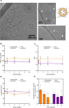
Fig. 2.. Morphology and stability of nanoGLA.
(A) Morphology of nanoGLA, analyzed by cryo-TEM, 1-week after production. White arrows highlight interactions between the enzyme and the nanoliposome. (B to E) Stability of nanoGLA stored at 2° to 8°C in terms of: (B) size, (C) PDI, (D) ζ-potential for 10 weeks. For each system, three independent batches per time point are represented (symbols) and the average (continuous line; SD represented by dotted lines). (E) Stability of specific enzymatic activity (EA) for 12 weeks, average of three independent batches. Values expressed in relation to reference GLA (agalsidase alfa).
Fig. 3.
Fig. 3.. In vitro efficacy in primary cultures of FD model.
Gb3 reduction of the nanoGLA in comparison with the free rhGLA, and the free agalsidase alfa in primary endothelial cells derived from Fabry KO mice. Incubation of GLA (0.25 μg ml−1) (or at the equivalent liposome concentration for empty liposomes) at 37°C for 48 hours. Empty liposomes refer to the nanoformulation without GLA (at the equivalent concentration), used as control. The assay corresponds to a single representative experiment, replicated in three independent assays.
Fig. 4.
Fig. 4.. PK profile of empty liposomes.
PK of MKC-containing nanoliposomes versus free MKC (n = 6 animals per group, values correspond to the means ± SEM). Both curves have been normalized to the maximum concentration (Cmax) for better comparison. # denotes values below the limit of detection.
Fig. 5.
Fig. 5.. PK profile of nanoGLA.
Mean (± SEM) concentration of total rhGLA in male rat plasma following a single intravenous administration of free rhGLA or nanoGLA (GLA dosing of 1 mg kg−1) (n = 3 animals per group). # denotes value below the limit of quantification.
Fig. 6.
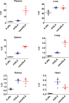
Fig. 6.. GLA EA in Fabry mice 30 min after administration.
Animals (n = 8 per group) received intravenous nanoGLA, rhGLA or agalsidase alfa (A.alfa) (1 mg kg−1). Activity was measured in plasma and tissues 30 min after administration (n = 4) and referred to activity at 1 min (n = 4). Values in the y axis correspond to the % of injected dose (% ID) and are expressed as the means ± SEM.
Fig. 7.
Fig. 7.. In vivo efficacy of nanoGLA in Fabry mice.
(A) Loss of Gb3 in Fabry mice with single intravenous administration of nanoGLA, free rhGLA, and free agalsidase alfa (A.alfa) (n = 8 animals per group). (B) Loss of Gb3 in Fabry mice receiving eight doses (every other day) of the same treatments (n = 6 animals per group. All administration were at 1 mg of GLA kg−1 dose. WT animals are included as controls (100% of Gb3 loss). (C) Comparative efficacy of the nanoGLA treatment in the brain of Fabry mice after a single or repeated dosing, as percentage of Gb3 loss. Results are compared with the nontreated WT and KO animals, as well as with the efficacy of the free non-nanoformulated enzymes (rhGLA and A.alfa) after repeated dosing. Analysis of variance (ANOVA) test and Tukey’s multiple comparison test were performed to compare results. Statistical differences that are not obvious and are relevant for understanding the behavior of nanoGLA are shown [n.s. (nonsignificant), P ≤ 0.05; **P ≤ 0.01, ***P ≤ 0.001, or ****P ≤ 0.0001].

References

    1. Zarate Y. A., Hopkin R. J., Fabry’s disease. Lancet 372, 1427–1435 (2008). - PubMed
    1. Carnicer-Cáceres C., Arranz-Amo J. A., Cea-Arestin C., Camprodon-Gomez M., Moreno-Martinez D., Lucas-Del-pozo S., Moltó-Abad M., Tigri-Santiña A., Agraz-Pamplona I., Rodriguez-Palomares J. F., Hernández-Vara J., Armengol-Bellapart M., Del-Toro-riera M., Pintos-Morell G., Biomarkers in fabry disease. Implications for clinical diagnosis and follow-up. J. Clin. Med. 10, 1664 (2021). - PMC - PubMed
    1. Bodary P. F., Shayman J. A., Eitzman D. T., α-Galactosidase A in Vascular Disease. Trends Cardiovasc. Med. 17, 129–133 (2007). - PubMed
    1. Shen J. S., Meng X. L., Moore D. F., Quirk J. M., Shayman J. A., Schiffmann R., Kaneski C. R., Globotriaosylceramide induces oxidative stress and up-regulates cell adhesion molecule expression in Fabry disease endothelial cells. Mol. Genet. Metab. 95, 163–168 (2008). - PMC - PubMed
    1. Kok K., Zwiers K. C., Boot R. G., Overkleeft H. S., Aerts J. M. F. G., Artola M., Fabry disease: Molecular basis, pathophysiology, diagnostics and potential therapeutic directions. Biomolecules 11, 271 (2021). - PMC - PubMed