. 2024 Dec 13;10(50):eadr9364.
doi: 10.1126/sciadv.adr9364. Epub 2024 Dec 13.

Regulation of volume-regulated anion channels alters sensitivity to platinum chemotherapy

Affiliations

Regulation of volume-regulated anion channels alters sensitivity to platinum chemotherapy

Lily Elizabeth R Feldman et al. Sci Adv. .

Abstract

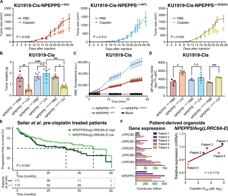
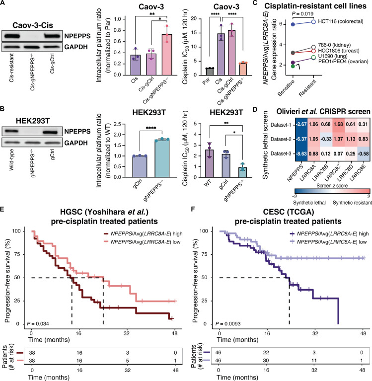
Cisplatin-based chemotherapy is used across many common tumor types, but resistance reduces the likelihood of long-term survival. We previously found the puromycin-sensitive aminopeptidase, NPEPPS, as a druggable driver of cisplatin resistance in vitro and in vivo and in patient-derived organoids. Here, we present a general mechanism where NPEPPS interacts with the volume-regulated anion channels (VRACs) to control cisplatin import into cells and thus regulate cisplatin response across a range of cancer types. We also find the NPEPPS/VRAC gene expression ratio is a predictive measure of cisplatin response in multiple cancer cohorts, showing the broad applicability of this mechanism. Our work describes a specific mechanism of cisplatin resistance, which, given the characteristics of NPEPPS as a drug target, has the potential to improve cancer patient outcomes. In addition, we describe an intracellular mechanism regulating VRAC activity, which is critical for volume regulation in normal cells - a finding with functional implications beyond cancer.


Figures

Fig. 1.
Fig. 1.. NPEPPS protein interaction partners.
(A) The heatmap represents LC-MS/MS analysis of NPEPPSFLAG pull-down using protein lysates from BCa cell lines (KU1919 and T24; n = 3 independent lysates per cell line). The relative strength of interaction is scaled from 0 to 1. Genes annotated as playing a role in platinum drug resistance by Huang et al. (34) are highlighted. Results from a CRISPR screen reported in Jones et al. (12) identifying synthetic gene-to-drug interactions against cisplatin-based chemotherapy in treatment-resistant cell lines are shown. *FDR < 0.05. (B) Proteins identified in the NPEPPSFLAG screen as black dots and the VRAC subunits (LRRC8A-E) as red dots are mapped onto the CRISPR screen results. NPEPPS is highlighted in orange. (C) Affinity tag (FLAG), or IgG control, IP of protein lysates from NPEPPSFLAG in KU1919 and T24 cells was immunoblotted for NPEPPS, LRRC8A, and LRRC8D.
Fig. 2.
Fig. 2.. NPEPPS alters platinum import and DNA damage by modulating VRACs.
(A) Untargeted metabolomics in KU1919 cells with shRNA-mediated NPEPPS suppression or control shRNA. Taurine levels are reported in control (PBS) and cisplatin (Cis) treatment conditions. Targeted metabolomic measured levels of taurine are reported in T24 cells in control (PBS) and cisplatin (Cis) treatment conditions. hr, hours. (B) CyTOF in KU1919 cells shows intracellular cisplatin levels after 4 hours of 10 μM cisplatin with siRNA-mediated suppression of VRAC subunits LRRC8A-E compared to control (scramble) siRNA. Median intracellular cisplatin measurements across biological triplicates were normalized to the siRNA control and compared using a one-way ANOVA (**P < 0.01; ****P < 0.001). (C) Intracellular cisplatin levels for KU1919, T24, and 5637 cells with siRNA-mediated knockdown of NPEPPS alone, LRRC8A alone, or the combination of NPEPPS and LRRC8A knockdown. All samples were normalized to siRNA control samples and compared using a one-way ANOVA (*P < 0.05; **P < 0.01; ****P < 0.0001). n.s., not significant. Immunoblot validation of the knockdowns is reported with native NPEPPS and LRRC8A antibodies. (D) KU1919, 5637, and T24 cells made resistant to GemCis were treated with cisplatin (10 μM) or PBS for 48 hours. Immunoblots with NPEPPS, LRRC8A, and phospho-γ-H2AX antibodies are shown, comparing shRNA-mediated knockdown of NPEPPS to shRNA scramble controls.
Fig. 3.
Fig. 3.. Subcellular localization of NPEPPS protein and VRAC.
(A) Confocal microscopy images illustrating the cellular localization of NPEPPS and LRRC8A via NPEPPSFLAG or LRRC8AFLAG in both KU1919 and T24 cells. Shown with and without differential interference contrast (DIC). Scale bars, 20 μm. (B) Immunoblots for NPEPPS and LRRC8A after FLAG affinity pull-down from NPEPPSFLAG in cytosolic and membranal fractions of T24 cell lysates. (C) Dual staining of LRRC8A and NPEPPS in LRRC8AFLAG KU1919 and T24 cells using anti-FLAG primary and Alexa Fluor (AF) 594 secondary antibodies for LRRC8A and anti-NPEPPS primary and AF647 secondary antibodies for NPEPPS. Scale bars, 20 μm [for nonzoomed images (top row)] and 2 μm [for zoomed images (bottom row)]. (D) Confocal microscopy showing colocalization of NPEPPS and LRRC8A in LRRC8A-mCherry reporter-containing T24 cells stained with anti-NPEPPS primary and AF647 secondary antibodies. Scale bars, 20 μm [for nonzoomed images (top row)] and 2 μm [for zoomed images (bottom row)]. NPEPPS-LRRC8A colocalization was calculated in ImageJ by PCC (r or Correlation), and statistical significance was evaluated by unpaired t test.
Fig. 4.
Fig. 4.. NPEPPS enzymatic function is critical for the NPEPPS-LRRC8A interaction.
(A) Quantification of NPEPPS enzymatic activity on the H-Leu-AMC substrate with vehicle (PBS, 0 μM) or tosedostat (20 μM) treatment in NPEPPSFLAG KU1919 parental cells. a.u., arbitrary units. (B) Immunoblot of NPEPPS and LRRC8A in KU1919 parental and GemCis-resistant cells after IP with native NPEPPS or LRRCA antibodies following 72 hours of PBS control (0 μM) or tosedostat (10 μM) treatment. (C) Quantification of NPEPPS-LRRC8A colocalization by FRET-AB in LRRC8AFLAG KU1919 parental cells with vehicle (PBS, 0 μM) or tosedostat (20 μM) treatment. The relative quantification is reported as FRET efficiency (%) and statistical comparisons were made using the Mann-Whitney U test. (D) Immunofluorescence confocal microscopy of NPEPPSFLAG constructs [NPEPPS−/−(WT-FLAG) and NPEPPS−/−(E353V-FLAG)] in cisplatin-resistant KU1919 cells. Scale bars, 20 μm. (E) Quantification of NPEPPS enzymatic activity on the H-Leu-AMC substrate in KU1919-Cis cells expressing NPEPPS−/−(WT-FLAG) or NPEPPS−/−(E353V-FLAG). (F) Immunoblots of NPEPPS and LRRC8A following anti-FLAG IP in parental and cisplatin-resistant KU1919 cells expressing NPEPPS−/−(WT-FLAG) or NPEPPS−/−(E353V-FLAG). (G) Intracellular cisplatin concentrations were measured by CyTOF in triplicate experiments in WT KU1919-Cis cells or KU1919-Cis cells expressing NPEPPS−/−(WT-FLAG) or NPEPPS−/−(E353V-FLAG). Cisplatin concentrations were normalized to the unmodified cisplatin-resistant KU1919 cells, and comparisons were made using one-way ANOVA (****P < 0.0001). (H) Cisplatin IC50 was measured in technical and biological triplicate by IncuCyte Zoom analysis over 120 hours of treatment in WT KU1919-Cis cells or KU1919-Cis cells expressing NPEPPS−/−(WT-FLAG) or NPEPPS−/−(E353V-FLAG). Results are reported as the mean IC50 ± SD. Comparisons were made using one-way ANOVA (*P < 0.05).
Fig. 5.
Fig. 5.. NPEPPS enzymatic function regulates cisplatin response in vivo.
(A) Mice were injected with 4 × 10⁶ cells in each flank to generate tumors using KU1919-Cis cells with NPEPPS−/−(WT-FLAG) or NPEPPS−/−(E353V-FLAG) expression. When engrafted tumors reached 100 mm3, mice were randomized to control or treatment groups and received either cisplatin (2 mg/kg; by intraperitoneal injection three times per week) or PBS control (equal volume of saline by intraperitoneal injection three times per week). Tumor size was measured with calipers every 2 to 3 days throughout the duration of treatment. Differences in growth rates by tumor genotype were evaluated by a two-way ANOVA mixed effects model. (B) Wet tumor weights were measured for each group. Comparisons were made using a one-way ANOVA (*P < 0.05; **P < 0.01; ***P < 0.001; ****P < 0.0001). (C) Fluorescence-based enzyme assay to quantify the catalytic activity of NPEPPS across the cells isolated from mouse tumors using H-Leu-AMC as the substrate. (D) DNA damage was measured by flow cytometry–based quantification of phosphorylated histone H2AX. Comparisons were made using a one-way ANOVA (*P < 0.05; ***P < 0.001). (E) Stratification of patient survival by above-median or below-median expression of the ratio of NPEPPS/Avg(LRRC8A-E) in BCa tumor samples before cisplatin treatment. Analysis of survival differences was conducted in R, and statistical significance was evaluated by Cox proportional hazard ratios and the log-rank test. Time to median survival is indicated by a dashed line. (F) Patient-derived organoids were evaluated for mRNA expression and cisplatin IC50. Left: Average counts per million mRNA expression of VRAC subunits LRRC8A-E and NPEPPS in each patient-derived organoid. Right: The correlation of NPEPPS/Avg(LRRC8A-E) expression with cisplatin sensitivity is plotted (r2 = 0.7172). Organoids from patient 1 were derived from a cystectomy sample, and the patient was not given neoadjuvant chemotherapy (NAC). Organoids from patients 2 to 5 were derived from the transurethral resection of bladder tumor samples before the patients received NAC.
Fig. 6.
Fig. 6.. NPEPPS regulates platinum chemotherapy import and efficacy beyond BCa.
(A) Immunoblots of NPEPPS in cisplatin-resistant Caov-3 cells (Caov-3-Cis) with WT NPEPPS (Caov-3-Cis-gCtrl) or KO of NPEPPS (Caov-3-Cis-gNPEPPS−/−). Intracellular cisplatin concentrations were measured by CyTOF in triplicate experiments in WT, chemotherapy-sensitive Caov-3-Par cells, WT, chemotherapy-resistant Caov-3-Cis cells, or engineered Caov-3-Cis cells with control (-gCtrl) or NPEPPS KO (-NPEPPS−/−). Cisplatin concentrations were normalized to unmodified cisplatin-sensitive Caov-3-Par cells, and comparisons were made using one-way ANOVA (*P < 0.05; **P < 0.01). Cisplatin IC50 was measured in technical and biological triplicate in WT Caov-3-Par, Cis, Cis-gCtrl, or Cis-gNPEPPS−/− cells. Results are reported as the mean IC50 ± SD. Comparisons were made using one-way ANOVA (****P < 0.0001). (B) Immunoblots of NPEPPS in WT HEK293T cells, HEK293T-gCtrl cells, or HEK293T-gNPEPPS−/− cells. Intracellular cisplatin concentrations were measured by CyTOF, and comparisons were made using one-way ANOVA (****P < 0.0001). Cisplatin IC50 was measured in technical and biological triplicate and reported as the mean IC50 ± SD. Comparisons were made using one-way ANOVA (*P < 0.05; **P < 0.01). (C) GE data from paired cisplatin-sensitive and cisplatin-resistant cancer cell lines representing colorectal, kidney, breast, lung (48), and ovarian (49) cancers were evaluated for the expression ratio of NPEPPS/Avg(LRRC8A-E). The comparison was made using a paired t test. (D) Synthetic lethal and synthetic resistant z scores of NPEPPS and LRRC8A-E across three cisplatin datasets were evaluated by CRISPR screening in the RPE1 cell line and retrieved from the Olivieri et al. CRISPR screen repository (50). (E) Stratification of patient survival by above-median or below-median expression of the ratio of NPEPPS/Avg(LRRC8A-E) in tumor samples from HGSC [Yoshihara et al. (51)] and CESC [TCGA (46)] prior to platinum-based treatment. Time to median survival is indicated by a dashed line (omitted where survival is >50% at all time points), and comparisons were made using the log-rank test.

References

    1. Aldossary S. A., Review on pharmacology of cisplatin: Clinical use, toxicity and mechanism of resistance of cisplatin. Biomed. Pharmacol. J. 12, 7–15 (2019).
    1. Alassadi S., Pisani M. J., Wheate N. J., A chemical perspective on the clinical use of platinum-based anticancer drugs. Dalton Trans. 51, 10835–10846 (2022). - PubMed
    1. Galanski M. S., Jakupec M. A., Keppler B. K., Update of the preclinical situation of anticancer platinum complexes: Novel design strategies and innovative analytical approaches. Curr. Med. Chem. 12, 2075–2094 (2005). - PubMed
    1. Xiao X., Oswald J. T., Wang T., Zhang W., Li W., Use of anticancer platinum compounds in combination therapies and challenges in drug delivery. Curr. Med. Chem. 27, 3055–3078 (2020). - PubMed
    1. van der Heijden M. S., Sonpavde G., Powles T., Necchi A., Burotto M., Schenker M., Sade J. P., Bamias A., Beuzeboc P., Bedke J., Oldenburg J., Chatta G., Ürün Y., Ye D., He Z., Valderrama B. P., Ku J. H., Tomita Y., Filian J., Wang L., Purcea D., Patel M. Y., Nasroulah F., Galsky M. D., CheckMate 901 Trial Investigators , Nivolumab plus gemcitabine-cisplatin in advanced urothelial carcinoma. N. Engl. J. Med. 389, 1778–1789 (2023). - PubMed

LinkOut - more resources