. 2024 Dec 13;10(50):eadr5188.
doi: 10.1126/sciadv.adr5188. Epub 2024 Dec 13.

An alternate route for cellulose microfibril biosynthesis in plants

Affiliations

An alternate route for cellulose microfibril biosynthesis in plants

Eric M Roberts et al. Sci Adv. .

Abstract

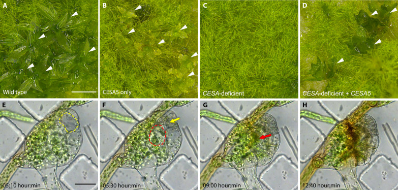
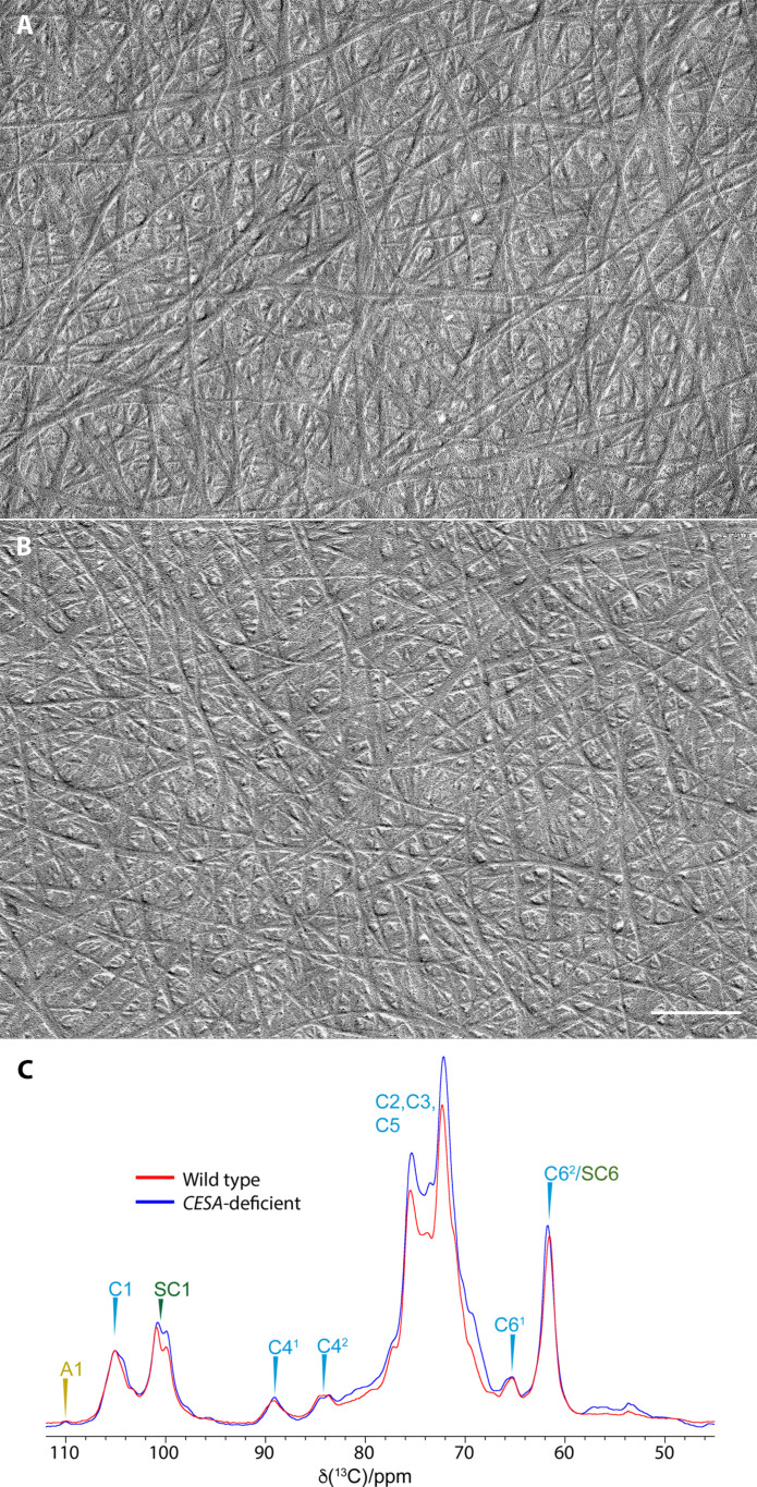
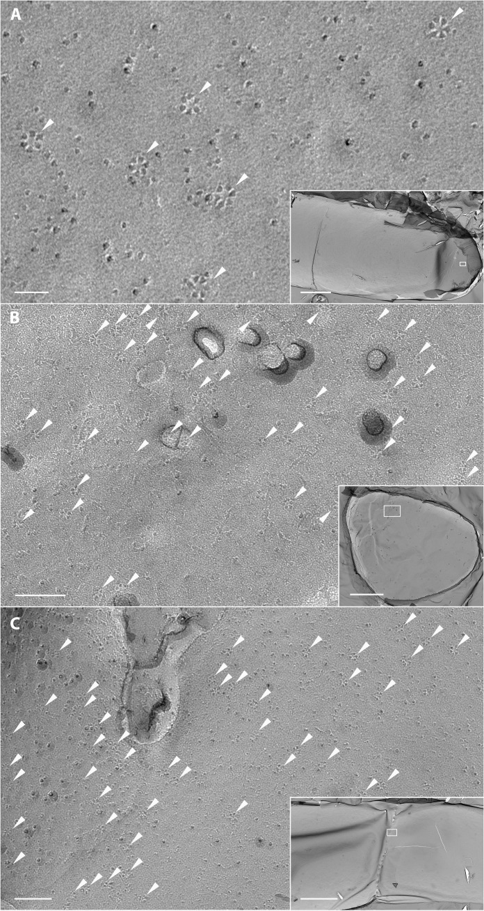
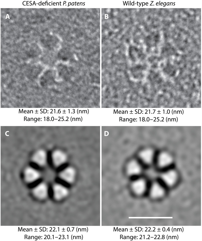
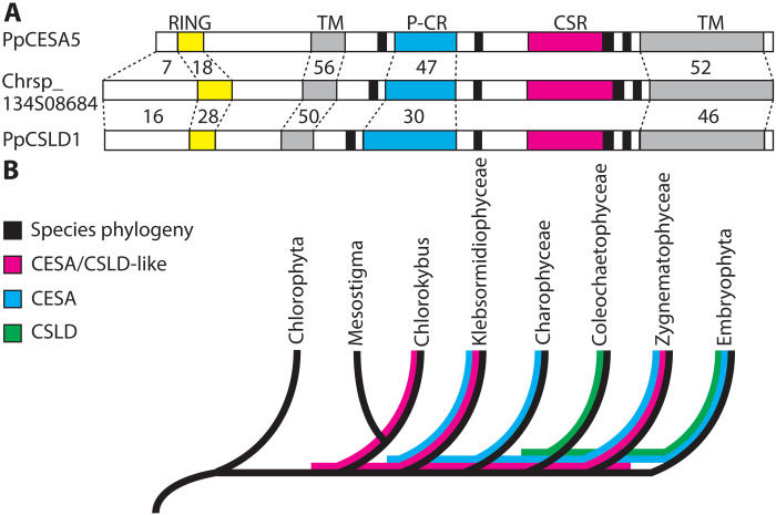
Similar to cellulose synthases (CESAs), cellulose synthase-like D (CSLD) proteins synthesize β-1,4-glucan in plants. CSLDs are important for tip growth and cytokinesis, but it was unknown whether they form membrane complexes in vivo or produce microfibrillar cellulose. We produced viable CESA-deficient mutants of the moss Physcomitrium patens to investigate CSLD function without interfering CESA activity. Microscopy and spectroscopy showed that CESA-deficient mutants synthesize cellulose microfibrils that are indistinguishable from those in vascular plants. Correspondingly, freeze-fracture electron microscopy revealed rosette-shaped particle assemblies in the plasma membrane that are indistinguishable from CESA-containing rosette cellulose synthesis complexes (CSCs). Our data show that proteins other than CESAs, most likely CSLDs, produce cellulose microfibrils in P. patens protonemal filaments. The data suggest that the specialized roles of CSLDs in cytokinesis and tip growth are based on differential expression and different interactions with microtubules and possibly Ca2+, rather than structural differences in the microfibrils they produce.


Figures

Fig. 1.
Fig. 1.. Viable CESA-deficient P. patens.
(A) Wild-type P. patens has filamentous protonemata and leafy gametophores (arrowheads). (B) Septuple CESA knockouts expressing only PpCESA5 have morphologically normal protonemata and stunted gametophores (arrowheads) as described previously (15). (C) CESA-deficient octuple knockouts are viable with morphologically normal protonemata, but no gametophores. (D) Expression of PpCESA5 under the control of the constitutive rice Actin1 promoter partially rescues gametophore development (arrowheads) in CESA-deficient octuple knockouts. (E to H) Time-lapse imaging of gametophore buds in CESA-deficient P. patens reveals cell rupture [(E) and (F), yellow and red outlines mark fully expanded cells; (F) and (G), yellow and red arrows indicate the positions of the respective cells after rupture] and (H) areas of early senescence marked by accumulation of brown pigment. Scale bar in (A) = 1 mm and applies to (A) to (D). Scale bar in (E) = 50 μm and applies to (E) to (H). Time-lapse interval = 10 min.
Fig. 2.
Fig. 2.. Cellulose microfibrils in wild-type and CESA-deficient P. patens.
Cell walls were extracted from protonemal filaments of (A) wild-type and (B) CESA-deficient P. patens with 1 N NaOH and acetic-nitric reagent before air-drying and shadowing to reveal microfibrils. (C) Neutral carbohydrate region of 1D 13C CP-MAS-NMR spectra of CESA-deficient and wild-type P. patens. Identifiable 13C NMR shifts of cellulose (C), arabinose (A), and starch (SC) are labeled. The spectra were recorded at a 13C Larmor frequency of 150.7 MHz and a MAS frequency of 12 kHz. ppm, parts per million.
Fig. 3.
Fig. 3.. TEM imaging of rosettes in freeze-fracture replicas of CESA-deficient P. patens.
(A) Plasma membrane region from the apex of a protonemal filament (box in inset) with rosettes (arrowheads). Scale bars, 40 nm and (inset) 3 μm. (B) Plasma membrane region from a protonemal filament (box in inset) viewed tip down with numerous rosettes (arrowheads). Scale bars, 100 nm and (inset) 2 μm. (C) Plasma membrane region of a dividing cell with fusing cell plate (box in inset) with numerous rosettes (arrowheads). Scale bars, 100 nm and (inset) 3 μm.
Fig. 4.
Fig. 4.. Original images and corresponding image averages of rosettes from two cell types with diameter measurements.
(A) Original FFTEM images and data from hand measurement of 543 rosettes from nine CESA-deficient P. patens protonemal cells (three cells from each of three independent genetic lines) frozen while synthesizing primary cell walls. (B) Original TEM image and data from hand measurement of 380 rosettes from five differentiating tracheary elements frozen while synthesizing secondary walls via CESAs. (C and D) Representative image averages of the rosettes measured in (A) and (B). The contrast of the original images was reversed before reference-free image averaging to accommodate the design of the EMAN2 program. Data for each cell type are from hand measurement of the 42 image averages (six class averages within each of seven refinements). Scale bar, 20 nm.
Fig. 5.
Fig. 5.. Sequence comparison and lineage sorting of CESAs, CSLDs, and CESA/CSLD-like proteins.
(A) Graphical comparison between P. patens CSLD and CESA protein sequences and a representative CESA/CSLD-like protein from C. atmophyticus (Chrsp_134508684). Numbers indicate the percent amino acid identity between PpCESA5 or PpCSLD1 and the C. atmophyticus sequence in different regions (dashed lines). CSLDs share greater identity with CESA/CSLD-like sequences in the N-terminal region and RING domain (yellow), but they are more similar to CESAs in the plant-conserved region (P-CR; cyan). Gray = TM regions, black = catalytic “D” and “QxxRW” domains, and magenta = class-specific region (CSR). (B) Evolutionary relationships of chlorophyte green algae, five classes of charophyte green algae, and land plants (Embryophyta) depicting incongruence of the CESA, CSLD, and CESA/CSLD-like sequence trees. Evolutionary relationships (topology only) are from (68). Sequence distributions are from (–47).

References

    1. Purushotham P., Ho R., Zimmer J., Architecture of a catalytically active homotrimeric plant cellulose synthase complex. Science 369, 1089–1094 (2020). - PubMed
    1. Nixon B. T., Mansouri K., Singh A., Du J., Davis J. K., Lee J. G., Slabaugh E., Vandavasi V. G., O’Neill H., Roberts E. M., Roberts A. W., Yingling Y. G., Haigler C. H., Comparative structural and computational analysis supports eighteen cellulose synthases in the plant cellulose synthesis complex. Sci. Rep. 6, 28696 (2016). - PMC - PubMed
    1. Penttila P. A., Paajanen A., Critical comment on the assumptions leading to 24-chain microfibrils in wood. Nat. Plants 10, 1064–1066 (2024). - PubMed
    1. Cosgrove D., Dupree P., Gomez E. D., Haigler C. H., Kubicki J. D., Zimmer J., How many glucan chains form plant cellulose microfibrils? A mini review. Biomacromolecules 25, 6357–6366 (2024). - PMC - PubMed
    1. Tai H. C., Tsao C. S., Lin J. H., Reply to: Critical comment on the assumptions leading to 24-chain microfibrils in wood. Nat. Plants 10, 1067–1070 (2024). - PubMed

LinkOut - more resources