. 2025 Jun;33(6):727-737.
doi: 10.1038/s41431-024-01768-8. Epub 2024 Dec 13.

Variants in the AGBL5 gene are responsible for autosomal recessive Retinitis pigmentosa with hearing loss

Affiliations

Variants in the AGBL5 gene are responsible for autosomal recessive Retinitis pigmentosa with hearing loss

Marianthi Karali et al. Eur J Hum Genet. 2025 Jun.

Abstract

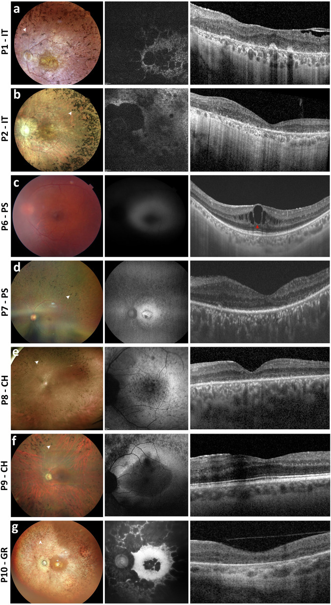
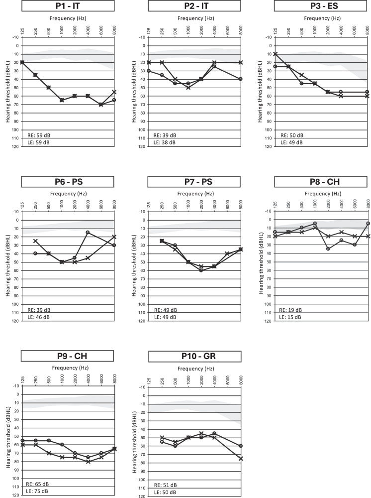
The AGBL5 gene encodes for the Cytoplasmic Carboxypeptidase 5 (CCP5), an α-tubulin deglutamylase that cleaves the γ-carboxyl-linked branching point of glutamylated tubulin. To date, pathogenic variants in AGBL5 have been associated only with isolated retinitis pigmentosa (RP). Hearing loss has not been reported in AGBL5-caused retinal disease. In this study, we performed exome sequencing in probands of eight unrelated families from Italy, Spain, Palestine, Switzerland, and Greece. All subjects had a clinical diagnosis of (suspected) Usher syndrome type II for the concurrent presence of RP and post-verbal sensorineural hearing loss (SNHL) that ranged from mild to moderate.We identified biallelic sequence variants in AGBL5 in all analysed subjects. Four of the identified variants were novel. The variants co-segregated with the retinal and auditory phenotypes in additional affected family members. We did not detect any causative variants in known deafness or Usher syndrome genes that could explain the patients' hearing loss. We therefore conclude that SNHL is a feature of a syndromic presentation of AGBL5 retinopathy. This study provides the first evidence that mutations in AGBL5 can cause syndromic RP forms associated with hearing loss, probably due to dysfunction of sensory cilia in the retina and the inner ear.


Conflict of interest statement

Competing interests: The authors declare no competing interests. Ethical approval: All procedures adhered to the tenets of the Declaration of Helsinki and were approved by the Ethics Committees of the participating institutes. An informed consent was obtained by all patients.

Figures

Fig. 1
Fig. 1. Pedigree structure and variants identified in the AGBL5 cohort.
a–h Pedigrees of the families included in this study and segregation analysis of the identified disease-causing variants. Probands are indicated by arrows. Filled symbols denote affected individuals and color indicates phenotype (black, RP and post-verbal SNHL; blue, profound prelingual deafness; green, isolated deafness). Variant positions refer to the RefSeq transcript sequences NM_021831.6 (AGBL5), NM_016239.4 (MYO15A) and NM_004004.6 (GJB2). i Schematic representation of the relative position of the variants identified in this study (in green) across the AGBL5 gene structure and in the context of the AGBL5 protein domains (dotted boxes). Novel variants are shown in bold. Variants already reported in the literature in cases with non-syndromic RP are shown in red font.
Fig. 2
Fig. 2. Multimodal ophthalmological imaging findings in the probands of this cohort.
Fundus photograph (left side of each panel), fundus autofluorescence imaging (FAF, central image of each panel), and macular spectral domain optical coherence tomography (SD-OCT, right side of each panel) of the left eye of the probands included in this study. Bone-spicule pigment (BSP) in fundus photographs and cystoid macular edema (CME) in SD-OCT scans are indicated with white and red arrowheads, respectively. Stretches of retinal pigment epithelium (RPE) dystrophy or atrophy in SD-OCT scans are represented by bright areas of increased light transmission beneath the RPE. a Patient P1-IT: Fundus photograph showing RP fundus with diffuse dystrophy of the RPE, BSP in mid-periphery and macular atrophy. FAF imaging shows reduced autofluorescence at the posterior pole, with focal areas of absent autofluorescence surrounding the optic disc and at the macular area. SD-OCT scan shows RPE dystrophy with epiretinal membranes. b Patient P2-IT: Fundus photograph showing waxy optic nerve head (ONH) pallor, RP fundus with diffuse RPE dystrophy and BSP in mid-periphery. FAF imaging shows reduced autofluorescence at the posterior pole, with focal areas of absent autofluorescence surrounding the optic disc. OCT scan shows reduced retinal thickness and RPE atrophy at the macular area. c Patient P6-PS: Fundus photograph showing RPE dystrophy without BSP. FAF image suggesting reduced autofluorescence which could not be fully assessed due to lens opacities. OCT scan shows CME. d Patient P7-PS: Fundus photograph showing RPE dystrophy with BSP. FAF imaging showed focal areas of reduced autofluorescence in mid-periphery and in the macular area. OCT scan shows stretches of RPE dystrophy and atrophy. e Patient P8-CH: Fundus photograph showing RPE dystrophy and BSP. FAF image showing focal areas of reduced AF in mid-periphery and at the macular area. OCT shows RPE dystrophy. f Patient P9-CH: Fundus photograph showing waxy ONH pallor, typical RP fundus with diffuse RPE dystrophy and peripheral BSP. FAF imaging showing focal areas of reduced autofluorescence in mid-periphery and at the macular area. OCT scan showing RPE dystrophy sparing the fovea. g Patient P10-GR: Fundus photograph showing diffuse RPE dystrophy with BSP. FAF shows focal areas of absent autofluorescence in mid-periphery and in the macular area. Macular SD-OCT scan shows RPE atrophy.
Fig. 3
Fig. 3. Pure-tone audiometry assessments in the AGBL5 patients.
Air conduction hearing thresholds of patients with biallelic variants in AGBL5 in the frequency range 125 Hz–8000 Hz. Air conduction pure-tone average values for the right (RE) and left ear (LE) are reported at the bottom part of each audiogram. AGBL5 patients showed a moderate hearing loss. The gray-shaded areas represent the hearing threshold average (± 1 SD) in sex- and age (decade)-matched normal individuals, as reported in a large retrospective study by Wasano et al. [34]. In each proband, air conduction and bone conduction thresholds (not depicted) overlapped. Symbols: O, air conduction in right ear; X, air conduction in left ear, dBHL, decibels hearing level.

References

    1. Hartong DT, Berson EL, Dryja TP. Retinitis pigmentosa. Lancet. 2006;368:1795–809. - PubMed
    1. Chang S, Vaccarella L, Olatunji S, Cebulla C, Christoforidis J. Diagnostic challenges in retinitis pigmentosa: genotypic multiplicity and phenotypic variability. Curr Genom. 2011;12:267–75. - PMC - PubMed
    1. Hamel C. Retinitis pigmentosa. Orphanet J Rare Dis. 2006;1:40. - PMC - PubMed
    1. Tatour Y, Ben-Yosef T. Syndromic inherited retinal diseases: genetic, clinical and diagnostic aspects. Diagnostics. 2020;10(Oct):779. - PMC - PubMed
    1. Kastner S, Thiemann IJ, Dekomien G, Petrasch-Parwez E, Schreiber S, Akkad DA, et al. Exome sequencing reveals agbl5 as novel candidate gene and additional variants for retinitis pigmentosa in five turkish families. Invest Ophthalmol Vis Sci. 2015;56:8045–53. - PubMed

LinkOut - more resources