. 2025 Oct;43(10):1683-1693.
doi: 10.1038/s41587-024-02505-8. Epub 2024 Dec 13.

Targeting peptide antigens using a multiallelic MHC I-binding system

Affiliations

Targeting peptide antigens using a multiallelic MHC I-binding system

Haotian Du et al. Nat Biotechnol. 2025 Oct.

Abstract

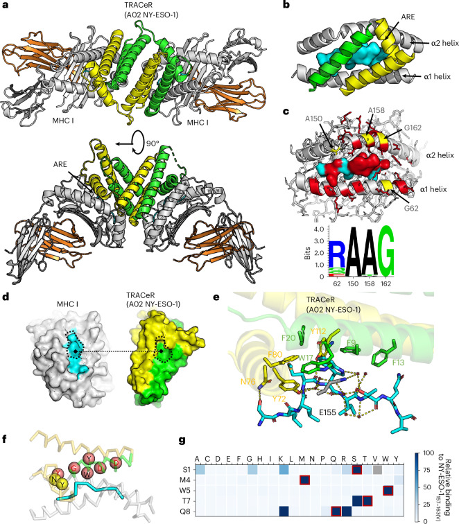
Identifying highly specific T cell receptors (TCRs) or antibodies against epitopic peptides presented by class I major histocompatibility complex (MHC I) proteins remains a bottleneck in the development of targeted therapeutics. Here, we introduce targeted recognition of antigen-MHC complex reporter for MHC I (TRACeR-I), a generalizable platform for targeting peptides on polymorphic HLA-A*, HLA-B* and HLA-C* allotypes while overcoming the cross-reactivity challenges of TCRs. Our TRACeR-MHC I co-crystal structure reveals a unique antigen recognition mechanism, with TRACeR forming extensive contacts across the entire peptide length to confer single-residue specificity at the accessible positions. We demonstrate rapid screening of TRACeR-I against a panel of disease-relevant HLAs with peptides derived from human viruses (human immunodeficiency virus, Epstein-Barr virus and severe acute respiratory syndrome coronavirus 2), and oncoproteins (Kirsten rat sarcoma virus, paired-like homeobox 2b and New York esophageal squamous cell carcinoma 1). TRACeR-based bispecific T cell engagers and chimeric antigen receptor T cells exhibit on-target killing of tumor cells with high efficacy in the low nanomolar range. Our platform empowers the development of broadly applicable MHC I-targeting molecules for research, diagnostic and therapeutic applications.


Conflict of interest statement

Competing interests: H.D., N.G.S. and P.-S.H. are listed as coinventors on provisional patent applications related to this work. The remaining authors declare no competing interests.

Figures

Fig. 1
Fig. 1. Overview of the TRACeR-I platform design scheme.
a. Summary of TCR and TCR-mimicking antibodies with pMHC-binding modes (PDB identifiers in Supplementary Appendix 1). TCRs, white ribbons; antibodies, magenta. Two edge-case TCRs are rendered as tan and purple surfaces. b, TRACeR-II platform. The N-terminal flexible region on TRACeR-II binders is defined as the ARE. The ARE loop and the scaffold form a concave surface for MHC II engagement. c, Comparison of the different peptide conformations and TRACeR (in yellow) orientations on MHC I and II. The bulged conformation of MHC I peptides requires a parallel binding mode while MHC II peptides can be engaged in a perpendicular orientation relative to the MHC. d, Schematic of our TRACeR-I development approach. In step 1, global docking was performed with PatchDock-seeded RifDock (Supplementary Appendix 2). In step 2, iterative RosettaDock and FastDesign cycles were performed to generate sequence diversity on the binding interface (Supplementary Appendices 3 and 4). Output models were evaluated by binding energy, binding energy to antigen, shape complementarity, buried unsatisfied hydrogen bonds and contact area (Supplementary Appendix 5). In step 3, the diverse sequence suggested from step 2 was implemented into a combinatorial library (library 1) for screening with FACS. In step 4, library 2 enabled the development of specific TRACeRs for different targets. Peptide-focused MHC I binders bind their cognate antigens but have minimal cross-reactivity with irrelevant pMHCs. Staining concentration, 50 nM tetramer concentration. e, Titration curves of TRACeRs with different monomeric pMHC targets under a concentration range from 0.1 nM to 50 μM. Binding signals are shown as the MFI ± s.d. (n = 3 technical replicates). f, Binding kinetics of TRACeRs determined by BLI. Binding kinetics and fitting quality are summarized in Supplementary Table 2.
Fig. 2
Fig. 2. Determination of the specificity of TRACeRMHCI,A02NY -ESO-1 against different peptides.
a, Structure of MHC-presented NY-ESO-1 peptide. Residue positions in yellow are solvent exposed and those in blue have side chains buried in the MHC I groove. T7 is partially solvent exposed. b, Alanine scan of the NY-ESO-1 peptide showing that TRACeR binding is sensitive to S1, M4, W5 and T7 substitutions, representing four of five solvent-exposed positions. The binding signal is shown as the MFI ± s.d. (n = 3 technical replicates). c, Binding signal for alanine scan substitutions on flow cytometry. d, MHC I tetramer SSM on the five key peptide residues responding to TRACeR (top) and 1G4 TCR (bottom). The S1V peptide failed to synthesize by the vendor (filled with gray). e, Normalized enrichment ratio showing enriched peptide sequences binding to TRACeR (top) and 1G4 TCR (bottom) from the randomized triplet library (n = 3 technical replicates). f, Clusters of the combined top 50 peptide sequences binding to TRACeR and 1G4 (based on enrichment ratio). Average distance of NY-ESO-1 peptide to each cluster: cluster 1, 33.5; cluster 2, 33.8; cluster 3, 24.9; cluster 4, 32.2; cluster 5,30.7. g, In vitro tumor cell cytotoxicity assays with TRACeRMHCI,A02NYESO1 anti-CD3 BiTEs. The percentage of live tumor cells was normalized to PBS-treated control cells (n = 6 replicates using T cells from two independent donors). Source data
Fig. 3
Fig. 3. Co-crystal structure of a TRACeR–MHC complex reveals an extended antigen recognition interface to guide specificity enhancement.
a, The asymmetric unit of TRACeRMHCI,A02NYESO1 engaging two pMHC I molecules. Domain-swapped TRACeR, yellow and green; MHC I heavy chain, gray; MHC I light chain, orange; NY-ESO-1 peptide, cyan. b, TRACeRMHCI,A02NYESO1 helices binding to pMHC I, with the location of the ARE. c, Footprint of TRACeRMHCI,A02NYESO1 on pMHC I. pMHC I interface atoms within 5-Å distance from TRACeR, red; small hydrophobic residues on MHC groove that TRACeR docks on, yellow. Sequence conservation for the TRACER-contacting MHC I residues on common HLA alleles are shown as a sequence logo. d, Open-book view of MHC I–TRACeR interface showing a high level of shape complementarity. e, The interacting residues between TRACeRMHCI,A02NYESO1 and NY-ESO-1 peptide. TRACeR residues, green and yellow; peptide, cyan. f, Refined set of interfacial residue positions to achieve better specificity for P7 and P8. Positions included in the original library containing AREs in library 2 are shown as yellow spheres; new positions are shown in salmon. g, SSM on the five key peptide residues corresponding to the refined TRACeR specificity. The S1V peptide failed to synthesize and was not included in the library (filled with gray). Source data
Fig. 4
Fig. 4. Engineered monomeric TRACeR-I shows multi-HLA allelic compatibility.
a, Rewiring scheme to connect domain-swapped TRACeR into a monomer. b, Model of rewired TRACeR on MHC I. c, In vitro tumor cell cytotoxicity assays with TRACeRMHCI,A02NYESO1CAR-T cells derived from primary CD8+ T cells from two donors. Percentage of live tumor cells cocultured with engineered T cells were normalized to those cocultured with untransduced T cells. Each data point represents technical replicates from five experiments, plotted as the mean ± s.d. d. BLI affinity measurements of the rewired TRACeRMHCI,A02NYESO1. e, Open-book view of the TRACeR interface with library positions colored in salmon and the peptide colored in cyan. f, TRACeR-I binding to a panel of diverse disease-relevant epitopic peptides presented by diverse HLAs. The final enrichment round before NGS is shown; multiple sequences were present in the pool. Staining concentration: on target, 25 nM tetramer concentration; off target, 100 nM tetramer concentration. g, Rosetta models of the most enriched TRACeR clone from NGS for each pMHC I target. Source data

References

    1. Spiegel, J. Y. et al. CAR T cells with dual targeting of CD19 and CD22 in adult patients with recurrent or refractory B cell malignancies: a phase 1 trial. Nat. Med.27, 1419–1431 (2021). - PMC - PubMed
    1. Xue, J. Y. et al. Rapid non-uniform adaptation to conformation-specific KRAS(G12C) inhibition. Nature577, 421–425 (2020). - PMC - PubMed
    1. Dang, C. V., Reddy, E. P., Shokat, K. M. & Soucek, L. Drugging the ‘undruggable’ cancer targets. Nat. Rev. Cancer17, 502–508 (2017). - PMC - PubMed
    1. Schumacher, T. N. & Schreiber, R. D. Neoantigens in cancer immunotherapy. Science348, 69–74 (2015). - PubMed
    1. Croft, N. P. et al. Most viral peptides displayed by class I MHC on infected cells are immunogenic. Proc. Natl Acad. Sci. USA116, 3112–3117 (2019). - PMC - PubMed

MeSH terms

Substances