Review
. 2025 Feb;28(2):234-247.
doi: 10.1038/s41593-024-01836-8. Epub 2024 Dec 13.

Amygdala intercalated cells form an evolutionarily conserved system orchestrating brain networks

Affiliations
Review

Amygdala intercalated cells form an evolutionarily conserved system orchestrating brain networks

Ayla Aksoy-Aksel et al. Nat Neurosci. 2025 Feb.

Abstract

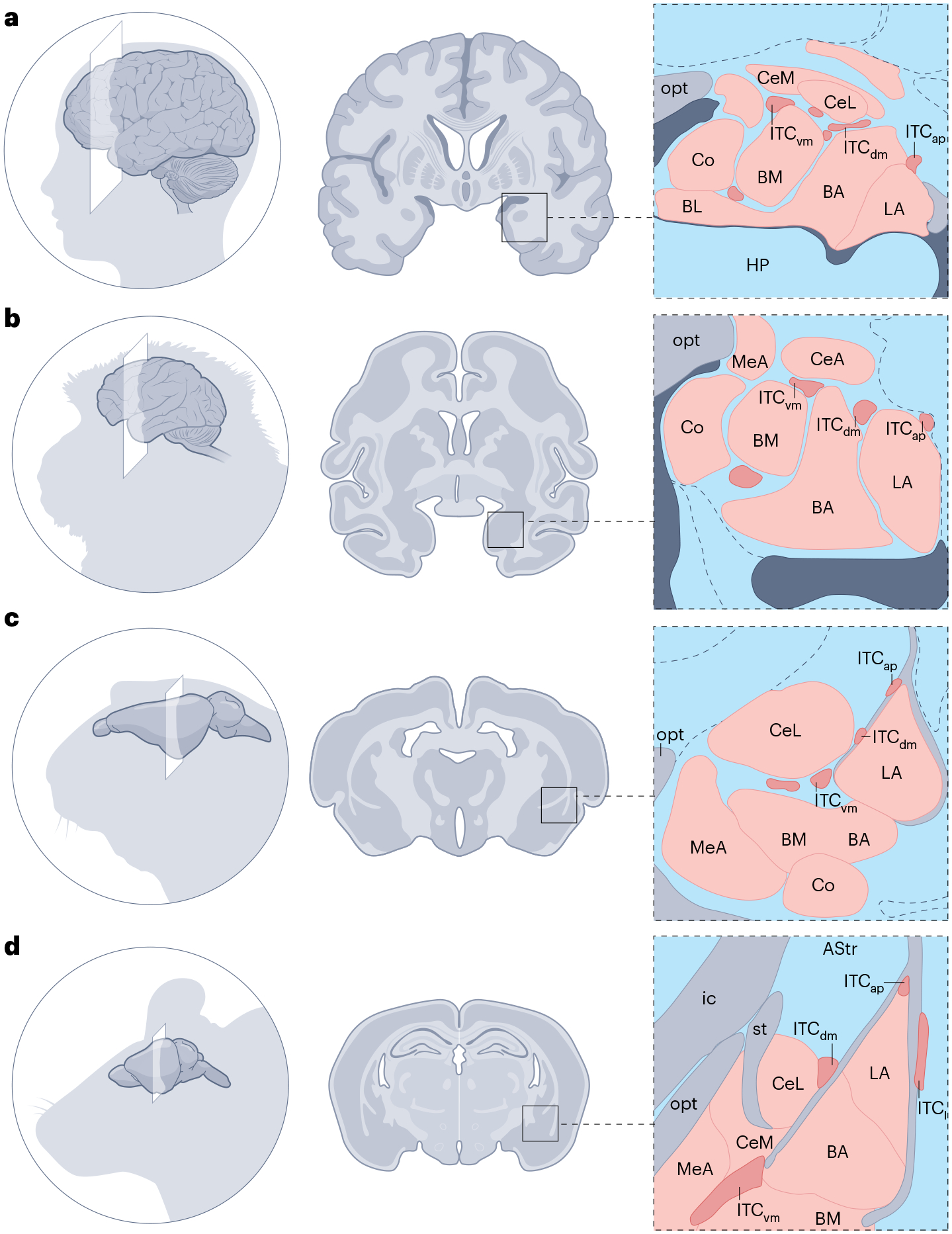
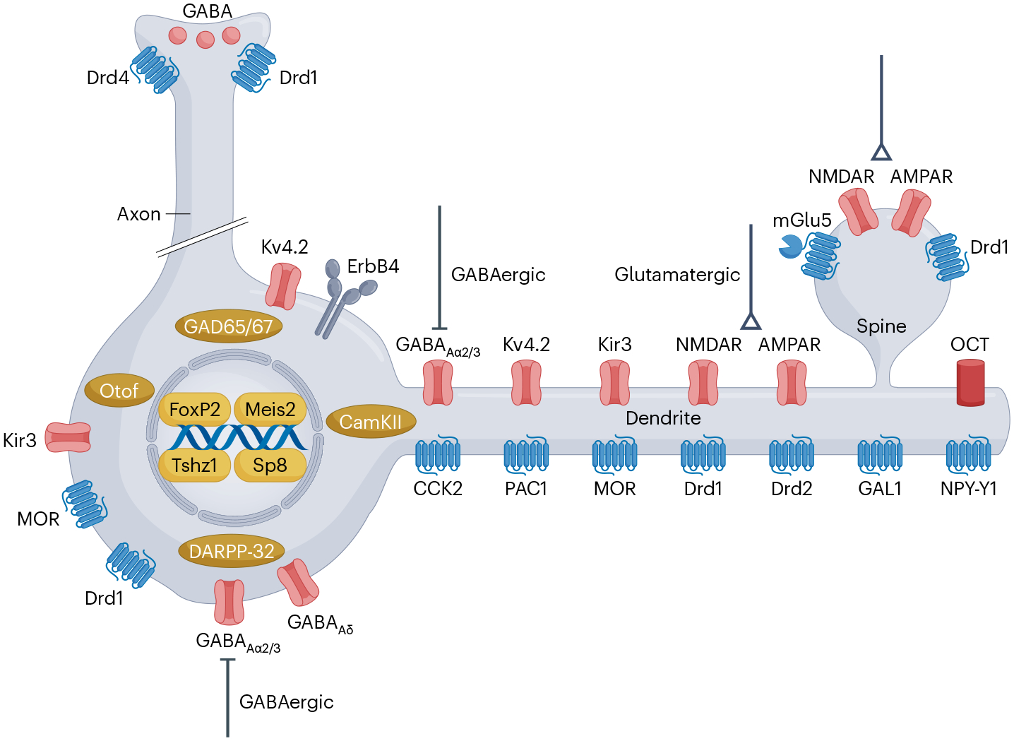
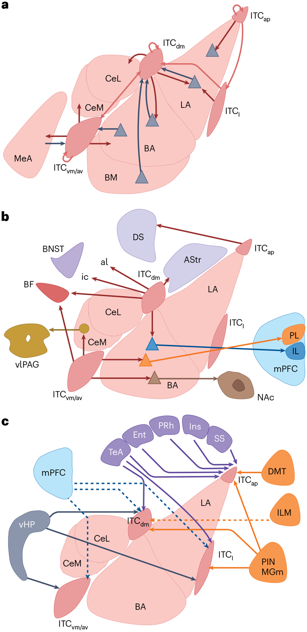
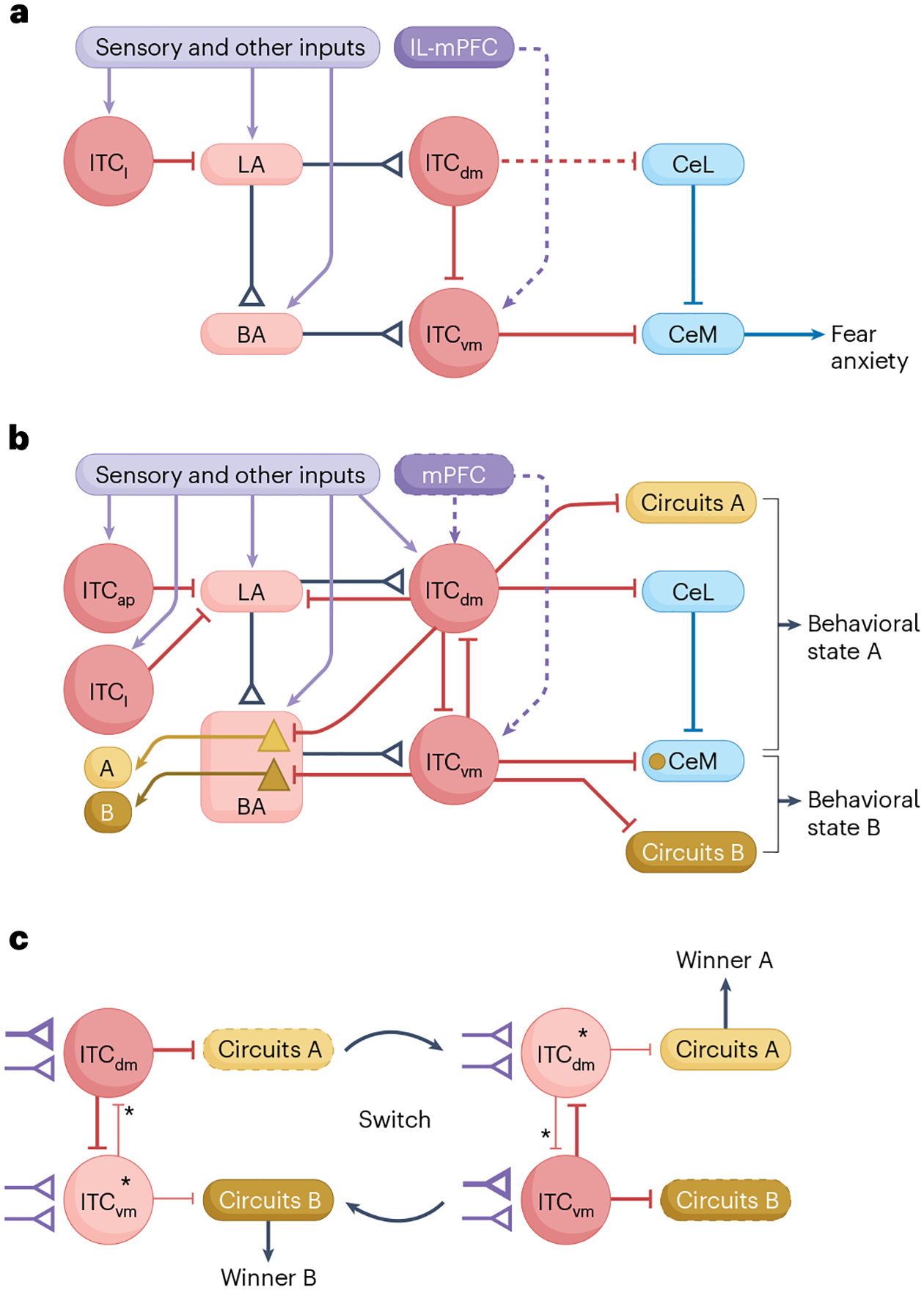
The amygdala attributes valence and emotional salience to environmental stimuli and regulates how these stimuli affect behavior. Within the amygdala, a distinct class of evolutionarily conserved neurons form the intercalated cell (ITC) clusters, mainly located around the boundaries of the lateral and basal nuclei. Here, we review the anatomical, physiological and molecular characteristics of ITCs, and detail the organization of ITC clusters and their connectivity with one another and other brain regions. We describe how ITCs undergo experience-dependent plasticity and discuss emerging evidence demonstrating how ITCs are innervated and functionally regulated by neuromodulatory systems. We summarize recent findings showing that experience alters the balance of activity between different ITC clusters, thereby determining prevailing behavioral output. Finally, we propose a model in which ITCs form a key system for integrating divergent inputs and orchestrating brain-wide circuits to generate behavioral states attuned to current environmental circumstances and internal needs.


Conflict of interest statement

Competing interests: The authors declare no competing interests.

Figures

Fig. 1 |
Fig. 1 |. ITCs gate information flow within the amygdala.
Canonical model of ITC function as inhibitory gates between the sensory input receiving BLA and the major CeA nuclei in controlling fear behavior. More dorsally versus more ventrally located ITCs act as an inhibitory relay between LA→CeL and BA→CeM. Additionally, ITCs are thought to receive, and are activated by, IL-mPFC afferents. During extinction (asterisk), activation of the IL-mPFC is thought to increase ITC inhibitory drive onto the CeM to reduce fear levels. IL-mPFC, infralimbic division of the mPFC.
Fig. 2 |
Fig. 2 |. Organization of the ITC network and conservation across species.
ad, Coronal sections containing the amygdala from human (a), macaque monkey (b), guinea pig (c) and mouse (d) brain together with an enlarged view of the arrangement of ITC clusters (red) among amygdala subnuclei (pink) at roughly comparable rostrocaudal levels. Across evolution, the amygdala undergoes a lateral to medial clockwise rotation and most of the ITC clusters follow the reorientation of the intermediate capsule. Not all clusters are shown, as they are localized at different rostrocaudal levels. Despite that the number and distribution of ITC clusters appears to vary between species, a fundamental common organization can be observed. We labeled the clusters that we believe are homologous across species based on their conserved relative location in relation to other amygdala nuclei: ITCap, ITCdm, ITCvm, and ITCl. A complete description and anatomical demarcation of the clusters in the mouse along the entire anteroposterior axis of the amygdala is provided in the text and in ref.. Co, cortical amygdala; HP, hippocampus; ic, internal capsule; opt, optic tract; st, stria terminalis.
Fig. 3 |
Fig. 3 |. Cellular and molecular features of ITCs.
ITCs express stereotypical markers of GABAergic neurons (GABA, and the GABA synthesizing enzymes GAD67 and GAD65) and a specific complement of GABAARs including the α3-subunit and δ-subunit. A unique combination of transcription factors (Meis2, FoxP2, Sp8, Tshz1) defines their ontogeny. Like GABAergic MSNs, ITCs express the intracellular signaling molecules DARPP-32 and CamKIIα, as well as the receptor tyrosine kinase ErbB4. Ionotropic glutamate receptors (AMPARs and NMDARs) are located on dendritic shafts and spines of ITCs, and spines also harbor the metabotropic glutamate receptor 5 (mGlu5). Dopaminergic signaling is supported by several DA receptors, most prominently the DA D1 receptor (Drd1). Drd1 is located perisomatically, on dendrites and spines, and on presynaptic terminals of ITCs. To a lesser extent, DA D2 receptors (Drd2) and D4 receptors (Drd4) are found postsynaptically and presynaptically, respectively. Dopaminergic signaling could be restrained by the organic cation transporter OCT3. ITCs also express a complement of G-protein-coupled receptors for neuropeptides such as CCK2 (CCK), PAC1 (PACAP), GAL2 (galanin), NPYY1 (NPY) and, most prominently, MOR (endogenous opioids). Among potassium channels, ITCs exclusively express Kv4.2 as the α-subunit of the A-type voltage-dependent potassium channel, and G-protein-coupled inwardly rectifying potassium channels of the Kir3 family, which mediate the hyperpolarization of ITCs upon Drd1 or MOR activation. Other differentially expressed genes include, for example, Otof.
Fig. 4 |
Fig. 4 |. Connectivity of the ITC network.
a, Local connectivity within the ITC network and with neighboring amygdala nuclei. Red arrows denote anatomically or electrophysiologically confirmed connectivity of ITCs within and between clusters (light red) and their inhibitory outputs (dark red) onto glutamatergic projection neurons (gray triangles) in the LA and BA nuclei, as well as confirmed connectivity with the CeL, CeM, and the MeA. Local excitatory inputs onto distinct ITC clusters from LA, BA, BM, and MeA are shown as gray arrows. b, Outputs of the ITC network (dark red arrows). Anatomically observed long-range connectivity though fiber tracts such as the Ansa lenticularis (al) and internal capsule (ic), to the basal forebrain (BF), amygdalostriatal transition area (AStr), dorsal striatum (DS) and bed nucleus of the stria terminalis (BNST) from different ITC clusters. Optophysiologically confirmed differential output of ITCdm and ITCvm,av clusters to CeM→ventrolateral periaqueductal gray (vlPAG) projection neurons, as well as BLA→PL and BLA→IL projection neurons and ITCvm/av inhibitory control of BLA→NAc projection neurons. c, Long-range excitatory inputs of the ITC network. Solid lines represent excitatory inputs determined by both tracing and optogenetic circuit mapping; dashed lines represent anatomically observed inputs. Dashed lines for mPFC inputs denote that the prevalence of these inputs is still controversially discussed. mPFC shown in blue, other cortical areas are shown in purple (TeA, temporal association; Ent, entorhinal; PRh, perirhinal; Ins, insular; SS, somatosensory). Thalamic regions are shown in orange (DMT, dorsomedial thalamus; ILM, intralaminar nuclei of the midline thalamus; PIN/MGm, posterior intralaminar and medial geniculate nuclei of the thalamus). The ventral hippocampus (vHP) is shown in gray.
Fig. 5 |
Fig. 5 |. Previous and extended models of ITC network function.
a, Previous model conceptualizing ITCs as inhibitory gates between the major input and output nuclei of the amygdala. ITCl gate inputs and plasticity in the LA. ITCdm and ITCvm act as a topographically organized inhibitory relay between LA→CeL and BA→CeM, respectively. Together with the unidirectional ITCdm→ITCvm and CeL→CeM inhibitory connections (capped lines) and the proposed activation of ITCvm by the IL-mPFC during extinction (dashed arrow), ITCs are thought to exert inhibitory or disinhibitory control over CeM to regulate fear and anxiety levels. b, Extended model based on novel functional and connectivity data, conceptualizing the ITC network as integrator and switch that orchestrates behavioral states. Multiple ITC clusters integrate converging information from sensory and other inputs, and potentially the mPFC, and shape LA and BA activity and plasticity via feedforward and feedback inhibition. Mutual inhibition between ITCdm and ITCvm can support their opposing activity patterns. ITC clusters differentially access defined output pathways within (for example, BLA and CeM) and beyond the amygdala (circuits A and B), enabling the ITC network to orchestrate opposing behavioral states (A and B) by engaging dedicated downstream circuitry. c, Network architecture that enables ITCs to orchestrate and rapidly switch between opposing behavioral states (A and B) by engaging dedicated downstream circuitry (circuits A and B). Differential and dynamic changes in the strength of selected excitatory inputs (purple, for example, by short-term plasticity, long-term synaptic plasticity or state-dependent modulation) onto specific ITC clusters, or the regulation of the activity of individual clusters or their inhibitory interaction strength (asterisks; for example, by neuromodulatory mechanisms, such as dopaminergic signaling), would allow reciprocally connected ITC clusters (for example, ITCdm and ITCvm) to perform functions such as winner-take-all ‘decisions’, and to orchestrate rapid switches between behavioral states. Stronger activity or input/output is denoted with thicker lines and weaker activity or input/output with thinner lines. Inhibitory outputs of ITC clusters are shown as capped red lines and excitatory inputs/outputs as lines with arrows or triangles.

Similar articles

References

    1. Duvarci S & Pare D Amygdala microcircuits controlling learned fear. Neuron 82, 966–980 (2014). - PMC - PubMed
    1. Janak PH & Tye KM From circuits to behaviour in the amygdala. Nature 517, 284–292 (2015). - PMC - PubMed
    1. Grundemann J & Luthi A Ensemble coding in amygdala circuits for associative learning. Curr. Opin. Neurobiol 35, 200–206 (2015). - PubMed
    1. Crosby EC & Humphrey T Studies of the vertebrate telencephalon. II. The nuclear pattern of the anterior olfactory nucleus, tuberculum olfactorium and the amygdaloid complex in adult man. J. Comp. Neurol 74, 309–352 (1941).
    1. Millhouse OE The intercalated cells of the amygdala. J. Comp. Neurol 247, 246–271 (1986). - PubMed

LinkOut - more resources