. 2024 Jan-Dec;16(1):2438828.
doi: 10.1080/19490976.2024.2438828. Epub 2024 Dec 14.

Hypusination in intestinal epithelial cells protects mice from infectious colitis

Affiliations

Hypusination in intestinal epithelial cells protects mice from infectious colitis

Alain P Gobert et al. Gut Microbes. 2024 Jan-Dec.

Abstract

Enteropathogenic Escherichia coli (EPEC) is a bacterium that causes attaching/effacing (A/E) lesions and serious diarrheal disease, a major health issue in developing countries. EPEC pathogenicity results from the effect of virulence factors and dysregulation of host responses. Polyamines, including spermidine, play a major role in intestinal homeostasis. Spermidine is the substrate for deoxyhypusine synthase (DHPS), which catalyzes the conjugation of the amino acid hypusine to eukaryotic translation initiation factor 5A (EIF5A); hypusinated EIF5A (EIF5AHyp) binds specific mRNAs and initiates translation. Our aim was to determine the role of hypusination during infection with A/E pathogens. We found that DHPS and EIF5AHyp levels are induced in i) a colonic epithelial cell line and human-derived colon organoids infected with EPEC, and ii) the colon of mice infected with Citrobacter rodentium, the rodent equivalent of EPEC. Specific deletion of Dhps in intestinal epithelial cells worsened clinical, histological, and pro-inflammatory parameters in C. rodentium-infected mice. These animals also exhibited an exacerbated pathogenic transcriptome in their colon. Furthermore, infected mice with specific Dhps deletion exhibited reduced levels of proteins involved in detoxification of tissue-damaging reactive aldehydes and consequently increased electrophile adducts in the colon. Thus, hypusination in intestinal epithelial cells protects from infectious colitis mediated by A/E pathogens.

Keywords: Citrobacter rodentium; Enteropathogenic Escherichia coli; Polyamines; attaching and effacing pathogen; colitis; host–pathogen interactions; hypusine; mucosal immune response; translation.


Conflict of interest statement

No potential conflict of interest was reported by the author(s).

Figures
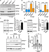
Figure 1.
Figure 1.
Induction of the DHPS/EIF5AHyp pathway by A/E pathogens. (a,b) Dhpsfl/fl and Dhps∆epi mice were infected or not with C. rodentium for 14 days. Colons (n = 4 mice per group) were removed and proteins were extracted from the whole tissue. DHPS and EIF5AHyp levels were assessed by Western blot (a) followed by densitometric analyses (b). (c,d) the human colon cell line HCT 116 was infected for 6 h with EPEC, and the level of hypusination and EIF5A was determined by Western blotting (c) and densitometry (d). (e,f) the level of EIF5AHyp and EIF5A in the normal human colonoid line DoD022, infected or not with EPEC for 6 h, was assessed by Western blotting (e) and densitometry (f). The level of EIF5AHyp and EIF5A was assessed by Western blotting (g) followed by densitometry (h) in the normal human colonoid line DoD022 infected or not with EPEC E2348/69, ΔnleA, Δtir, or ΔescN for 6 h. Representative data of 3 independent experiments. *p < 0.05, **p < 0.01, ***p < 0.001 by one-way ANOVA and Tukey test (b, g, h) or by unpaired t test; C, E, and G are representative data of 3 independent experiments.
Figure 2.
Figure 2.
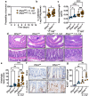
C. rodentium colitis in Dhpsfl/fl and Dhps∆epi mice. Animals were infected or not with C. rodentium (C. rod) and survival was monitored daily (a); there was no death in the uninfected groups. P was calculated by the Log-rank (mantel-cox) test; n = 5 Dhpsfl/fl and 7 Dhps∆epi mice uninfected mice, and n = 19 Dhpsfl/fl and 18 Dhps∆epi infected mice. Animals were sacrificed after 14 days, and colonization was determined by plating serial dilutions of ground colon biopsies (b). The colon weight/length ratio was assessed (c). Colons were Swiss-rolled, stained with H&E (d), and scored for histologic injury (e); scale bar, 50 μm. Proliferation was assessed by IHC for ki-67 ((f); scale bar represents 50 μm) and quantification of ki-67-positive nuclei in CECs (g); positive epithelial cells were counted in 100 crypts along the Swiss roll from n = 3 Dhpsfl/fl; n = 3 Dhps∆epi mice; n = 5 Dhpsfl/fl + C. rodentium; n = 5 Dhpsfl/fl + C. rodentium. in panels with dot plots, *p < 0.05, **p < 0.01, ***p < 0.001, ****p < 0.0001 by one-way ANOVA and Newman–Keuls test (b, c, e) or by one-way ANOVA and Tukey test (G); each dot represents a mouse.
Figure 3.
Figure 3.
Inflammation in colonic tissues. The expression of the genes Cxcl1, Nos2, Tnf, and Il17 analyzed by rt-real-time PCR (a) and the measurement of cytokine concentration assessed by luminex (b) were determined in the colon of Dhpsfl/fl and Dhps∆epi mice, infected or not with C. rodentium. *p < 0.05, **p < 0.01 by Dunnett’s multiple comparisons test (a) and Holm-šídák’s multiple comparisons test (b).
Figure 4.
Figure 4.
Transcriptomic changes orchestrated by DHPS in the infected mucosa. RNA extracted from the colon from Dhpsfl/fl and DhpsΔepi (n = 3 uninfected and 5 infected mice per genotype) at 14 days post-inoculation with C. rodentium, was sequenced, and the volcano plots corresponding to the different comparisons were generated (a-d); red dots correspond to genes significantly (p < 0.05) upregulated or downregulated by 1.5-fold or more. Genes with values outside the X and/or Y axis limits are given with their (x;y) coordinates. All the DEGs are provided in Table S1. The DEGs identified in (d) were used for the identification of the “canonical” pathways affected by dhps deletion in infected mice using IPA (e); the complete list of pathways is shown in Table S2.
Figure 5.
Figure 5.
Effect of C. rodentium infection on the proteome of Dhpsfl/fl and DhpsΔepi mice. Label-free quantitative analysis was used to determine the differential expression of the proteins of isolated CECs from Dhpsfl/fl and DhpsΔepi mice infected or not with C. rodentium (n = 4 mice per group) for 14 days. The number of proteins significantly upregulated or downregulated in infected mice in both genotypes is depicted as a venn diagram (a). The proteome of CECs isolated from infected Dhpsfl/fl (b) and Dhps∆epi (c) mice was compared to naïve animals. These figures depict the 50 proteins that are the most significantly upregulated or downregulated for each genotype. The complete list of proteins is given in Table S3.
Figure 6.
Figure 6.
Effect of hypusination on the proteome of C. rodentium-infected CECs. Dhpsfl/fl and DhpsΔepi mice were infected or not with C. rodentium (n = 4 mice per group). Proteins were extracted from isolated CECs. Label-free quantitative analysis was performed and the 40 proteins that are the most significantly upregulated (A) or downregulated (B) in infected DhpsΔepi CECs compared to infected Dhpsfl/fl mice are shown. The complete list of proteins identified is provided in table S3. The proteins involved in aldehyde detoxification have been also identified in this analysis (C); *p < 0.05, **p < 0.01, ***p < 0.001. Proteins were also used to assess GSTO1 level by Western blot (D) followed by densitometric analyses (E); *p < 0.05, **p < 0.01 by ANOVA and Tukey test.
Figure 7.
Figure 7.
Levels of bifunctional electrophile adducts in infected mice. Colon from Dhpsfl/fl and DhpsΔepi mice infected with C. rodentium (n = 3 per genotype) or not (n = 5 per genotype) were immunostained with the D11 ab that detects isoLG-lysyl adducts. Representative images are shown. Scale bars represent 50 μm.

References

    1. Kaper JB, Nataro JP, Mobley HL.. Pathogenic Escherichia coli. Nat Rev Microbiol. 2004. Feb;2(2):123–17. doi:10.1038/nrmicro818. - DOI - PubMed
    1. Ochoa TJ, Contreras CA. Enteropathogenic Escherichia coli infection in children. Curr Opin Infect Dis. 2011. Oct;24(5):478–483. doi:10.1097/QCO.0b013e32834a8b8b. - DOI - PMC - PubMed
    1. Lanata CF, Fischer-Walker CL, Olascoaga AC, Torres CX, Torres MJ, Black, RE. Child Health Epidemiology Reference Group of the World Health Organization and Unicef . Global causes of diarrheal disease mortality in children <5 years of age: a systematic review. PLOS ONE. 2013;8(9):e72788. doi:10.1371/journal.pone.0072788. - DOI - PMC - PubMed
    1. Nataro JP, Kaper JB. Diarrheagenic Escherichia coli. Clin Microbiol Rev. 1998. Jan;11(1):142–201. doi:10.1128/CMR.11.1.142. - DOI - PMC - PubMed
    1. Donnenberg MS, Giron JA, Nataro JP, Kaper JB. A plasmid-encoded type IV fimbrial gene of enteropathogenic Escherichia coli associated with localized adherence. Mol Microbiol. 1992. Nov;6(22):3427–3437. doi:10.1111/j.1365-2958.1992.tb02210.x. - DOI - PubMed

MeSH terms