. 2024 Dec 14;151(1):9.
doi: 10.1007/s00432-024-06034-4.

Identification of a distinctive immunogenomic gene signature in stage-matched colorectal cancer

Affiliations

Identification of a distinctive immunogenomic gene signature in stage-matched colorectal cancer

Pankaj Ahluwalia et al. J Cancer Res Clin Oncol. .

Abstract

Background: Colorectal cancer (CRC) remains one of the leading causes of cancer-related mortality worldwide. Despite advances in diagnosis and treatment, including surgery, chemotherapy, and immunotherapy, accurate clinical markers are still lacking. The development of prognostic and predictive indicators, particularly in the context of personalized medicine, could significantly improve CRC patient management.

Method: In this retrospective study, we used FFPE blocks of tissue samples from CRC patients at Augusta University (AU) to quantify a custom 15-gene panel. To differentiate the tumor and adjacent normal regions (NAT), H&E staining was utilized. For the quantification of transcripts, we used the NanoString nCounter platform. Kaplan-Meier and Log-rank tests were used to perform survival analyses. Several independent datasets were explored to validate the gene signature. Orthogonal analyses included single-cell profiling, differential gene expression, immune cell deconvolution, neoantigen prediction, and biological pathway assessment.

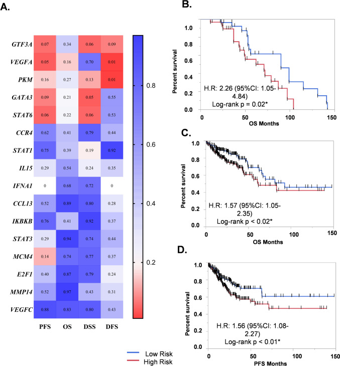
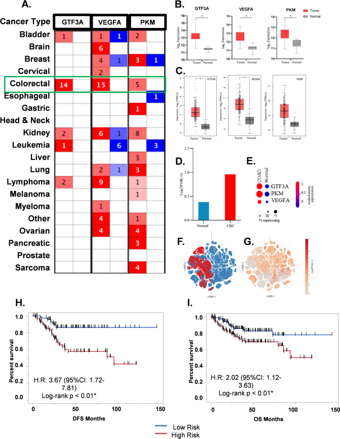
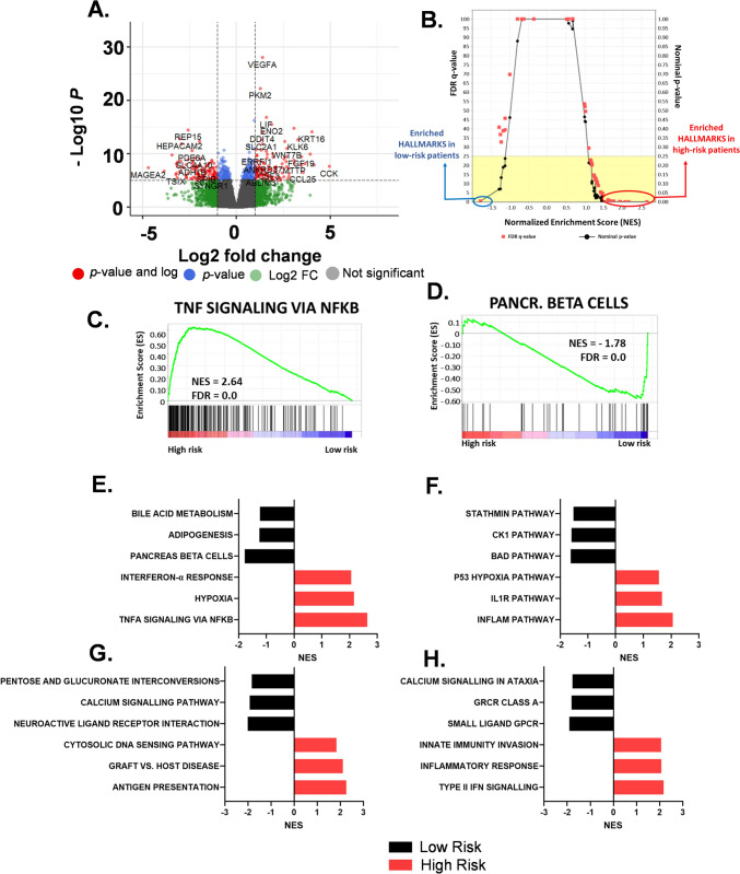
Results: A 3-gene signature (GTF3A, PKM, and VEGFA) was found to be associated with overall survival in the AU cohort (HR = 2.26, 95% CI 1.05-4.84, p = 0.02, 93 patients), TCGA cohort (HR = 1.57, 95% CI 1.05-2.35, p < 0.02, 435 patients) and four other GEO datasets. Independent single-cell analysis identified relatively higher expression of the 3-gene signature in the tumor region. Differential analysis revealed dysregulated tissue inflammation, immune dysfunction, and neoantigen load of cell cycle processes among high-risk patients compared to low-risk patients.

Conclusion: We developed a 3-gene signature with the potential for prognostic and predictive clinical assessment of CRC patients. This gene-based stratification offers a cost-effective approach to personalized cancer management. Further research using similar methods could identify therapy-specific gene signatures to strengthen the development of personalized medicine for CRC patients.

Keywords: Colon; Colorectal cancer; Gene signature; Immune infiltration; Immunotherapy responsiveness; Personalized medicine; Precision medicine; Prognostic genes; Stratified medicine.


Conflict of interest statement

Declarations. Conflcit of interest: The authors declare no competing interests. Ethical Standards: The study was approved by the Institutional Review Board (IRB-HAC # 611298) of Augusta University. Consent to participate: The research was conducted using de-identified samples and no consent was required. Consent for publication: N/A.

Figures

Fig. 1
Fig. 1
Flowchart of the study
Fig. 2
Fig. 2
Genomic Analysis of the TCGA dataset (A) Waterfall plot depicting the frequency of mutations in CRC in context of other tumors (B) Heatmap depicting the percentage of Single Nucleotide Variations (SNVs) across different samples in the TCGA CRC dataset. (C) Copy Number Variation (CNV) analysis of the TCGA-COAD dataset, showing the amplification and deletion of specific genes in different samples
Fig. 3
Fig. 3
Comparative analysis of internal and TCGA-COAD cohort (A) Volcano plot to depict the differential expression of the genes in the AU cohort. (B) Hierarchal clustering of the genes in the AU dataset. (C) Principal component analysis (PCA) of genes in the TCGA dataset. (D) Higher expression of tumor enriched genes in TCGA dataset. (E) Percentage variance explained by the top 3 PCA components. (F) The higher expression of NAT-enriched genes in the TCGA dataset
Fig. 4
Fig. 4
Survival analysis of 3-gene signature (A) The prognostic gene signature of GTF3A, VEGFA, and PKM showed significant survival differences in TCGA-COAD datasets. (B) The overall survival distribution of the gene signature in the internal dataset. (C) The overall survival distribution in the TCGA dataset. (D) The Progression-free survival distribution in the TCGA dataset
Fig. 5
Fig. 5
External validation of 3-gene signature (A) The 3 genes showed tumorigenic roles in multiple GEO datasets In the Oncomine database. (B) The distribution of gene expression between tumor and normal in an independent dataset. (C) The distribution of gene expression between tumor and normal in the TCGA dataset. (D and E) The higher expression of the gene signature in single cell dataset (F & G) t-SNE mapping of the prognostic gene signature. (H) The DFS distribution in the GSE17536 dataset. (I.) The overall survival distribution in an GSE14333
Fig. 6
Fig. 6
Analysis of Biological Pathway Variation in High-Risk and Low-Risk Patients (A) Volcano plot depicting differential expression of genes in two survival groups. (B) The differential enrichment of hallmarks in cancer in two risk groups (C) The hallmark with the highest enrichment in the high-risk group. (D) The hallmark with the highest enrichment in the low-risk group. The distribution of hallmarks in cancer (E), Biocarta (F), KEGG (G), and Wikipathways (H) in two-survival groups
Fig. 7
Fig. 7
Comparative analysis of immunogenomic characterization in high-risk and low-risk patients: (A) Pathway analysis showing neoantigen distribution in high-risk colorectal cancer patients, and (B) low-risk colorectal cancer patients (C) Immune deconvolution analysis of the gene signature. (D) Immune dysfunction analysis using the TIDE algorithm in the high-risk group and (E) in the low-risk group

References

    1. Ahluwalia P, Kolhe R, Gahlay GK (2021) The clinical relevance of gene expression based prognostic signatures in colorectal cancer. Biochim Biophys Acta Rev Cancer 1875(2):188513. 10.1016/j.bbcan.2021.188513 - PubMed
    1. Ahluwalia P, Mondal AK, Ahluwalia M, Sahajpal NS, Jones K, Jilani Y, Kolhe R (2022) Clinical and molecular assessment of an onco-immune signature with prognostic significance in patients with colorectal cancer. Cancer Med, 11(6): 1573–1586. 10.1002/cam4.4568 - PMC - PubMed
    1. Al Obeed OA., Alkhayal KA, Al Sheikh A, Zubaidi AM, Vaali-Mohammed MA, Boushey R, Abdulla MH (2014) Increased expression of tumor necrosis factor-alpha is associated with advanced colorectal cancer stages. World J Gastroenterol 20(48): 18390–18396. 10.3748/wjg.v20.i48.18390 - PMC - PubMed
    1. Anderson CA, Patel P, Viney JM, Phillips RM, Solari R, Pease JE (2020) A degradatory fate for CCR4 suggests a primary role in Th2 inflammation. J Leukoc Biol 107(3):455–466. 10.1002/JLB.2A0120-089RR - PMC - PubMed
    1. Anuraga G, Tang WC, Phan NN, Ta HDK, Liu YH, Wu YF, Wang CY (2021) Comprehensive analysis of prognostic and genetic signatures for general transcription factor III (GTF3) in clinical colorectal cancer patients using bioinformatics approaches. Curr Issues Mol Biol, 43(1): 2–20. 10.3390/cimb43010002 - PMC - PubMed

Substances