Observational Study
. 2025 Feb 4;19(2):jjae189.
doi: 10.1093/ecco-jcc/jjae189.

Open Label Vancomycin in Primary Sclerosing Cholangitis-Inflammatory Bowel Disease: Improved Colonic Disease Activity and Associations With Changes in Host-Microbiome-Metabolomic Signatures

Affiliations
Observational Study

Open Label Vancomycin in Primary Sclerosing Cholangitis-Inflammatory Bowel Disease: Improved Colonic Disease Activity and Associations With Changes in Host-Microbiome-Metabolomic Signatures

Mohammed Nabil Quraishi et al. J Crohns Colitis. .

Abstract

Background: We conducted a single-arm interventional study, to explore mucosal changes associated with clinical remission under oral vancomycin (OV) treatment, in primary sclerosing cholangitis-associated inflammatory bowel disease (PSC-IBD); NCT05376228.

Methods: Fifteen patients with PSC and active colitis (median fecal calprotectin 459 µg/g; median total Mayo score 5) were treated with OV (125 mg QID) for 4 weeks and followed-up for a further 4 weeks of treatment withdrawal (8 weeks, end-of-study). Colonic biopsies were obtained at baseline and Week 4. Clinical assessments, and serum and stool samples (metagenomics, metatranscriptomics, and metabolomics) were collected at Weeks 0, 2, 4, and 8. The primary efficacy outcome measure was the induction of clinical remission.

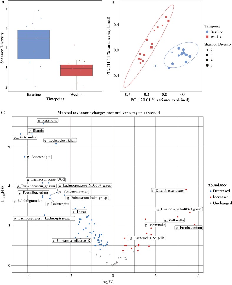
Results: Oral vancomycin resulted in clinical remission in 12/15 patients and significant reductions in fecal calprotectin. Oral vancomycin was associated with reduced abundances of Lachnospiraceae, genera Blautia and Bacteroides; and enrichment of Enterobacteriaceae, and genera Veillonella, Akkermansia, and Escherichia. Oral vancomycin treatment was associated with the downregulation of multiple metatranscriptomic pathways (including short-chain fatty acid [SCFA] metabolism and bile acid [BA] biotransformation), along with host genes and multiple pathways involved in inflammatory responses and antimicrobial defence; and an upregulation of genes associated with extracellular matrix repair. Oral vancomycin use resulted in the loss of specific fecal SCFAs and secondary BAs, including lithocholic acid derivatives. Colitis activity relapsed following OV withdrawal, with host mucosal and microbial changes trending toward baseline.

Conclusions: Four weeks of OV induces remission in PSC-IBD activity, associated with a reduction in gut bacterial diversity and compositional changes relating to BA and SCFA homeostasis.

Keywords: Primary sclerosing cholangitis; inflammatory bowel disease; vancomycin.


Conflict of interest statement

MNQ has received speaker fees from Pfizer, Janssen, Takeda, Tillotts, Falk, and Nordic Pharma and consultancy fees from Nordic Pharma. BM has no conflicts of interest to declare. JRM has received consultancy fees from Cultech Ltd. and EnteroBiotix Ltd. PJT receives institutional funding from the NIHR Birmingham BRC. This article presents independent research supported by the Birmingham NIHR BRC, based at the University Hospitals Birmingham NHS Foundation Trust and the University of Birmingham. The views expressed are those of the author(s) and not necessarily those of the NHS, the NIHR, or the Department of Health. PJT has received advisory board/consultancy fees from Chemomab, Cymabay, Albireo/Ipsen Pharma, Advanz Pharma/Intercept, Dr. Falk Pharma, GlaxoSmithKline, Gilead Sciences, Mirum, and Pliant Pharma. PJT has received independent grant funding from Albireo/Ipsen, Advanz Pharma/Intercept, Bristol Myers Squibb, GlaxoSmithKline, Gilead Sciences, Guts UK, the European Association for Study of the Liver (EASL), Innovate UK, LifeArc, the Medical Research Foundation, the NIHR, Mirum, PSC Support, Regeneron, and the Wellcome Trust.

Figures

Figure 1
Figure 1
Study schematic. Fifteen patients with active PSC-IBD participated in an 8-week study where they received open-label 125 mg QDS (quater die sumendum) of oral vancomycin for the first 4 weeks (Week 4), followed by withdrawal of OV for the subsequent 4 weeks (Week 8). Lower gastrointestinal endoscopies were performed at baseline and Week 4. Clinical assessments, serum, biopsies for transcriptomics, and stool for calprotectin, metagenomics, metatranscriptomics, and bile acid metabolomics were collected at Baseline, Weeks 2, 4, and 8. Abbreviation: OV, oral vancomycin.
Figure 2
Figure 2
Changes in clinical parameters following treatment and withdrawal of oral vancomycin in patients with active PSC-IBD. A, Significant reductions in fecal calprotectin, and partial Mayo and Mayo endoscopic subscores were observed following 4 weeks of oral vancomycin therapy. Both fecal calprotectin and the partial Mayo score appeared to increase significantly following the subsequent 4 weeks of withdrawal of oral vancomycin. B, Both ALP and ALT appeared to significantly decrease following oral vancomycin, however, no changes in total serum bilirubin levels were observed. Abbreviations: ALP, alkaline phosphatase; ALT, alanine transaminase; PSC-IBD, primary sclerosing cholangitis-associated inflammatory bowel disease.
Figure 3
Figure 3
Metagenomic analysis of fecal microbiome at different study timepoints. A, A significant reduction in alpha diversity (Shannon Index) is observed following oral vancomycin by the second week of treatment (Week 2) compared with baseline. This diversity loss remains stable in the subsequent 2 weeks of treatment (Week 4) with some restoration of diversity following 4 weeks of vancomycin withdrawal (Week 8). B, Bray–Curtis PCoA analysis demonstrates distinct clustering of microbiota as early as Week 2 compared with baseline. The microbial compositional cluster appears to move back toward baseline at Week 8 following withdrawal of OV. C, Changes within the phylum are observed as early as Week 2 with depletion of Bacteroidetes and Firmicutes and enrichment of Proteobacteria and Fusobacteria. D, At the species level, a significant decrease in Faecalibacterium prausnitzii, Anaerostipes hadrus, and multiple short-chain fatty acid (SCFA) producing species was observed when compared with baseline. Conversely, a significant increase in Fusobacterium nucleatum, Enterobacter hormaechei, Escherichia coli, and multiple species belonging to the Veillonella and Klebsiella genera were increased at Week 4 compared with baseline. Vancomycin withdrawal appeared to reverse some of these changes with an increase in Roseburia hominis, Prevotella buccae, Lachnospiraceae bacterium, Blautia hansenii, and Bacteroides thetaiotaomicron and a decrease in species belonging to the Proteobacterium phylum at Week 8 compared with baseline. E, Negative correlations were seen between fecal calprotectin levels and multiple species including those belonging to Fusobacterium, Klebsiella, Veillonella, and positive correlations with Lachnospira, Roseburia, and Bacteriodes. Abbreviations: OV, oral vancomycin; PCoA, Principal Coordinates Analysis.
Figure 3
Figure 3
Metagenomic analysis of fecal microbiome at different study timepoints. A, A significant reduction in alpha diversity (Shannon Index) is observed following oral vancomycin by the second week of treatment (Week 2) compared with baseline. This diversity loss remains stable in the subsequent 2 weeks of treatment (Week 4) with some restoration of diversity following 4 weeks of vancomycin withdrawal (Week 8). B, Bray–Curtis PCoA analysis demonstrates distinct clustering of microbiota as early as Week 2 compared with baseline. The microbial compositional cluster appears to move back toward baseline at Week 8 following withdrawal of OV. C, Changes within the phylum are observed as early as Week 2 with depletion of Bacteroidetes and Firmicutes and enrichment of Proteobacteria and Fusobacteria. D, At the species level, a significant decrease in Faecalibacterium prausnitzii, Anaerostipes hadrus, and multiple short-chain fatty acid (SCFA) producing species was observed when compared with baseline. Conversely, a significant increase in Fusobacterium nucleatum, Enterobacter hormaechei, Escherichia coli, and multiple species belonging to the Veillonella and Klebsiella genera were increased at Week 4 compared with baseline. Vancomycin withdrawal appeared to reverse some of these changes with an increase in Roseburia hominis, Prevotella buccae, Lachnospiraceae bacterium, Blautia hansenii, and Bacteroides thetaiotaomicron and a decrease in species belonging to the Proteobacterium phylum at Week 8 compared with baseline. E, Negative correlations were seen between fecal calprotectin levels and multiple species including those belonging to Fusobacterium, Klebsiella, Veillonella, and positive correlations with Lachnospira, Roseburia, and Bacteriodes. Abbreviations: OV, oral vancomycin; PCoA, Principal Coordinates Analysis.
Figure 4
Figure 4
Metatranscriptomic analysis of fecal microbiome at different study timepoints. A, Bray–Curtis PCoA analysis demonstrates distinct clustering of metatranscriptome following OV therapy at Weeks 2 and 4 compared with baseline (both p < 0.01) with restoration of microbial function back to baseline at Week 8 following withdrawal of OV. B, Horizontal bar plot highlighting changes in the top 30 KEGG and MetaCyc metatranscriptomic pathways (FDR adjusted <0.1) following OV therapy (Week 4 compared with baseline). Abbreviations: KEGG, Kyoto Encyclopaedia of Genes and Genomes; OV, oral vancomycin; PCoA, Principal Coordinates Analysis.
Figure 5
Figure 5
Changes in mucosally adherent microbiota following treatment with oral vancomycin. A, Significant reduction in alpha diversity (Shannon Index) is observed following oral vancomycin at Week 4. B, Bray–Curtis PCoA analysis demonstrates distinct clustering of microbiota at Week 4 clusters compared with baseline (p < 0.001). C, Oral vancomycin is associated with to significant depletion of relative abundances of several genera including Roseburia, Faecalibacterium, Ruminococcus, Bacteriodes, and an enrichment of Fusobacterium, Enterobacteria, and Veillonella. Abbreviation: PCoA, Principal Coordinates Analysis.
Figure 6
Figure 6
Impact of vancomycin upon fecal bile acid profiles. Analysis of reversed-phase (RP) ultra-high performance liquid chromatography–mass spectrometry (UHPLC–MS), performed on OmniMet stool-preservative mixture, for bile acid profiling. A, Principal components analysis (PCA) scores plot, analyzing stool bile acid profiling data (with log-transformation and Pareto scaling), with samples annotated by a week of collection; B, orthogonal partial least squares-discriminant analysis (OPLS-DA) scores plot of baseline versus Week 2 samples; C, S-plot derived from OPLS-DA of (B); the further that bile acids are toward the bottom left of the plot then the higher they are in baseline samples, while those nearer to the top right of the plot are higher in Week 2 samples. Additional abbreviations: CA, cholic acid; CDCA, chenodeoxycholic acid; GCDCA, glycochenodeoxycholic acid; GUDCA, glycoursodeoxycholic acid.
Figure 7
Figure 7
Changes in host colonic mucosal transcriptomic profiles following treatment with oral vancomycin. A, MDS (multidimensional scaling) plot analysis demonstrates distinct clustering of colonic mucosal gene expression profiles following OV therapy at Week 4 compared with baseline (p = 0.004). B, Heatmap plot of differentially expressed genes highlighting clear clusters of downregulated and upregulated genes that appear to correlate with fecal calprotectin levels following OV therapy. C, Volcano plot that demonstrating significant differential expression of 843 genes with an increase in 629 genes and a decrease in 214 genes (FDR adjusted p < 0.1). Abbreviation: OV, oral vancomycin.

References

    1. Karlsen TH, Folseraas T, Thorburn D, Vesterhus M.. Primary sclerosing cholangitis—a comprehensive review. J Hepatol 2017;67:1298–323. - PubMed
    1. Weismuller TJ, Trivedi PJ, Bergquist A, et al.; International PSC Study Group. Patient age, sex, and inflammatory bowel disease phenotype associate with course of primary sclerosing cholangitis. Gastroenterology 2017;152:1975–84.e8. - PMC - PubMed
    1. Culver EL, Bungay HK, Betts M, et al.Prevalence and long-term outcome of sub-clinical primary sclerosing cholangitis in patients with ulcerative colitis. Liver Int 2020;40:2744–57. - PubMed
    1. Khrom M, Long M, Dube S, et al.Comprehensive association analyses of extraintestinal manifestations in inflammatory bowel disease. Gastroenterology 2024;167:315–32. - PMC - PubMed
    1. Trivedi PJ, Hirschfield GM.. Recent advances in clinical practice: epidemiology of autoimmune liver diseases. Gut 2021;70:1989–2003. - PubMed

Publication types

MeSH terms

Associated data