. 2025 Mar;69(3):133-147.
doi: 10.1111/1348-0421.13193. Epub 2024 Dec 15.

Lonidamine, a Novel Modulator for the BvgAS System of Bordetella Species

Affiliations

Lonidamine, a Novel Modulator for the BvgAS System of Bordetella Species

Natsuko Ota et al. Microbiol Immunol. 2025 Mar.

Abstract

The Gram-negative bacteria Bordetella pertussis, B. parapertussis, and B. bronchiseptica cause respiratory diseases in various mammals. They share the BvgAS two-component system, which regulates the phenotypic conversion between the virulent Bvg+ and avirulent Bvg- phases. In the BvgAS system, the sensor kinase BvgS senses environmental cues and transduces a phosphorelay signal to the response regulator BvgA, which leads to the expression of Bvg+ phase-specific genes, including virulence factor genes. Bacteria grown at 37°C exhibit the Bvg+ phenotype. In contrast, at lower than 26°C or in the presence of modulators, such as MgSO4 and nicotinic acid, the BvgAS system is inactivated, leading bacteria to the avirulent Bvg- phase. Therefore, effective modulators are expected to provide a therapeutic measure for Bordetella infection; however, no such modulators are currently available, and the mechanism by which modulators inactivate the BvgAS system is poorly understood. In the present study, we identified lonidamine as a novel modulator after screening an FDA-approved drug library using bacterial reporter systems with the Bvg+-specific and Bvg--specific promoters. Lonidamine directly bound to the VFT2 domain of BvgS and inactivated the BvgAS system at concentrations as low as 50 nM, which was at least 2000- to 20,000-fold lower than the effective concentrations of known modulators. Lonidamine significantly reduced the adherence of B. pertussis to cultured cells but unexpectedly exacerbated bacterial colonization of the mouse nasal septum. These results provide insights into the structural requirements for BvgAS modulators and the role of Bvg phenotypes in the establishment of infection.

Keywords: Bordetella; BvgAS; lonidamine; modulator; phase conversion.


Figures

Figure 1
Figure 1
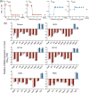
GFP‐expressing Bordetella pertussis reporter strains to indicate Bvg states. GFP reporter strains with Bvg+ phase‐dependent promoters (fhaB, cya, dnt, prn, ptx, and vag8; red) and Bvg phase‐dependent promoters (vrgX, vrg6, vrg73, bp1618, bp1738, and kpsM; blue) were incubated in SS medium (a, b) and on BG‐BSA agar (c) in the presence (dark color) or absence (light color) of 50 mM MgSO4 for 3 days, and the fluorescence intensity of expressed GFP was measured. The tac promoter was used as the BvgAS‐independent control. The wild type (Wt) does not carry the reporter plasmid. Note that the Y‐axis scale is different for each panel. Values represent the mean ± SD (n = 3 for a and b, and n = 24 for c). The significance of differences was analyzed by an unpaired t‐test on each row with Holm−Šídák's multiple comparisons test.
Figure 2
Figure 2
Screening of chemical compounds that inactivate the BvgAS system of Bordetella pertussis. (a) An FDA‐approved drug library consisting of 1134 chemical compounds was screened for activity stimulating the expression of GFP using the Thm/P vrgX gfp strain. Eleven compounds that increased GFP expression levels by more than 1.5‐fold the average of the negative control (mock, M) are indicated as red spots. (b–d) Candidate compounds after the first screening were further examined using Thm/P vrgX gfp (b–d), Thm/P vrg73 gfp (c, d), Thm/P fhaB gfp (c, d), and Thm/P ptx gfp (c, d). Reporter strains were grown on BG‐BSA agar (a–c) or in SS medium (d). Each compound was applied at 10 µM (a and b), 30 µM (c), or under bactericidal concentrations (d; 10 µM [#5, #8], and 30 µM [#3, #6]). Numbered compounds are etoposide (#1), artemisinin (#2), fludarabine (#3), econazole nitrate (#4) lonidamine (#5), dydrogesterone (#6), auranofin (#7), and otilonium bromide (#8). As the control, 50 mM MgSO4 was applied (b−d). Values represent the mean ± SD (n = 4 for b, n = 8 for c, and n = 3 for d). Data were statistically analyzed by a one‐way analysis of variance (ANOVA) with Tukey's multiple comparisons test (b).
Figure 3
Figure 3
Lonidamine converts classical Bordetella species to the Bvg phase. (a) The GFP reporter assay using Thm/P fhaB gfp, Thm/P ptx gfp, Thm/P vrgX gfp, and Thm/P vrg73 gfp strains incubated in SS medium containing 0.03−10 µM lonidamine, 1.5% DMSO (mock, M), or 50 mM MgSO4. Representative data are shown after three independent experiments under similar conditions. Values represent the mean ± SD (n = 3). Some error bars that are shorter than the symbol size are not depicted. (b) The relative expression levels of Bvg+ phase‐specific genes (fhaB, prn, dnt, fim2, fim3, bscN, cyaA, ptxA, and vag8; red) and Bvg phase‐specific genes (vrgX, vrg73, and flaA; blue) of B. pertussis (Tohama, 18323, BP140, and BP142), B. parapertussis 12822, and B. bronchiseptica RB50. Bacteria were grown in SS medium containing 50 mM MgSO4 (dark color) or 1 µM lonidamine (light color) and subjected to qPCR, as described in the Materials and Methods. Data represent fold changes in the expression of each gene compared to bacteria grown in SS medium containing 1.5% DMSO (n = 3).
Figure 4
Figure 4
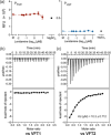
Lonidamine targets BvgS but not BvgA. (a) The GFP reporter assay using the Bvg+ phase‐locked mutants of Thm/P fhaB gfp and Thm/P vrgX gfp incubated in SS medium containing 0.03−10 µM lonidamine, 1.5% DMSO (mock, M), or 50 mM MgSO4. Data are shown from a single experiment with three independent samples. Values represent the mean ± SD (n = 3). Some error bars that are shorter than the symbol size are not depicted. (b, c) The interaction of the VFT domains of BvgS and lonidamine analyzed by ITC. The upper panels show the rate of heat released by 2 µL injections of 300 µM lonidamine into a cell containing 20 µM VFT1 (b) or VFT2 (c). The lower panels show the integrated areas of the respective peaks in the upper panel plotted against the molar ratio of lonidamine to VFT1 (b) or VFT2 (c).
Figure 5
Figure 5
Putative interaction mode of lonidamine with VFT2. (a) Docking simulation of lonidamine (cyan) and VFT2 (green) in the dimeric state. The structure of VFT2 is represented as a ribbon diagram. Lonidamine and the amino acid residues involved in the interaction are shown as sticks. The magenta and yellow dotted lines represent hydrogen and halogen bonds, respectively. The blue dotted lines represent the π–π interaction. The glide score of the model is −7.862. (b) The P fhaB ‐ and P vrgX ‐based reporter assay using Bordetella pertussis bvgS mutants (F375A, R380A, T462A + S465A, and quadA). Bacteria were incubated in SS medium containing 0.03−10 µM lonidamine, 1.5% DMSO (mock, M), or 50 mM MgSO4, and expressed GFP was estimated as described in the Materials and Methods. Values represent the mean ± SD (n = 3).
Figure 6
Figure 6
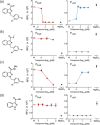
Effects of lonidamine and its analogs on Bvg states of Bordetella pertussis. Thm/P fhaB gfp and Thm/P vrgX gfp were incubated in SS medium containing 1.5% DMSO (mock, M) or the indicated concentrations of lonidamine and its analogs or 50 mM MgSO4, and expressed GFP was estimated as described in the Materials and Methods. The chemical structural formulas of lonidamine (1‐(2,4‐dichlorobenzyl)‐1H‐indazole‐3‐carboxylic acid (a), methyl 1‐(2,4‐dichlorobenzyl)‐1H‐indazole‐3‐carboxylic acid (b), adjudin (1‐(2,4‐dichlorobenzyl)‐1H‐indazole‐3‐carbohydrazide) (c), and indazole‐3‐carboxylic acid (d) are shown on the left of each panel. Experiments for (a−c) were performed at least twice and representative data are shown. For (d), data from a single experiment are shown. Values represent the mean ± SD (n = 3 for each panel). Some error bars that are shorter than the symbol size are not depicted.
Figure 7
Figure 7
Effects of lonidamine on Bordetella pertussis infection. (a) Microscopic images of A549 cells infected with mCherry Thm/P fhaB gfp (mC/P fhaB gfp, upper panel) or mCherry Thm/P vrgX gfp (mC/P vrgX gfp, lower panel). Bacteria were precultured with (“pre−post”) or without (“mock” and “post”) 1 µM lonidamine (Lon) and added to a culture of A549 cells as described in the Materials and Methods. A549 cells with bacteria were incubated for 24 h in the presence (“post” and “pre−post”) or absence (“mock”) of 1 µM lonidamine. In the mock control, 0.5% DMSO was applied. In the “post” culture, 1 µM lonidamine was applied after bacteria adhered to cells for the first hour. Bar, 10 µm. (b) Numerical data on mCherry‐positive bacteria adhering to A549 cells. Microscopic images were captured from at least eight independent fields and the number of adhering bacteria was counted. A single plot symbol represents a single microscopic field. (c) The correlation between luminescence intensity and the number of P tac ‐Akaluc Thm (P tac ‐Ak) and P vrgX ‐Akaluc Thm (P vrgX ‐Ak) strains. Serially diluted bacterial samples precultured with or without 1 µM lonidamine (Lon) were mixed with 100 µM akalumine in a 96‐well black plate. The luminescence intensity in each well was measured as described in the Materials and Methods and expressed as relative luminescence units (RLU). (d) Effects of lonidamine on Bordetella pertussis infection in mice. P tac ‐Akaluc Thm was precultured with (“pre−post”) or without (“mock” and “post”) 1 µM lonidamine and intranasally inoculated into anesthetized mice at 1 × 107 CFU/25 µL/mouse (Day 0). Mice were intranasally injected with (“post” and “pre−post”) or without (“mock”) 10 mg/kg body weight of lonidamine every day from Day 1. On Days 1, 4, and 8 of infection, mice were intraperitoneally injected with Akalumine‐HCl, and bioluminescence images of bacteria were acquired. On Day 1, images were acquired 7 h before the lonidamine injection. In the mock control, 4.6% DMSO was applied. (e) Quantitative data on luminescence levels in the nasal septum estimated by Living Image 4.7 software. Values represent the mean ± SD (b, c and n = 5 for e). The significance of differences was analyzed by a two‐way ANOVA with Šídák's multiple comparisons test (b, e).

Similar articles

References

    1. Melvin J. A., Scheller E. V., Miller J. F., and Cotter P. A., “ Bordetella pertussis Pathogenesis: Current and Future Challenges,” Nature Reviews Microbiology 12 (2014): 274–288. - PMC - PubMed
    1. Hamidou Soumana I., Linz B., and Harvill E. T., “Environmental Origin of the Genus Bordetella ,” Frontiers in Microbiology 8 (2017): 28. - PMC - PubMed
    1. Goodnow R. A., “Biology of Bordetella Bronchiseptica ,” Microbiological Reviews 44 (1980): 722–738. - PMC - PubMed
    1. Park J., Zhang Y., Buboltz A. M., et al., “Comparative Genomics of the Classical Bordetella Subspecies: The Evolution and Exchange of Virulence‐Associated Diversity Amongst Closely Related Pathogens,” BMC Genomics 13 (2012): 545. - PMC - PubMed
    1. Diavatopoulos D. A., Cummings C. A., Schouls L. M., Brinig M. M., Relman D. A., and Mooi F. R., “ Bordetella pertussis, the Causative Agent of Whooping Cough, Evolved From a Distinct, Human‐Associated Lineage of B. bronchiseptica ,” PLoS Pathogens 1 (2005): e45. - PMC - PubMed

MeSH terms

LinkOut - more resources