. 2025 Jan;301(1):108088.
doi: 10.1016/j.jbc.2024.108088. Epub 2024 Dec 13.

An SH3-binding allosteric modulator stabilizes the global conformation of the AML-associated Src-family kinase, Hck

Affiliations

An SH3-binding allosteric modulator stabilizes the global conformation of the AML-associated Src-family kinase, Hck

Ari M Selzer et al. J Biol Chem. 2025 Jan.

Abstract

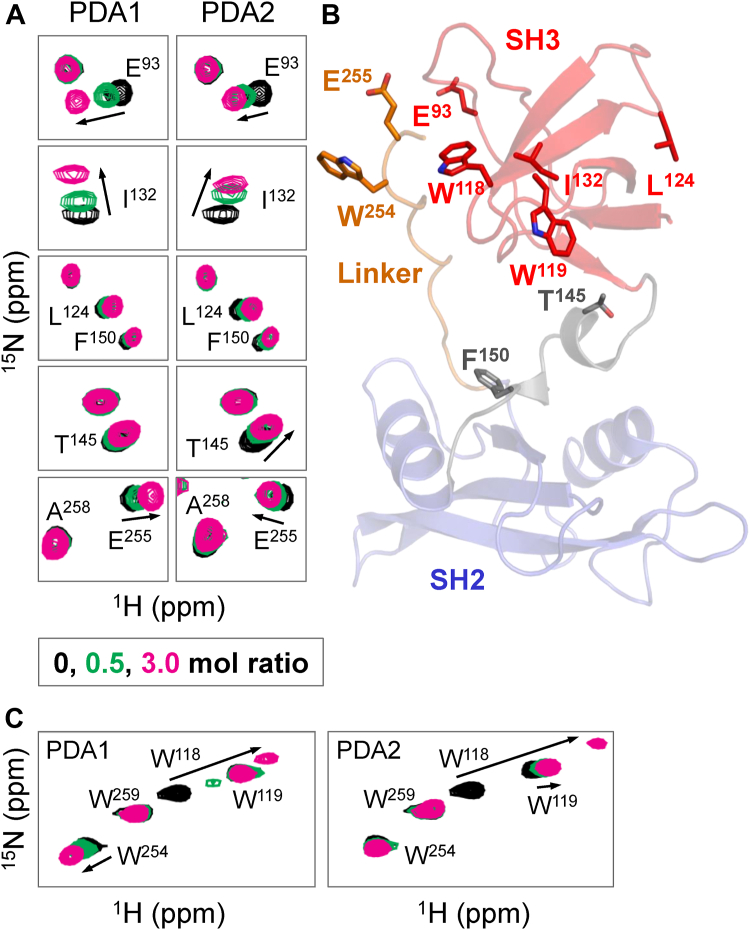
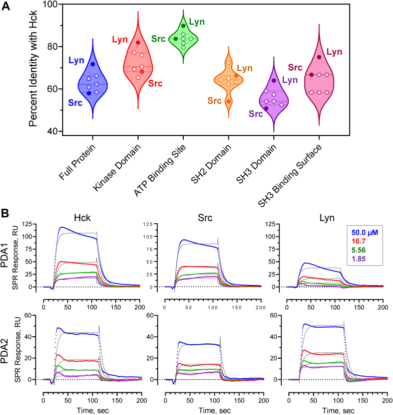
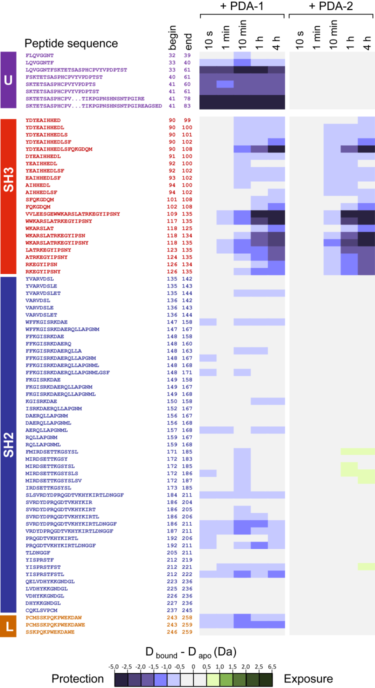
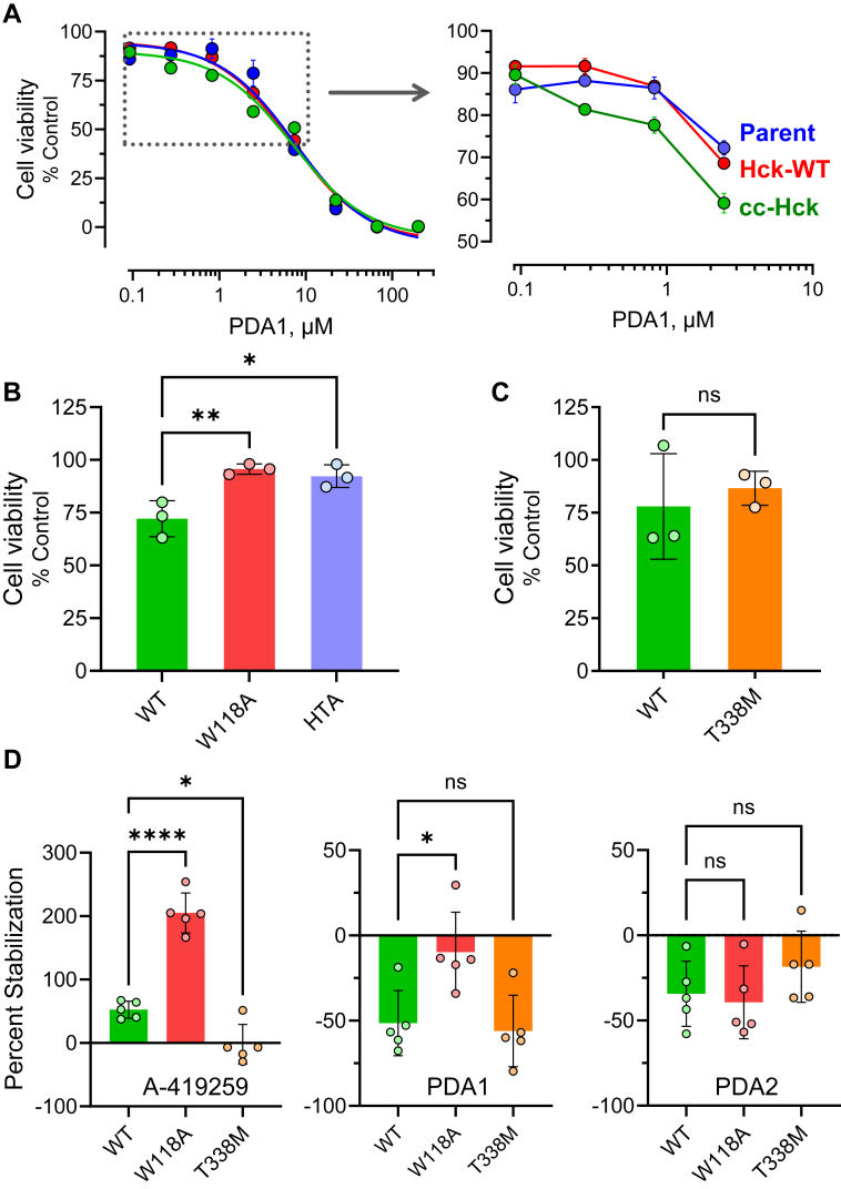
While ATP-site inhibitors for protein-tyrosine kinases are often effective drugs, their clinical utility can be limited by off-target activity and acquired resistance mutations due to the conserved nature of the ATP-binding site. However, combining ATP-site and allosteric kinase inhibitors can overcome these shortcomings in a double-drugging framework. Here we explored the allosteric effects of two pyrimidine diamines, PDA1 and PDA2, on the conformational dynamics and activity of the Src-family tyrosine kinase Hck, a promising drug target for acute myeloid leukemia. Using 1H-15N HSQC NMR, we mapped the binding site for both analogs to the SH3 domain. Despite the shared binding site, PDA1 and PDA2 had opposing effects on near-full-length Hck dynamics by hydrogen-deuterium exchange mass spectrometry, with PDA1 stabilizing and PDA2 disrupting the overall kinase conformation. Kinase activity assays were consistent with these observations, with PDA2 enhancing kinase activity while PDA1 was without effect. Molecular dynamics simulations predicted selective bridging of the kinase domain N-lobe and SH3 domain by PDA1, a mechanism of allosteric stabilization supported by site-directed mutagenesis of N-lobe contact sites. Cellular thermal shift assays confirmed SH3 domain-dependent interaction of PDA1 with WT Hck in myeloid leukemia cells and with a kinase domain gatekeeper mutant (T338M). These results identify PDA1 as a starting point for Src-family kinase allosteric inhibitor development that may work in concert with ATP-site inhibitors to suppress the evolution of resistance.

Keywords: NMR; SH2 domain; SH3 domain; Src-family kinase; hydrogen-deuterium exchange mass spectrometry; kinase inhibitors; tyrosine kinases.


Conflict of interest statement

Conflict of interest The authors declare that they have no conflicts of interest with the contents of this article.

Figures

Figure 1
Figure 1
Crystal structure of near-full-length Hck and chemical structures of pyrimidine diamine allosteric modulators.A, crystal structure of Hck bound to the ATP-site inhibitor, A-419259 (A-419; cyan). In the presence of this inhibitor, Hck adopts the closed conformation with the SH3 domain (red) bound to the SH2-kinase linker (orange) and the SH2 domain (blue) engaging the phosphorylated C-terminal tail (cyan; pTyr527). Highlighted features of the kinase domain include the C-helix (purple) in the N-lobe as well as the activation loop (green) and autophosphorylation site (Tyr416). The SH2-kinase linker forms a polyproline type II helix involving prolines 250 and 253 (side chains shown) which engages the SH3 domain; SH3 Trp118 (green) is essential for this interface to form. Model produced using PyMol (Schrödinger) and Hck crystal coordinates from PDB entry 9BYJ. B, chemical structures of PDA1 and PDA2. The common pyrimidine diamine scaffold is highlighted in red.
Figure 2
Figure 2
HSQC NMR analysis of Hck SH3-SH2-linker protein interaction with PDA analogs. PDA1 and PDA2 were titrated with 15N-labeled Hck-SH3-SH2-linker protein and 1H-15N HSQC NMR spectra were recorded. Backbone amide resonances were identified from a previous assignment (28). Tryptophan indole amide resonances were assigned by mutagenesis (Figs. S2 and S3). Significant chemical shift perturbations (CSPs) observed in a subset of (A) backbone amides and (C) tryptophan indole amides are mapped on the crystal structure of the Hck SH3-SH2-linker region in (B). CSPs induced by PDA binding observed in the SH3 domain include E93, W118, I132. PDA1 shows more pronounced CSPs of amides, compared to PDA2, in the connector between SH3 and SH2 (E144 and T145) and W254 and E255 in the SH2-kinase linker. SH3-SH2-linker protein concentration in this experiment was 60 μM.
Figure 3
Figure 3
Sequence identity among SFK domains and binding selectivity of PDAs to Hck, Src, and Lyn.A, violin plot showing distribution of sequence identities among Src family members and their domains relative to Hck. Lyn is the closest relative to Hck, while Src is among the most distant with the SH3 domains showing the least sequence conservation. B, SPR analysis of PDA1 and PDA2 binding to near-full-length Hck, Src, and Lyn. Each kinase protein was immobilized on the SPR chip, and the PDA analogs were injected over the range of concentrations shown in triplicate. Representative sensorgrams are shown in color with fitted curves overlaid in black. KD values are reported in the text.
Figure 4
Figure 4
HDX-MS difference maps for Hck-U32L protein in the presence of PDA1 and PDA2. Hck-U32L protein was equilibrated in the presence and absence of PDA1 and PDA2, followed by exposure to D2O-based buffer for the timepoints shown. Peptic peptides were identified by LC-MS/MS, and changes in deuterium uptake are shown as colored squares according to the scale. Peptide locations within the protein are indicated at left. All values used to make these heat maps are provided in the HDX Supplemental Data File.
Figure 5
Figure 5
Deuterium uptake plots for Hck-U32L peptic peptides. The Hck-U32L protein was equilibrated in the absence or presence of PDA1 or PDA2 followed by HDX-MS analysis as shown in Figure 4. Deuterium uptake curves are shown for peptides derived from the SH3 domain (A), the N-terminal unique domain (B), the SH2 domain (C), and the SH2-kinase linker (D). All values used to make these heat maps are provided in the HDX Supplemental Data File.
Figure 6
Figure 6
PDA1 and PDA2 have opposing effects on Hck dynamics and kinase activity. Recombinant near-full-length Hck was equilibrated with PDA1 or PDA2 or left untreated followed by HDX-MS analysis. Complete HDX-MS peptide deuterium uptake difference maps are presented in Fig. S6 and in the HDX Supplemental Data File. A, peptides showing changes in protection from (blue) or exposure to (green) deuterium uptake in response to PDA treatment are mapped on a crystal structure of Hck (PDB: 9BYJ). HDX data from the U32L protein and the kinase domain from near-full-length Hck were combined to show the overall impacts of each PDA analog. Any peptide showing a significant difference in deuterium uptake in the presence of ligand at the 4 h time point is highlighted as shown. Individual deuterium uptake curves are shown for peptides derived from the SH2-kinase linker (B), the N-lobe of the kinase domain (C), and the SH3 domain (D). Kinetic kinase assays were performed with the same preparation of Hck used for the HDX-MS analysis (E). Reaction velocities were measured over the range of PDA1 and PDA2 concentrations shown. Each data point represents the mean velocity from four technical replicates ± SE.
Figure 7
Figure 7
Molecular dynamics simulations of PDA ligand binding to near-full-length Hck and verification of the binding site by kinetic kinase assay. PDA1 and PDA2 were docked with the crystal structure of near-full-length Hck followed by 600 ns MD simulations as described under Experimental procedures. Movies of the complete simulations are provided in the Supporting information. A, molecular models from the final frame of the simulation show that PDA1 forms a bridge between the SH3 domain and the N-lobe of the kinase domain while PDA2 perturbs the structure of the SH3 domain RT loop to disrupt the SH3-linker interface. B, kinetic kinase assays were performed with recombinant Hck proteins over the range of PDA1 and PDA2 concentrations shown. The assays compared PDA effects on WT Hck versus a double mutant in which kinase domain N-lobe residues H289 and T290 were replaced with alanine (HTA mutant). This mutant is based on the MD simulations which predicted a key role for these N-lobe amino acids in PDA1-mediated bridging with the SH3 domain as shown in part A.
Figure 8
Figure 8
Interaction of PDA1 with Hck in TF-1 myeloid leukemia cells.A, TF-1 erythroleukemia cells were transduced with wild-type (WT) Hck or with an active coiled-coil fusion protein (cc-Hck). Cells were cultured in the presence of PDA1 over the range of concentrations shown or the dimethyl sulfoxide (DMSO) carrier solvent alone (0.1%) as a control. After 96 h, cell viability was assessed using the CellTiter-Blue assay. Data were normalized to the DMSO control and each point represents the mean of three replicates ± SE. The curves were best-fit by nonlinear regression and IC50 values are presented in the text. The boxed area of the main curve is expanded on the right to show enhanced sensitivity of cc-Hck cells to PDA1. B, TF-1 cells expressing cc-Hck with WT kinase domains or mutations in the SH3 domain (W118A) or kinase domain N-lobe (HTA) were treated with PDA1 (1 μM) and viability was assessed 96 h later. C, As in (B) with TF-1 cells expressing cc-Hck or Hck with a mutation of the kinase domain gatekeeper residue, T338M. In (B) and (C), each data point represents a single biological replicate, and the bar heights indicate mean cell viability relative to the DMSO control ± SE. Significant differences were determined by one-way ANOVA (B) or unpaired Student’s t test (C); ∗, p < 0.05; ∗∗, p < 0.01; ns, not significant. D, cellular thermal shift assay (CETSA) in TF-1 cells expressing WT Hck or the SH3 (W118A) and gatekeeper (T338M) mutants. In-cell interaction of Hck with A-419259, PDA1, or PDA2 was assessed as the difference in soluble Hck protein recovery by immunoblot following incubation of the cells at 35 °C versus 48 °C prior to lysis. Each data point represents a single biological replicate of the mean difference (stability) for five independent determinations. Bar heights show the mean stabilization ± SE and significant differences were evaluated by unpaired Student’s t test. ∗, p < 0.05; ∗∗∗∗, p < 0.0001; ns, not significant.

References

    1. Amatya N., Lin D.Y., Andreotti A.H. Dynamic regulatory features of the protein tyrosine kinases. Biochem. Soc. Trans. 2019;47:1101–1116. - PMC - PubMed
    1. Engen J.R., Wales T.E., Hochrein J.M., Meyn M.A., III, Banu O.S., Bahar I., et al. Structure and dynamic regulation of Src-family kinases Cell. Mol. Life Sci. 2008;65:3058–3073. - PMC - PubMed
    1. Boggon T.J., Eck M.J. Structure and regulation of Src family kinases. Oncogene. 2004;23:7918–7927. - PubMed
    1. Chong Y.P., Ia K.K., Mulhern T.D., Cheng H.C. Endogenous and synthetic inhibitors of the Src-family protein tyrosine kinases. Biochim. Biophys. Acta. 2005;1754:210–220. - PubMed
    1. Xu W., Doshi A., Lei M., Eck M.J., Harrison S.C. Crystal structures of c-Src reveal features of its autoinhibitory mechanism. Mol. Cell. 1999;3:629–638. - PubMed

Publication types

MeSH terms

LinkOut - more resources