. 2025 Jan;301(1):108077.
doi: 10.1016/j.jbc.2024.108077. Epub 2024 Dec 13.

Redirecting E3 ubiquitin ligases for targeted protein degradation with heterologous recognition domains

Affiliations

Redirecting E3 ubiquitin ligases for targeted protein degradation with heterologous recognition domains

Huan Yang et al. J Biol Chem. 2025 Jan.

Abstract

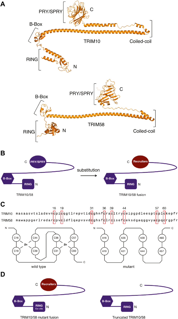
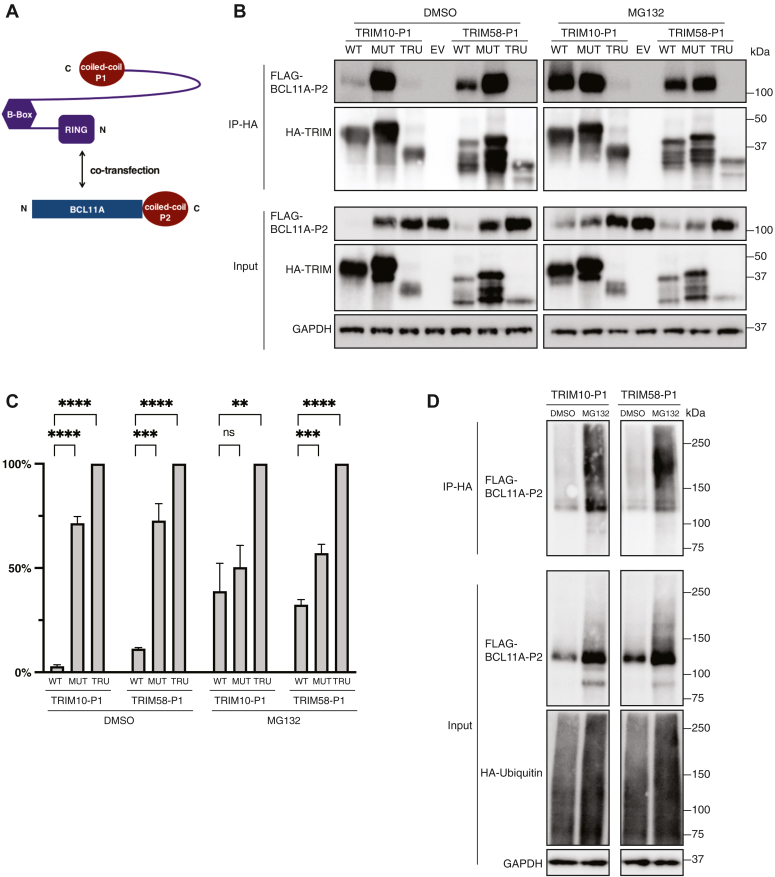
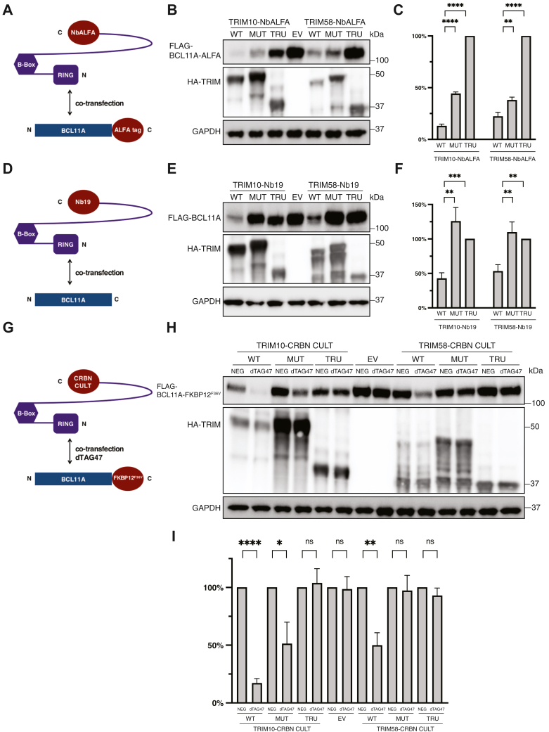
Targeted protein degradation (TPD) mediated by proteolysis targeting chimeras or molecular glues is an emerging therapeutic strategy. Despite greater than 600 E3 ligases and their associated components, a limited number have been deployed in TPD. Those commonly used include cereblon and von Hippel-Lindau tumor suppressor (VHL), which is expressed widely and for which high affinity ligands are available. Limiting TPD to specific cells or tissues would be desirable in many settings. To this goal we have determined the potential of two erythroid cell-enriched E3 ligases, TRIM10 and TRIM58, to degrade a protein of interest, BCL11A, a validated therapeutic target for the β-hemoglobinopathies. We established a general strategy in which heterologous recognition domains replace the PRY-SPRY domain of TRIM10 and TRIM58. Recruitment of TRIM10 or TRIM58 to BCL11A by coiled-coil peptides, nanobodies, or the substrate recognition domain of cereblon led to its degradation. Our findings illustrate a strategy that may be widely useful in evaluating the TPD potential of other E3 ubiquitin ligases and provide a rationale for discovery of ligands for TRIM10 and TRIM58 for erythroid-selective depletion of proteins of interest.

Keywords: BCL11A; E3 ubiquitin ligases; PROTACs; TRIM10; TRIM58; targeted protein degradation.


Conflict of interest statement

Conflict of interest S. H. O. is an Investigator of the Howard Hughes Medical Institute. The other authors declare that they had no conflicts of interest with the contents of this article.

Figures

Figure 1
Figure 1
TRIM10 and TRIM58, modular TRIM proteins, are selectively expressed in the erythroid lineage.A, heat map representing relative RNA expression of E3 ligases (or components) that are upregulated during erythroid differentiation. Genevestigator dataset HS_mRNASeq_HUMAN_GL-0 was analyzed. Colors from white to burgundy indicate from the minimum to the maximum expression in the dataset. B, RNA expression kinetics of TRIM10, TRIM58, CRBN, VHL, and HBB during in vitro erythroid differentiation of CD34+ cells. The values represent means ± SD, n = 3 independent repeats. C, subcellular localization of TRIM10 and TRIM58. 3xHA-tagged full-length TRIM10 and TRIM58 were exogenously expressed in HUDEP2 cells. Anti-HA primary antibody and FITC-conjugated secondary antibody were used for Immunofluorescence. Nuclei were stained with 4′,6-diamidino-2-phenylindole. The scale bar represents 10 μm. D, full-length E3 ligase-P1 fusion proteins tagged at C or N terminus. E, BCL11A-P2 levels in the presence of full-length E3 ligase-P1 fusion proteins. Constructs of BCL11A-P2 and full length E3 ligase-P1 were cotransfected into HEK293T cells. 24 h later, cells were lysed for Western blot. Molecular weight markers are indicated. Full-length E3 ligase-P1 proteins were tagged with 3xHA to monitor their expression. CRBN, cereblon; HBB, hemoglobin beta; VHL, von Hippel–Lindau tumor suppressor.
Figure 2
Figure 2
Substitution and mutational strategies based on the modular structures of TRIM10 and TRIM58.A, domain structures of TRIM10 and TRIM58 predicted by AlphaFold2 (38). Structures were retrieved from AlphaFold Protein Structure Database. TRIM10, A0A2K6ARI5 (A0A2K6ARI5_MACNE). TRIM58, A0A2K6F3G9 (A0A2K6F3G9_PROCO). B, general strategy of replacing the TRIM PRY-SPRY domain with a heterologous-recruiting domain. C, the sequences of conserved RING domain and mutations introduced to inactive TRIM proteins. The eight conserved cysteine residues which form the C3HC4 zinc finger are highlighted with red boxes. The eight conserved cysteine residues were mutated to alanine residues to disrupt the zinc-finger structure. D, schematics of the two controls: TRIM mutant fusion protein and TRIM truncated protein without recruiters.
Figure 3
Figure 3
Replacement of the PRY-SPRY domain of TRIM10 and TRIM58 with coiled-coil peptide allows recruitment to and degradation of BCL11A.A, schematic for cotransfection of TRIM10/58-P1 and BCL11A-P2. B, Western Blot of cotransfection, MG132 treatment and coimmunoprecipitation. Twenty-four hours post cotransfection, 10 μM MG132 or equivalent volume of DMSO was added. Sixteen hours later, cells were lysed for coimmunoprecipitation. TRIM10/58-P1 proteins were tagged with 3xHA. BCL11A-P2 protein was tagged with 3xFLAG. WT fusion proteins; MUT, mutant fusion proteins; TRU, truncated TRIM proteins; EV, empty vector–expressing mCherry. C, quantitation of Western blot in B (input of BCL11A-P2). The protein level of BCL11A-P2 in cotransfection with the truncated protein construct was set as 100%. D, ubiquitylation assay with coexpression of HA-ubiquitin, 3xFLAG-BCL11A-P2, and 6xHis-TRIM-P1 constructs. The values in statistical graphs represent means ± SD, n = 3 independent repeats, unpaired t test was used for the calculation of p values, ∗∗∗∗p < 0.0001; ∗∗∗p < 0.001; and ∗∗p < 0.01, ns not significant.
Figure 4
Figure 4
Nanobodies and the CRBN-CULT domain recruit TRIM10 and TRIM58 to BCL11A for TPD.A, cotransfection of TRIM10/58-NbALFA and BCL11A-ALFA into HEK293T cells. B, Western blot of lysates prepared at 24 hours after cotransfection, as illustrated in A. TRIM10/58-NbALFA proteins were tagged with 3xHA. C, quantitation of Western blot in B. The protein level of BCL11A-ALFA in cotransfection with the mutant construct was set as 1. D, cotransfection of TRIM10/58-Nb19 and BCL11A into HEK293T cells. E, Western blot of lysates prepared at 24 h after cotransfection, as illustrated in D. TRIM10/58-Nb19 proteins were tagged with 3xHA. F, quantitation of Western blot in D. The protein level of BCL11A in cotransfection with the mutant construct was set as 1. G, cotransfection of TRIM10/58-CULT and BCL11A-FKBP12F36V into HEK293T cells. 0.5 μM dTAG-47 and an inactive analog (dTAG-47-NEG) were added at the time of cotransfection. HEK293T cells were lysed for Western blot 24 h post transfection. H, Western blot of lysates prepared at 24 h after cotransfection, as illustrated in G. TRIM10/58-CULT proteins were tagged with 3xHA. I, quantitation of Western blot in H. The protein level of BCL11A-FKBP12F36V in cotransfection with mutant construct was set as 1. The values in the statistical graphs represent means ± SD, n = 3 independent repeats, unpaired t test was used for the calculation of p values, ∗∗∗∗p < 0.0001; ∗∗∗p < 0.001; ∗∗p < 0.01; and ∗p < 0.05, ns not significant. CRBN, cereblon; TPD, targeted protein degradation.
Figure 5
Figure 5
TRIM10/58-NbALFA fusion proteins degraded endogenous BCL11A-ALFA proteins, leading to reactivation of γ-globin expression.A, infection of biallelic BCL11A-ALFA knock-in HUDEP2 cells with lentiviral TRIM10/58-NbALFA constructs. Transduction was monitored by IRES-mCherry. After infection, mCherry+ cells were purified by FACS, and then cultured in erythroid differentiation medium for 7 days before analysis. B, Western blot of experiment in A. TRIM10/58-NbALFA proteins were tagged with 3xHA. BCL11A-ALFA was blotted with BCL11A antibody. MTA2, CHD4 and GATA1 were blotted with their antibodies, respectively. C, quantitation of BCL11A-ALFA levels in B. The protein level of BCL11A-ALFA in the truncated protein construct infected cells was set as 100%. D, percent of γ-globin versus γ + β globin RNA transcripts. E, schematic depicting the experiments with endogenously expressed TRIM10-NbAFLA. The TRIM10-NbAFLA knockin is monoallelic. F, Western blot of experiments illustrated in E. G, quantitation of Western blot in F. The values in the statistical graphs represent means ± SD, n = 3 independent repeats, unpaired t test was used for the calculation of p values, ∗∗∗∗p < 0.0001; ∗∗∗p < 0.001; and ∗p < 0.05; ns not significant. FACS, fluorescence-activated cell sorting.

References

    1. Chamberlain P.P., Hamann L.G. Development of targeted protein degradation therapeutics. Nat. Chem. Biol. 2019;15:937–944. - PubMed
    1. Schapira M., Calabrese M.F., Bullock A.N., Crews C.M. Targeted protein degradation: expanding the toolbox. Nat. Rev. Drug Discov. 2019;18:949–963. - PubMed
    1. Erb M.A., Scott T.G., Li B.E., Xie H., Paulk J., Seo H.S., et al. Transcription control by the ENL YEATS domain in acute leukaemia. Nature. 2017;543:270–274. - PMC - PubMed
    1. Huang H.T., Seo H.S., Zhang T., Wang Y., Jiang B., Li Q., et al. MELK is not necessary for the proliferation of basal-like breast cancer cells. Elife. 2017;6 - PMC - PubMed
    1. Bekes M., Langley D.R., Crews C.M. PROTAC targeted protein degraders: the past is prologue. Nat. Rev. Drug Discov. 2022;21:181–200. - PMC - PubMed

Publication types

MeSH terms

LinkOut - more resources