

Case report: Non-invasive cyto-salivary sampling and biomarker detection via ELISA versus histopathology for diagnosing oral potentially malignant disorders - Insights from a case-control study
- PMID: 39676869
- PMCID: PMC11638211
- DOI: 10.3389/fimmu.2024.1477477
Case report: Non-invasive cyto-salivary sampling and biomarker detection via ELISA versus histopathology for diagnosing oral potentially malignant disorders - Insights from a case-control study
Abstract
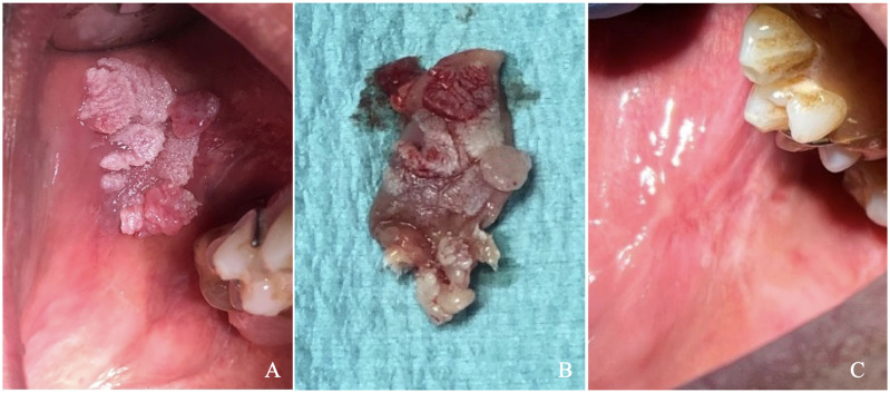
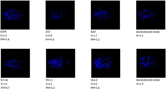
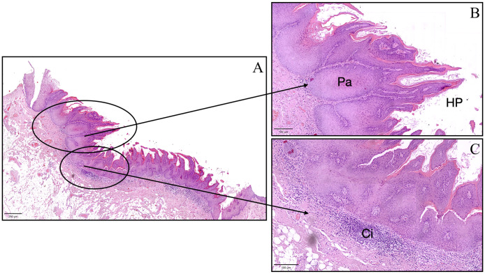
Oral leukoplakia is classified among oral potentially malignant disorders (OPMDs) by the World Health Organization (WHO). The visual oral examination (VOE) is the most used method for identifying lesions in their early stages. Given that the diagnosis of oral cancer is often late, there is an urgent need for early detection and examination of oral lesions. Surgical biopsy represents the gold standard as a diagnostic method, but because it is invasive, it cannot be repeated for periodic checks. We report the case of a lesion on the buccal mucosa of a 65-year-old male patient with a malignant appearance. The patient underwent a novel non-invasive cyto-salivary sampling and ELISA immunoassay for tumor biomarker detection and biopsy with histopathological analysis. The rapid ELISA test results excluded signs of malignancy, providing valuable insights into the lesion's immunophenotypic profile, which were consistent with the histopathological examination findings. This case report highlights the clinical and histopathological characteristics of a lesion with the aspect of Proliferative Verrucous Leukoplakia (PVL), emphasizing its challenging diagnosis and management. The integration of non-invasive cytobrush sampling with biomarker analysis proved valuable in detecting specific tumor biomarkers, potentially indicating ongoing tumor transformation. Monitoring these markers over time could enhance early detection and management strategies, thereby improving patient outcomes. This approach underscores the utility of non-invasive techniques in phenotyping oral lesions and supporting clinical decision-making in oral medicine.
Keywords: biomarker analysis; cytobrush sampling; histopathology; immune checkpoints; oral potentially malignant disorders.
Copyright © 2024 Rebaudi, Rebaudi, De Rosa, Rebaudi, Pesce, Greppi, Roghi, Boggio, Candiani and Marcenaro.
Conflict of interest statement
The authors declare that the research was conducted in the absence of any commercial or financial relationships that could be construed as a potential conflict of interest. The author(s) declared that they were an editorial board member of Frontiers, at the time of submission. This had no impact on the peer review process and the final decision.
Figures
References
-
- Warnakulasuriya S, Kujan O, Aguirre-Urizar JM, Bagan JV, González-Moles MÁ, Kerr AR, et al. Oral potentially Malignant disorders: A consensus report from an international seminar on nomenclature and classification, convened by the WHO Collaborating Centre for Oral Cancer. Oral Dis. (2021) 27:1862–80. doi: 10.1111/odi.13704 - DOI - PubMed
-
- Campisi G, Giovannelli L, Ammatuna P, Capra G, Colella G, Di Liberto C, et al. Proliferative verrucous vs conventional leukoplakia: no significantly increased risk of HPV infection. Oral Oncol. (2004) 40:835–40. https://linkinghub.elsevier.com/retrieve/pii/S1368837504000673. - PubMed
-
- Upadhyaya JD, Fitzpatrick SG, Cohen DM, Bilodeau EA, Bhattacharyya I, Lewis JS, et al. Inter-observer variability in the diagnosis of proliferative Verrucous leukoplakia: clinical implications for oral and maxillofacial surgeon understanding: A collaborative pilot study. Head Neck Pathol. (2019) 14:156–65. https://europepmc.org/articles/PMC7021885. - PMC - PubMed
Publication types
MeSH terms
Substances
LinkOut - more resources
Full Text Sources
Medical