. 2024 Dec 12;3(12):e70025.
doi: 10.1002/jex2.70025. eCollection 2024 Dec.

Characterization of Spirulina-derived extracellular vesicles and their potential as a vaccine adjuvant

Affiliations

Characterization of Spirulina-derived extracellular vesicles and their potential as a vaccine adjuvant

Mohammad Farouq Sharifpour et al. J Extracell Biol. .

Abstract

Spirulina is an edible cyanobacterium that increasingly gaining recognition for it untapped potential in the biomanufacturing of pharmaceuticals. Despite the rapidly accumulating information on extracellular vesicles (EVs) from most other bacteria, nothing is known about Spirulina extracellular vesicles (SPEVs). This study reports the successful isolation, characterization and visualization of SPEVs for the first time and it further investigates the potential therapeutic benefits of SPEVs using a mouse model. SPEVs were isolated using ultracentrifugation and size-exclusion-chromatography. Cryo-Transmission Electron Microscopy revealed pleomorphic outer-membrane-vesicles and outer-inner-membrane-vesicles displaying diverse shapes, sizes and corona densities. To assess short- and long-term immune responses, mice were injected intraperitoneally with SPEVs, which demonstrated a significant increase in neutrophils and M1 macrophages at the injection site, indicating a pro-inflammatory effect induced by SPEVs without clinical signs of toxicity or hypersensitivity. Furthermore, SPEVs demonstrated potent adjuvanticity by enhancing antigen-specific IgG responses in mice by over 100-fold compared to an unadjuvanted model vaccine antigen. Mass-spectrometry identified 54 proteins within SPEVs, including three protein superfamily members linked to the observed pro-inflammatory effects. Our findings highlight the potential of SPEVs as a new class of vaccine adjuvant and warrant additional studies to further characterize the nature of the immune response.

Keywords: adjuvant; extracellular vesicle; immune response; spirulina; vaccine.


Conflict of interest statement

The authors declare no competing interests.

Figures

FIGURE 1
FIGURE 1
Culture and axenisation of Spirulina organism, and extracellular vesicle isolation. (a) The original xenic culture of the Spirulina stock was axenised by sonication and cultivating on agar medium in the presence of kanamycin. The axenic culture was then propagated in Zarrouk's medium in photobioreactors. (b) When Spirulina reached the lag phase in growth, the cell suspension went through a series of low‐velocity centrifugation steps; the supernatant was collected and passed through several filters and concentrated with 100 kDa cut‐off filters. Ultracentrifugation precipitated EVs in a pellet. The EV pellet was resuspended in water (or PBS) and ultracentrifuged several times in order to remove any soluble proteins. Fractionation was then performed by size exclusion chromatography. EV, extracellular vesicle.
FIGURE 2
FIGURE 2
Cryogenic transmission electron micrographs of Spirulina extracellular vesicles (a) and (b). Wide‐field CryoTEM images (6000 × magnification) showcase SPEVs, specifically OMVs enclosed with a single bilayer phospholipid membrane, along with OIMVs indicated by black arrows, featuring a distinctive double bilayer phospholipid membrane (c—j). Narrow‐field CryoTEM images (60,000 × magnification) provide detailed views of SPEVs, revealing a range of sizes, predominantly comprising OMVs and a smaller fraction of OIMVs (black arrows). The white arrows indicate non‐spherical EVs. EV, extracellular vesicle; OMIV, outer‐inner membrane vesicles; OMV, Outer membrane vesicles; SPEV, Spirulina EVs.
FIGURE 3
FIGURE 3
Size distribution and concentration of the isolated SPEV using two different technologies (a) Unfractionated SPEV suspension versus fractionated EVs (fractions 1–7) (b) Results for size distribution and concentration of SPEVs using NTA technology (c) Results for size distribution and concentration of SPEVs using TRPS technology. EV, extracellular vesicle; NTA, nanoparticle tracking analysis; SPEV, Spirulina EVs; TRPS, tuneable resistive pulse sensing.
FIGURE 4
FIGURE 4
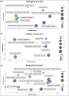
Sequences identified from SPEVs were analysed and annotated with Blast2GO to identify GO terms and quantify node scores. Results were summarised and visualised with ReviGO. Semantically similar GO terms will cluster more closely. The colour value indicates the Blast2GO node score. Circle size represents the Log10 number of annotations for a GO term in the entire Uniprot‐to‐GO database. EV, extracellular vesicle; GO, gene ontology; SPEV, Spirulina EVs.
FIGURE 5
FIGURE 5
Sequences identified from SPEVs were analysed with InterPro to identify and quantify Pfam features. The top seven most common features across all sequences are displayed in the columns on the left, while the number of unique sequences with the corresponding Pfam feature is displayed on the right. SPEV, Spirulina EVs.
FIGURE 6
FIGURE 6
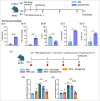
Spirulina EVs activate pro‐inflammatory cells and enhance anti‐TSP‐2 IgG production. Experimental design for short‐term (a) and long‐term immune response in mice (b). There was persistent neutrophilia (Live CD11b+ Ly6G+) (a.i and b.i), early higher M1 macrophage (Live CD11b+ F4/80+ CD11c+) (a.ii) and lower cDC1 (Live CD11c+ CD103+) (a.iii) infiltration and IFNγ+ Th1 cells (of Live CD3+ CD4+ CD44+) (b.ii) locally in the peritoneal cavity. Injection schedule for adjuvanticity experiment in BALB/c mice (c). TSP2 antigen‐specific total IgG (d) and IgG2a (e) response at Day 40. Error bars represent standard errors of the mean. Statistical analysis by T‐test, *p < 0.05, **p < 0.01, ***p < 0.001, ****p < 0.0001. EV, extracellular vesicle; TSP‐2, tetraspanin‐2.

Similar articles

References

    1. Adamo, G. , Fierli, D. , Romancino, D. P. , Picciotto, S. , Barone, M. E. , Aranyos, A. , Božič, D. , Morsbach, S. , Raccosta, S. , Stanly, C. , Paganini, C. , Gai, M. , Cusimano, A. , Martorana, V. , Noto, R. , Carrotta, R. , Librizzi, F. , Randazzo, L. , Parkes, R. , … Bongiovanni, A. (2021). Nanoalgosomes: Introducing extracellular vesicles produced by microalgae. Journal of Extracellular Vesicles, 10(6), e12081. 10.1002/jev2.12081 - DOI - PMC - PubMed
    1. Ausiello, C. M. , Cerquetti, M. , Fedele, G. , Spensieri, F. , Palazzo, R. , Nasso, M. , Frezza, S. , & Mastrantonio, P. (2006). Surface layer proteins from Clostridium difficile induce inflammatory and regulatory cytokines in human monocytes and dendritic cells. Microbes and Infection, 8(11), 2640–2646. 10.1016/j.micinf.2006.07.009 - DOI - PubMed
    1. Baumann, U. (2019). Structure‐function relationships of the repeat domains of RTX toxins. Toxins, 11(11), 657. 10.3390/toxins11110657 - DOI - PMC - PubMed
    1. Bharat, T. A. M. , von Kügelgen, A. , & Alva, V. (2021). Molecular logic of prokaryotic surface layer structures. Trends in Microbiology, 29, 405–415. - PMC - PubMed
    1. Biller, S. J. , Muñoz‐Marín, M. D. C. , Lima, S. , Matinha‐Cardoso, J. , Tamagnini, P. , & Oliveira, P. (2022). Isolation and characterization of cyanobacterial extracellular vesicles. Journal of Visualized Experiments : JoVE, 00(180), 00–00. 10.3791/63481 - DOI - PubMed

LinkOut - more resources