Case Reports
. 2024 Nov 14;16(11):e73656.
doi: 10.7759/cureus.73656. eCollection 2024 Nov.

A Salivary Clear Cell Tumor With an Unclear Diagnosis: A Report of a Rare Case

Affiliations
Case Reports

A Salivary Clear Cell Tumor With an Unclear Diagnosis: A Report of a Rare Case

Svyat Strokov et al. Cureus. .

Abstract

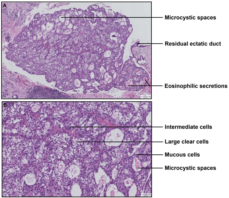
The histopathological diagnosis of salivary tumors is considered complex, due to their histological, phenotypic, and genotypic diversity. There are numerous tumors with morphological and/or immunohistochemical aspects that are superimposable but require very different treatment. In this context, salivary lesions containing clear cells are numerous and form part of the diagnostic challenges. We present the case of a 63-year-old woman with a tumor of the accessory salivary glands of the palate, with a predominant clear cell contingent. The aim of this paper is to report a case of a clear cell salivary tumor and to detail the difficulties of differential diagnosis, highlighting new data in the literature and the role of molecular biology.

Keywords: clear cells; malignant salivary gland tumor; minor salivary gland; oral cavity; salivary gland tumor.


Conflict of interest statement

Human subjects: Consent for treatment and open access publication was obtained or waived by all participants in this study. Conflicts of interest: In compliance with the ICMJE uniform disclosure form, all authors declare the following: Payment/services info: All authors have declared that no financial support was received from any organization for the submitted work. Financial relationships: All authors have declared that they have no financial relationships at present or within the previous three years with any organizations that might have an interest in the submitted work. Other relationships: All authors have declared that there are no other relationships or activities that could appear to have influenced the submitted work.

Figures

Figure 1
Figure 1. Clinical photograph showing a bluish nodule 5-6 mm in diameter, sessile, located on the right side of the hard palate
Figure 2
Figure 2. Histological section showing an intra-cystic tumoral proliferation
(A) Microphotograph (hematoxylin-eosin stain, original magnification ×175) showing a tumor proliferation hollowed out by glandular microcystic spaces and bordered by clear cells, a few intermediate cells, and very rare mucus cells (legends). (B) Microphotograph (hematoxylin-eosin stain, original magnification ×60) showing in situ tumor proliferation developed at the expense of an ectatic salivary duct.
Figure 3
Figure 3. Histological section showing the three cellular contingents of the tumor
(A) Microphotograph (hematoxylin-eosin stain, original magnification ×60) showing a tumoral nodule with microcystic spaces, sometimes filled by an eosinophilic pseudocolloidal secretory substance. (B) Microphotograph (hematoxylin-eosin stain, original magnification ×150) showing the morphology of the cellular contingents with a majority population of cells with large clear cytoplasm, intermediate cells with amphophilic cytoplasm and rounded nuclei, and very rare mucous cells.
Figure 4
Figure 4. Positive immunohistochemical markers
(A) CK7 (original magnification ×60): homogeneous and diffuse cytoplasmic positivity within the tumor, suggestive of salivary differentiation. (B) p63 (original magnification ×60): heterogeneous nuclear positivity within the tumor. (C) p63 (original magnification ×140): heterogeneous nuclear positivity, rather peripheral but indicative of basal differentiation.
Figure 5
Figure 5. Negative immunohistochemical staining
(A) Antibody anti-SOX-10, negative staining (original magnification ×60), indicating the absence of pure acinar differentiation. (B) Antibody anti-S-100 protein, negative staining (original magnification ×60), indicating the absence of myoepithelial differentiation and ruling out secretory carcinoma. (C) Antibody anti-smooth muscle actin, negative staining (original magnification ×60), indicating the absence of myoepithelial differentiation and ruling out biphasic or pure myoepithelial tumors (epithelial-myoepithelial carcinoma, myoepithelial carcinoma, etc.). (D) Antibody anti-CD10, negative staining (original magnification ×60), an argument to rule out metastasis of clear cell renal cell carcinoma.

References

    1. Bishop JA, Thompson LD, Wakely PE, Weinreb I. Arlington (VA): American Registry of Pathology; 2021. Tumors of the salivary glands.
    1. Epidemiology of primary epithelial salivary gland tumors in Southern Poland-a 26-year, clinicopathologic, retrospective analysis. Gontarz M, Bargiel J, Gąsiorowski K, Marecik T, Szczurowski P, Zapała J, Wyszyńska-Pawelec G. J Clin Med. 2021;10:1663. - PMC - PubMed
    1. Head and neck tumours: WHO classification of tumours. Geneva, Switzerland: World Health Organization; 2024.
    1. Distribution and frequency of salivary gland tumours: an international multicenter study. Alsanie I, Rajab S, Cottom H, et al. Head Neck Pathol. 2022;16:1043–1054. - PMC - PubMed
    1. The evolving role of molecular pathology in the diagnosis of salivary gland tumours with potential pitfalls. Kaur K, Mehta S, Vanik S, et al. Eur Arch Otorhinolaryngol. 2022;279:3769–3783. - PubMed

Publication types

LinkOut - more resources