

This is a preprint.
Genomic Landscape of Thrombosis Recurrence Risk Across Venous Thromboembolism Subtypes
- PMID: 39677447
- PMCID: PMC11643180
- DOI: 10.1101/2024.12.02.24317788
Genomic Landscape of Thrombosis Recurrence Risk Across Venous Thromboembolism Subtypes
Update in
-
Molecular Determinants of Thrombosis Recurrence Risk Across Venous Thromboembolism Subtypes.Blood. 2025 Aug 14:blood.2024027879. doi: 10.1182/blood.2024027879. Online ahead of print. Blood. 2025. PMID: 40811855
Abstract
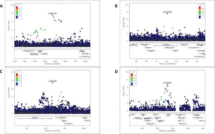
Venous thromboembolism (VT) is a frequent (annual incidence of 1 to 2 per 1,000) and potentially life-threatening (case-fatality rate up to 10%) disease. VT is associated with serious short-term and long-term complications including a recurrence rate of approximately 20% within five years. Anticoagulant therapy, the mainstay of VT treatment, drastically reduces the risk of early VT recurrence, but it exposes patients to a substantial risk of bleeding. We analysed the genomic architecture of VT recurrence using data from 6,571 patients across eight cohorts, 1,816 of whom experienced recurrence, with a particular focus on the clinical manifestation of the type of first VT event. Through genome-wide association studies (GWAS), we identified three loci significantly associated (P<5×10⁻⁸) with VT recurrence in the general VT population: GPR149/MME, L3MBTL4, and THSD7B. Protein Quantitative Trait Locus and Mendelian Randomization analyses further identified elevated plasma levels of coagulation factor XI and GOLM2 as risk factors for recurrence, while decreased levels of PCSK9 and pro-IL16 were linked to reduced VT recurrence risk. Subgroup analyses revealed 18 loci associated with VT recurrence, with notable differences between pulmonary embolism (PE) and deep vein thrombosis (DVT). For example, the exonic variant SLC4A1 p.Glu40Lys was significantly associated with recurrence in PE patients (Hazard Ratio (HR)=3.23, P=9.7×10⁻¹²) but showed no effect in DVT (HR=1.00, P=0.98). These findings emphasize the role of specific genetic loci and protein pathways in influencing VT recurrence and provide valuable insights into potential therapeutic targets. Further research is needed to clarify the biological mechanisms driving these associations.
Keywords: genetics; genome-wide association study; meta-analysis; venous thromboembolism recurrence.
Conflict of interest statement
Disclosures The authors have no conflict of interest to declare.
Figures


References
-
- Raskob GE, Angchaisuksiri P, Blanco AN, et al. Thrombosis: A Major Contributor to Global Disease Burden. ATVB. 2014;34(11):2363–2371. - PubMed
-
- Cohen AT, Agnelli G, Anderson FA, et al. Venous thromboembolism (VTE) in Europe. The number of VTE events and associated morbidity and mortality. Thromb Haemost. 2007;98(4):756–764. - PubMed
-
- Stevens SM, Woller SC, Kreuziger LB, et al. Antithrombotic Therapy for VTE Disease: Second Update of the CHEST Guideline and Expert Panel Report. Chest. 2021;160(6):e545–e608. - PubMed
-
- Delluc A, Tromeur C, Le Ven F, et al. Current incidence of venous thromboembolism and comparison with 1998: a community-based study in Western France. Thromb Haemost. 2016;116(11):967–974. - PubMed
-
- Couturaud F, Sanchez O, Pernod G, et al. Six Months vs Extended Oral Anticoagulation After a First Episode of Pulmonary Embolism: The PADIS-PE Randomized Clinical Trial. JAMA. 2015;314(1):31. - PubMed
Publication types
Grants and funding
LinkOut - more resources
Full Text Sources
Research Materials
Miscellaneous