[Preprint]. 2024 Dec 5:2024.12.05.627040.
doi: 10.1101/2024.12.05.627040.

Genetic Analysis of Flagellar-Mediated Surface Sensing by Pseudomonas aeruginosa PA14

Affiliations

Genetic Analysis of Flagellar-Mediated Surface Sensing by Pseudomonas aeruginosa PA14

Sherry Kuchma et al. bioRxiv. .

Update in

Abstract

Surface sensing is a key aspect of the early stage of biofilm formation. For P. aeruginosa, the type IV pili (TFP), the TFP alignment complex and PilY1 were shown to play a key role in c-di-GMP signaling upon surface contact. The role of the flagellar machinery in surface sensing is less well understood in P. aeruginosa. Here we show, consistent with findings from other groups, that a mutation in the gene encoding the flagellar hook protein (ΔflgK) or flagellin (ΔfliC) results in a strain that overproduces the Pel exopolysaccharide (EPS) with a concomitant increase in c-di-GMP levels. We use a candidate gene approach and genetic screens, combined with phenotypic assays, to identify key roles for the MotAB and MotCD stators and the FliG protein, a component of the flagellar switch complex, in stimulating the surface-dependent, increased c-di-GMP level noted for these flagellar mutants. These findings are consistent with previous studies showing a role for the stators in surface sensing. We also show that mutations in the genes coding for the diguanylate cyclases SadC and RoeA as well as SadB, a protein involved in early surface colonization, abrogate the increased c-d-GMP-related phenotypes of the ΔflgK mutant. Together, these data indicate that bacteria monitor the status of flagellar synthesis and/or function during surface sensing as a means to trigger the biofilm program.

Keywords: Pseudomonas aeruginosa; biofilm; flagella; stators; surface sensing.


Figures

Figure 1.
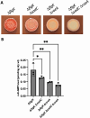
Figure 1.. Mutations that eliminate stator production or impact stator occupancy suppress the ΔflgK mutant hyper-signaling phenotype.
A and D. Representative CR images of the indicated strains cultured on M8 medium solidified with 1% agar for 16 h at 37° C followed by 72 hrs at room temperature. B. Quantification of c-di-GMP levels in the indicated strains grown either in M8 liquid or on M8 1% agar plates for extraction of nucleotides and measurement of c-di-GMP levels via mass spectrometry. C and E. Quantification of c-di-GMP levels in the indicated strains grown on M8 agar swarm plates for 16 h prior to harvest for extraction of nucleotides and measurement of c-di-GMP levels via mass spectrometry. Experiments (B, C and E) were performed in triplicate with three technical replicates per strain and analyzed by ANOVA with Dunnett’s multiple comparisons test. Significant differences are shown for comparisons to the ΔflgK strain. ns, non-significant difference; * P < 0.05 and ** P < 0.01.
Figure 2.
Figure 2.. Mutations that impact proton binding suppress hyper-signaling.
A. Top panel shows representative CR images of the indicated strains. The proton-binding aspartate residue MotB-D30, analogous to the E. coli MotB-D32, allele is mutated to alanine in the ΔflgK deletion strain. Middle panel shows the Western blot for the MotB-His6 WT and D30A variant epitope-tagged proteins detected in lysate samples from surface-grown strains using an anti-His antibody (α-His). MotB-His6 protein levels were quantified and normalized to a cross-reacting band (bottom panel, ctrl) detected in all samples and used as a loading control. Numbers below the middle panel show the mean and standard deviation (SD), in parentheses, from three independent experiments, normalized to the WT, which is set to 1.0. Statistical analysis was performed using ANOVA with Dunnett’s test for multiple comparisons. Significant differences are shown for comparisons to the ΔflgK motB+-His6 strain, with * P < 0.05 and **** P < 0.0001. B. Top panel shows representative CR images of the indicated strains. The proton-binding aspartate residue of MotD-D23 is mutated to alanine in the ΔflgK deletion strain. Middle panel shows the MotD WT and D23A His6-epitope tagged proteins detected and quantified as described in panel A. Significant differences are shown for comparisons to the ΔflgK motD+-His6 strain. *, P < 0.05; *** P < 0.0005. C. Quantification of c-di-GMP for the indicated strains grown on M8 swarm plates for 16 h. Experiments were performed in triplicate with three technical replicates per strain and analyzed by ANOVA with Tukey’s post-test comparison. Significant differences are shown for comparisons either to the ΔflgK motB+::His strain or to the ΔflgK motD+::His strain as indicated. ns, non-significant difference; significant differences noted as follows: ***, P < 0.001; ****, P < 0.0001.
Figure 3.
Figure 3.. The DGCs SadC and RoeA are required for hyper signaling in the flgK mutant.
A. Representative CR images of the indicated strains. B. Quantification of c-di-GMP extracted from swarm-grown strains. Experiments were performed in triplicate with three technical replicates per strain and analyzed by ANOVA with Dunnett’s post-test comparison. Significant differences are shown for comparisons to the ΔflgK mutant; *P < 0.05, **P < 0.005.
Figure 4.
Figure 4.. Testing candidate genes for their impact on the ΔflgK mutant hyper-signaling phenotype.
A. Representative CR plate images of the ΔfliL and ΔflhF mutations in the WT and the ΔflgK mutant background. B. Impact of mutations in the stators on the ΔfliF mutant phenotypes. C. Representative CR plate images of mutations the fliG and fliMN genes. D. Impact of mutations in the fimV, fimW, and sadB genes in the WT and ΔflgK mutant backgrounds on the CR plate phenotype. All CR assays were performed on M8 medium solidified with 1% agar for 16 h at 37° C followed by 72 hrs at room temperature.

References

    1. Henrici AT. 1933. Studies of freshwater bacteria: I. A direct microscopic technique. J Bacteriol 25:277–287. - PMC - PubMed
    1. Sharma S, Mohler J, Mahajan SD, Schwartz SA, Bruggemann L, Aalinkeel R. 2023. Microbial biofilm: a review on formation, infection, antibiotic resistance, control measures, and innovative treatment. Microorganisms 11:1614. - PMC - PubMed
    1. Zobell CE, Allen EC. 1935. The significance of marine bacteria in the fouling of submerged surfaces. J Bacteriol 29:239–251. - PMC - PubMed
    1. Belas R. 2014. Biofilms, flagella, and mechanosensing of surfaces by bacteria. Trends Microbiol 22:517–527. - PubMed
    1. Chawla R, Gupta R, Lele TP, Lele PP. 2020. A skeptic’s guide to bacterial mechanosensing. J Mol Biol 432:523–533. - PMC - PubMed

Publication types

LinkOut - more resources